Editorial: Mechanisms of ATP release – future therapeutic targets?
When Ferguson et al. [1] demonstrated ATP release from the rabbit bladder and concluded: ‘… ATP is released from the urothelium as a sensory mediator … ’, they opened a new field of research with focus on urothelial signaling mechanisms and afferent nerve functions in bladder control. Other investigators have shown, in several animal models, that ATP is released from urothelial cells during distention of the bladder and that the amount released is proportional to the extent of distention [2]. P2X3 purinergic receptors are present in the urothelium and specifically on suburothelial afferent nerve fibres. After release, ATP acts on these receptors to convey information to the CNS, where voiding can be initiated. P2X3 receptor knockout mice had marked urinary bladder hyporeflexia with reduced voiding frequency and increased voiding volume, suggesting that these receptors are involved in mechanosensory transduction underlying activation of afferent fibres that control voiding reflexes during bladder filling [3]. In the last decade the proposal of Ferguson et al. [1] has been well supported [4], making ATP release an essential step in the activation of the bladder.
Although release of ATP from bladder tissues has been studied extensively, there are still many unanswered questions. In a recent study, McLatchie and Fry [5] have used unique experimental approaches that allowed them to study some essential questions in a new way: i) from which urothelial cells is ATP released, ii) how is ATP stored, and iii) what release pathways are involved?
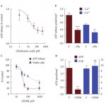
Previous studies have established that ATP comes from the urothelial cell layer, although they have not identified the actual cell type responsible. Using freshly isolated cells that could be separated into umbrella, intermediate and basal subtypes, McLatchie and Fry [5]showed that umbrella and basal/intermediate cells are equally effective in generating ATP release. The magnitude of ATP release from the urothelium was large compared with that from multicellular preparations.
ATP has for many years been known as a postjunctional contraction-producing transmitter stored in vesicles of cholinergic nerves [4], but whether the release from urothelial cells is vesicular or not has been unclear. Ferguson et al. [1] presented three types of argument against non-vesicular ATP release: i) rather than inhibiting ATP release, absence of calcium in the bathing medium actually potentiated the release, ii) tetrodotoxin in concentrations completely blocking field-stimulated smooth muscle contraction had no significant effect on electrically induced ATP, and iii) although the suburothelial sensory nerves are packed with secretory granules, there are no such granules to be seen within the urothelial cells. McLatchie and Fry [5] stimulated urothelial cells in suspension by imposing upon them a mild drag force stress and found that urothelial ATP release was reduced with 1.8 mm external calcium, and was increased approximately two-fold by increasing intracellular calcium. ATP release was reduced by agents blocking pannexin and connexin hemichannels. The calcium-dependence of ATP release and its influence by connexin/pannexin blockers suggested to the investigators that a major fraction (up to 50%) of release is through such channels. However, the conspicuous effect of N-ethylmaleimide, which has been proposed to reduce vesicular docking to the surface membrane of secretory cells, is consistent with a substantial fraction of release by vesicular exocytosis.
It is obvious that more than 15 years after the observation of urothelial ATP release, this remains a fruitful research field. As suggested by McLatchie and Fry [5], characterisation of the pathways involved may help to develop new therapeutics for disorders assumed to be characterised by increased ATP release, such as bladder pain and overactive bladder syndromes.