Tag Archive for: Article of the Month

Posts

Editorial: VEIL – is a new standard ready to be accepted?

In this interesting prospective study, Kumar and Sethia [1] provide further evidence that video endoscopic inguinal lymphadenectomy (VEIL) could be considered the procedure of choice for most patients with an indication for ILND. In their comparison with historical patients submitted to open ILND (OILND) in a reference centre in the UK, VEIL achieved reduced morbidity, shorter hospital stay, and equivalent oncological control. Also, we must consider that some cases of the VEIL learning curve were included in this study, suggesting that good results can be obtained with the endoscopic technique from its inception.

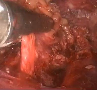
The idea for an endoscopic approach to ILND was conceived and developed in a cadaveric model by Bishoff et al. [2] in 2003. In 2005, our group reported the first successful experience in humans [3]. In 2006, in collaboration with the Brazilian National Institute of Cancer, we published a landmark study in the Journal of Urology comparing OILND in one limb and VEIL in the contralateral limb in the same patient to minimise comparative bias [4]. In all, 10 patients with impalpable nodes underwent bilateral ILND. We found that morbidity was reduced with VEIL (20% vs 70%) with the removal of the same number of nodes as with OILND [4]. In 2008, we reported on a series of bilateral VEIL obtaining reduced postoperative morbidity and a mean hospital stay of 1 day [5]. Several other small series have reported essentially the same results that we initially described.

Robot-assisted VEIL duplicates the procedure but is more ergonomic, with excellent freedom of movement and amplification of lymphatic visualisation. Matin et al. [6] reported that robot-assisted VEIL could remove the same number of nodes as the open procedure.

The present study [1] reports a large experience comparing bilateral procedures (VEIL and OILND). The follow-up data for OILND are longer but oncological indicators suggest equivalence. This is also the first report of VEIL after node cytology and dynamic sentinel node biopsy with no related adverse events.

To date, a few series have reported long-term oncological results [1, 7] and future studies will be important to evaluate and reproduce the oncological efficacy of VEIL.

We congratulate Kumar and Sethia [1] for these significant data to establish VEIL in the modern urological armamentarium. Indeed, I really have some doubt as whether a randomised controlled study would be necessary considering the accumulated worldwide data on VEIL in the last 10 years.

This year we present at the University of California meeting the promising results of our Latin America collaborative group with 150 VEIL in 110 patients. The mean (range) follow up was 6 (2–10) years, with an overall morbidity of 30% (Clavien–Dindo grade III–IV, 2%), mean lymph node removal of 8 nodes/groin, node positive disease rate of 25%, inguinal recurrence rate of 2%, cancer-specific survival of 90%, and overall survival of 85% (Tobias-Machado et al., 2016, unpublished data). Our data suggest that we can obtain a dramatic reduction in severe complications and also that VEIL is surviving the test of time.

In my opinion, as the reduction in morbidity is already confirmed, we only need larger series with long-term reports of oncological equivalence with OILND to designate VEIL as a ‘gold standard’ in centres with appropriately trained surgeons.

I speculate that in the near future the utilisation of robotic surgery associated with improvements in imaging methods and contrasts, which will improve the identification of lymph nodes to be removed and lymphatic channels to be clipped, will achieve further reductions in morbidity and optimisation of oncological control.

How to Cite this article:

Tobias-Machado, M. (2017), Video endoscopic inguinal lymphadenectomy (VEIL): is a new standard ready to be accepted?. BJU International, 119: 504–505. doi: 10.1111/bju.13723

Marcos Tobias-Machado
Department of Urology, ABC Medical School, Sao PauloBrazil


References


2 Bishoff JT, Basler JW, Teichman JM et al. Endoscopic subcutaneous modied inguinal lymph node dissection (ESMIL) for squamous cell carcinoma of the penis. J Urol (Suppl.) 2003; 169: 78 (abstract 301)


3 Tobias-Machado M, Tavares A, Molina WR Jr, Forseto PH Jr, Juliano RV, Wroclawski ER. Video endoscopic inguinal lymphadenectomy (VEIL): minimally invasive resection of inguinal lymph nodes. Int Braz Urol 2006; 32: 31621


4 Tobias-Machado M, Tavares A, Ornellas AA, Molina WR Jr, Juliano RVWroclawski ER. Video endoscopic inguinal lymphadenectomy: a new minimally invasive procedure for radical management of inguinal nodes in


7 SchwentneC, Todenhofer T, Seibold J e t al. Endoscopic inguinofemoral lymphadenectomy extended follow-up. JEndouro20 13; 27: 497503


Editorial: Is minimally invasive inguinal node dissection the way forward?

In this issue of BJUI, Kumar and Sethia report their experience of laparoscopic inguinal node dissection for patients with penile cancer [1]. The authors should be congratulated for what is a technically and feasibly difficult study to complete.

The step-wise spread of penile carcinoma first to the inguinal lymph nodes is well known. It is also recognized that accurate staging and treatment of inguinal metastases play a crucial part in a patient’s management pathway. Furthermore, evidence points to a survival advantage for those who undergo prophylactic inguinal lymph node dissection (ILND) and have positive nodes, compared with those treated with surveillance and delayed dissection when nodes become palpable or radiologically positive.

The overall postoperative complication rate after ILND has been reported to be nearly 50% or even up to 75%. Patients with comorbidities or locally advanced disease are at particular risk of postoperative morbidity [2]. High rates of wound dehiscence and skin necrosis after ILND have also been reported [3]. More recently, complication rates among men with no palpable adenopathy have been reported to be decreasing in comparison with historical experience, but overall complication rates still range from 10 to 40%.

Adoption of dynamic sentinel node biopsy has reduced the absolute number of prophylactic ILNDs, but in any high-volume unit (as is the case in the UK supra-network centres), a large number of inguinal node dissections will still be undertaken [4].

For these reasons it is desirable to minimize or modify the inguinal wound to reduce the complication rate, and the use of minimally invasive techniques has gradually progressed over the last 10 years.

The first case series reporting the use of laparoscopic node dissection (videoendoscopic inguinal node dissection [VEILND]) described 10 patients who underwent bilateral ILND using standard lymphadenectomy on one side and the VEILND technique on the contralateral side [5]. A more recent development has been the incorporation of robotic assistance as an enabling tool for performing ILND [6].

In these small series the technique appeared to be safe and oncologically comparable to open surgery, but the numbers were too small to give a meaningful assessment of complications. It is therefore welcome to read a paper on a high-volume unit’s experience of VEILND.

The authors describe their experience of VEILND on a sequential prospective group of patients and compared them with a historical control cohort who underwent open inguinal node dissection. The data appear to show a significant difference in length of stay and wound-related comorbidity in favour of the minimally invasive approach without compromising oncological outcome. Interestingly, the rate of lower limb lymphedema was also significantly reduced in the VEILND group.

The headline data are impressive but the comparison does have a number of potential weaknesses. Firstly, the rate of wound-related complications is quite high in the open group, with 23% of patients needing re-exploration or debridement and 37% some form of drainage of collection. The choice of incision position, length and preparation of skin flaps can influence the rate of wound breakdown and length of stay.

Secondly, the headline 6% complication rate for VEILND does not include the 27% lymphocoele rate which is still a troublesome complication in both the open and endoscopic groups.

Finally, there is no mention of the limitations of the technique with size and extent of the malignant node burden. The paper states that all patients were included, and yet in any high-volume centre there must be patients with grossly enlarged and sometimes fungating or fixed nodes and I would have thought that VEILND would not be appropriate in these instances.

In conclusion, there is much to commend this paper and the direction of travel may well be towards minimally invasive techniques; however, as with all comparisons between open and keyhole surgery, the focus should be on improving the open technique to its limit. Only then will the results be meaningful and adopted with confidence.

How to Cite this article:

Watkin, N. (2017), Is minimally invasive inguinal node dissection the way forward?. BJU International, 119: 505–506. doi: 10.1111/bju.13761

Nick Watkin
Department of Urology, St Geo rges Hospital, London, UK


References


Video: Comparing VEILND with OILND for Penile Cancer

Prospective study comparing video-endoscopic radical inguinal lymph node dissection (VEILND) with open radical ILND (OILND) for penile cancer over an 8-year period

Abstract

Objective

To compare the complications and oncological outcomes between video-endoscopic inguinal lymph node dissection (VEILND) and open ILND (OILND) in men with carcinoma of the penis.

Patients and methods

A prospectively collected institutional database was used to determine the outcomes in 42 consecutive patients undergoing ILND between 2008 and 2015 in a centre for treating penile cancer. Before 2013 all procedures were OILNDs. Since 2013 we have performed VEILND on all patients in need of ILND. The wound-related and non-wound-related complications, length of stay, and oncological safety between OILND and VEILND groups were compared. The mean duration of follow-up was 71 months for OILND and 16 months for the VEILND groups.

Results

In the study period 42 patients underwent 68 ILNDs (OILND 35, VEILND 33). The patients’ demographics, primary stage and grade, and indications were comparable in both groups. There were no intraoperative complications in either group. The wound complication rate was significantly lower in the VEILND group at 6% compared to 68% in the OILND group. Lymphocoele rates were similar in both the groups (27% and 20%). The VEILND group had a better or the same lymph node yield, mean number of positive lymph nodes, and lymph node density confirming oncological safety. There were no groin recurrences in either group of patients. VEILND significantly reduced the mean length of stay by 4.8 days (P < 0.001).

Conclusion

VEILND is an oncologically safe procedure with considerably low morbidity and reduced length of stay, at a mean (range) follow-up of 16 (4–35) months.

View more videos

Article of the Month: ProCare Trial

Every Month the Editor-in-Chief selects an Article of the Month from the current issue of BJUI. The abstract is reproduced below and you can click on the button to read the full article, which is freely available to all readers for at least 30 days from the time of this post.

In addition to the article itself, there is an accompanying editorial written by a prominent member of the urological community. This blog is intended to provoke comment and discussion and we invite you to use the comment tools at the bottom of each post to join the conversation.

If you only have time to read one article this week, it should be this one.

ProCare Trial: a phase II randomized controlled trial of shared care for follow-up of men with prostate cancer

Jon D. Emery*,,, Michael Jefford§,¶, Madeleine King**,††, Dickon Hayne‡‡,§§, Andrew Martin¶¶, Juanita Doorey, Amelia Hyatt, Emily Habgood*, Tee Lim***Cynthia Hawks‡‡,§§, Marie Pirotta*, Lyndal Trevena††† and Penelope Schoeld§,¶,‡‡‡


*Department of General Practice, University of Melbourne, Carlton, Western Health and the Victorian Comprehensive Cancer Centre, Melbourne, Vic., School of Primary Aboriginal and Rural Health Care, University of Western Australia, Crawley, WA, §Sir Peter MacCallum Department of Oncology, Faculty of Medicine, Dentistry and Health Sciences, The University of Melbourne, Parkville, Department of Cancer Experiences Research, Peter MacCallum Cancer Centre, East Melbourne, Vic., **Quality of Life Ofce, Psycho-oncology Co-operative Research Group, School of Psychology, University of Sydney, ††Sydney Medical School, University of Sydney, Sydney, NSW, ‡‡School of Surgery, University of Western Australia, Crawley,WA, §§Department of Urology, Fiona Stanley Hospital, Perth, WA, ¶¶NHMRC Clinical Trials Centre, University of Sydney, Sydney, NSW, ***Genesis Cancer Care, Department of Radiation Oncology, Fiona Stanley Hospital, Perth, WA, †††Primary Health Care, Sydney School of Public Health, University of Sydney, Sydney, NSW, and ‡‡‡Department of Psychology, Swinburne University of Technology, Melbourne, Vic., Australia


Read the full article

Abstract

Objectives

To test the feasibility and efficacy of a multifaceted model of shared care for men after completion of treatment for prostate cancer.

Patients and Methods

Men who had completed treatment for low- to moderate-risk prostate cancer within the previous 8 weeks were eligible. Participants were randomized to usual care or shared care. Shared care entailed substituting two hospital visits with three visits in primary care, a survivorship care plan, recall and reminders, and screening for distress and unmet needs. Outcome measures included psychological distress, prostate cancer-specific quality of life, satisfaction and preferences for care and healthcare resource use.

march-2017-aotm

Results

A total of 88 men were randomized (shared care n = 45; usual care n = 43). There were no clinically important or statistically significant differences between groups with regard to distress, prostate cancer-specific quality of life or satisfaction with care. At the end of the trial, men in the intervention group were significantly more likely to prefer a shared care model to hospital follow-up than those in the control group (intervention 63% vs control 24%; P<0.001). There was high compliance with prostate-specific antigen monitoring in both groups. The shared care model was cheaper than usual care (shared care AUS$1411; usual care AUS$1728; difference AUS$323 [plausible range AUS$91–554]).

Conclusion

Well-structured shared care for men with low- to moderate-risk prostate cancer is feasible and appears to produce clinically similar outcomes to those of standard care, at a lower cost.

Read more articles of the week

Editorial: Rethinking cancer surveillance with shared-care models and survivorship plans: the time is now!

Urologists are increasingly facing significant practice concerns related to timely access, surgeon availability, clinical throughput and rising cost of care, yet little has changed over the years regarding the routine postoperative surveillance of urological cancers. While urologists have appropriately focused evaluations on oncological outcomes and procedure-specific quality-of-life concerns, the ability to maintain this practice model in the setting of more new patients (and subsequently more cancer survivors) seems unrealistic. In addition, gaps exist with the current model related to timely and effective communication to the local care team and assurances that specialists comprehensively address all concerns raised by patients. Furthermore, the role of the local care team in cancer survivorship remains poorly defined. Recognising these and other unmet needs in cancer care survivorship, the American Cancer Society (ACS) and the American Society of Clinical Oncology (ASCO) recently published guidelines on cancer survivorship [1-3]. The guidelines recommend a standardised approach to follow-up with emphasis on quality, comprehensive patient assessments, value, and shared use of a multidisciplinary team. With prostate cancer survivorship, for instance, ASCO recommends PSA checks every 6–12 months for the first 5 years and then annually (higher-risk patients can have more frequent checks), adherence to ACS guidelines for early detection of prostate cancer, assessment of physical and psychological effects of prostate cancer and it’s treatments, and annual assessments for long-term or late side-effects [3]. To help with the coordination of care between the patient, the oncological specialist, and the local primary care provider, survivorship care plans have been developed. [4]. While use of survivorship care plans has been sparse in urology to date, new mandates will spur their use in the coming years and development will likely involve innovative healthcare delivery solutions.

Leading the way in this nascent field, Emery et al. [4] report, in this issue of BJUI, an innovative phase II prospective randomised study on the feasibility of a novel shared-care model for follow-up of patients with prostate cancer. Men who had completed treatment for low- and moderate-risk prostate cancer were randomised to undergo usual care or shared care with the assistance of the patient’s primary care team. The novel shared-care model substituted two postoperative urology visits with three postoperative visits in primary care, provided patients and primary care providers a survivorship care plan, included appointment reminders, and provided a novel mechanism to screen for distress and other unmet needs. Among the 88 men randomised in the prospective study, no significant differences were noted between delivery models for satisfaction of care, overall quality of life, incidence of distress, or compliance with serum PSA testing. Patients in the shared-care model were significantly more likely to prefer the new model compared to normal care (cases, 63% vs controls, 24%, P < 0.001). Importantly, the shared-care model was also more economical, saving 323 Australian dollars compared to usual care [4].

The authors should be congratulated for their well-designed study and early contribution to the field. Rethinking all aspects of care delivery will become increasingly important as the practice of urology responds to access limitations, the shortage of urologists, and financial pressures of value-based reimbursement. The report also engenders many questions about the ideal care model of the future, composition of the collaborative care team, and the importance of making evidence-based clinical recommendations. For instance, are already overburdened primary care providers ideal or realistic in shared-care models? Should care remain primarily under the control of urologist with assistance provided by other current (e.g. advance practice providers, urology nurses) or future team member roles (e.g. survivorship care coordinators)? What role can the patient alone play in a self-guided survivorship care plan under the watchful eye of the collaborative care team acting asynchronously? How can enabling technologies such as smartphones, mobile applications, wearables, and video-conferencing contribute to high-value cancer surveillance building upon the principles highlighted in the current article and further engaging patients in their cancer survivorship care? [5]. Lastly, what actually are the evidence-based imperatives of survivorship care (what risk groups, what testing intervals and duration of testing) that provide measurable value to the patient experience? In the current study [4], for instance, high risk patients were excluded but ultimately these patients may be best suited for comprehensive survivorship care. Future work on survivorship and care models will hopefully continue to advance ‘win-win’ situations where patients and providers alike experience increasingly high-value systems of healthcare delivery.

Read the full article
Matthew T. Gettman


Mayo Clinic Department of Urology, 200 First Street, SW, Rochester, MN 55905, USA


References


1 Mayer DK, Nekhlyudov L, Snyder CF, Merrill JK , Wollins DS, Shulman LN. American Society of Clinical Oncology clinical expert statement on cancer survivorship care planning. J Oncol Pract 2014; 10: 34551


2 Skolarus TA, Wolf AM, Erb NL et al. American Cancer Society prostate cancer survivorship care guidelines. CA Cancer J Clin 2014; 64: 22549


Article of the Month: 68Ga-PSMA PET/CT for LN staging in PCa

Every Month the Editor-in-Chief selects an Article of the Month from the current issue of BJUI. The abstract is reproduced below and you can click on the button to read the full article, which is freely available to all readers for at least 30 days from the time of this post.

In addition to the article itself, there is an accompanying editorial written by a prominent member of the urological community. This blog is intended to provoke comment and discussion and we invite you to use the comment tools at the bottom of each post to join the conversation.

If you only have time to read one article this week, it should be this one.

Prospective evaluation of 68Gallium-prostate-specific membrane antigen positron emission tomography/computed tomography for preoperative lymph node staging in prostate cancer

Pim J. van Leeuwen*, Louise Emmett,§, Bao Ho, Warick Delprado, Francis Ting*Quoc Nguyen† and Phillip D. Stricker*


*St Vincents Prostate Cancer Centre, St Vincents Clinic, Australian Prostate Cancer Research Centre, New South Wales, The Garvan Institute of Medical Research/The Kinghorn Cancer Centre, Department of Diagnostic Imaging, St Vincents Public Hospital, §University of New South Wales, Sydney, and University of Notre Dame, Darlinghurst, NSW, Australia


Read the full article

Abstract

Objectives

To assess the accuracy of 68Gallium-prostate-specific membrane antigen (68Ga-PSMA) positron emission tomography/computed tomography (PET/CT) for lymph node (LN) staging in intermediate- and high-risk prostate cancer (PCa).

Materials and Methods

From April to October 2015, 30 patients with intermediate- (n = 3) or high-risk (n = 27) PCa were prospectively enrolled. Patients underwent preoperative 68Ga-PSMA PET/CT. Both visual and semi-quantitative analyses were undertaken. Subsequently, all patients underwent radical prostatectomy (RP) with an extended pelvic lymph node dissection. The sensitivity, specificity, and positive (PPV) and negative predictive value (NPV) for LN status of 68Ga-PSMA were calculated using histopathology as reference.

aotmfeb2017-results

Results

Eleven patients (37%) had lymph node metastases (LNMs); 26 LNMs were identified in the 11 patients. Patient analysis showed that 68Ga-PSMA PET/CT had a sensitivity of 64% for the detection of LNMs, its specificity was 95%, the PPV was 88%, and the NPV was 82%. In total, 180 LN fields were analysed. In the LN-region-based analysis, the sensitivity of 68Ga-PSMA PET/CT for detection of LNMs was 56%, the specificity was 98%, the PPV was 90% and the NPV was 94%. The mean size of missed LNMs was 2.7 mm. Receiver-operating characteristic curve analysis showed a high accuracy of maximum standardized uptake value (SUVmax) for the detection of LNMs, with an area under the curve of 0.915 (95% confidence interval 0.847–0.983); the optimum SUVmax was 2.0.

Conclusions

In patients with intermediate- to high-risk PCa, 68Ga-PSMA PET/CT had a high specificity and a moderate sensitivity for LNM detection. 68Ga-PSMA PET/CT had the potential to replace current imaging for LN staging of patients with PCa scheduled for RP.

Read more articles of the week

info-feb-2017

Click on image for full infographic


Editorial: Bringing clarity or confusion? The role of prostate-specific membrane antigen positron-emission/computed tomography for primary staging in prostate cancer

The use of 68Ga-labelled prostate-specific membrane antigen (PSMA) positron-emission tomography (PET)/CT for staging prostate cancer in Australia has reached almost plague-like proportions. Despite what must be admitted is little high-level evidence to guide us in the accuracy or appropriateness of this imaging technique for either primary staging or prostate cancer recurrence, hundreds of these scans are being performed every week around Australia, and in many cases we simply do not know what to do with the results. We performed the first such scan at our centre in Melbourne in August 2014, and were soon receiving 10 requests per day, with patients waiting up to 3 months to be scanned. Fast-forward 2 years, and there are now eight centres offering PSMA PET/CT in Melbourne, a city of 4.5 million people. Scans can be obtained within 24 h of referral and costs have dropped to €500. A similar situation exists in Germany where this imaging method was pioneered [1], and interest is also growing in Belgium, Italy, India and a number of other countries (the USA being a notable exception). But do we really understand the impact of the decision to perform PSMA/PET scanning, and do we have enough evidence to guide us on the most appropriate setting for its use?

The current interest in PSMA PET/CT has been triggered by the development of small molecule ligands which bind to the extracellular domain of the PSMA molecule, leading to increased sensitivity and specificity when compared with conventional imaging [2]. Previously, the use of PET imaging for prostate cancer detection was greatly limited by the relatively poor performance characteristics of choline-based PET/CT, and limited availability and high costs associated with this type of imaging. The introduction of 68Ga-labelled PSMA PET/CT has addressed many of these concerns, although high-quality evidence is still lacking to help guide its most appropriate utility. The best data exist for identification of prostate recurrence in patients with biochemical recurrence (BCR) after previous definitive therapy. In our recent systematic review and meta-analysis of this topic, we reported pooled data on 1309 men with BCR undergoing PSMA PET/CT [3]. When stratified by PSA level post-radical prostatectomy, positive scans are reported in 42, 58, 76 and 95% of patients with PSA levels of 0–0.2, 0.2–1, 1–2, and >2 ng/mL, respectively. Fewer data exist for the role of PSMA PET/CT in the primary staging setting.

In this interesting paper from some of our Australian colleagues, van Leeuwen et al. [4] report their experience of PSMA PET/CT in the primary staging setting, in particular to evaluate the performance of PSMA PET/CT to evaluate lymph node positivity in patients with intermediate- and high-risk disease, scheduled for radical prostatectomy. A total of 30 patients underwent preoperative PSMA PET/CT, of which 27 were stratified as high risk, and all subsequently underwent radical prostatectomy and pelvic lymph node dissection. In total, 11 patients (37%) had histologically proven lymph node metastases. On a per-patient basis, PSMA PET had a sensitivity of 64%, specificity of 95%, positive predictive value of 88%, and negative predictive value of 82%. The average size of positive lymph nodes not detected by PSMA PET/CT was 2.7 mm; therefore, in this population of patients with predominately high-risk prostate cancer, PSMA PET/CT had very high specificity and moderate sensitivity for lymph node metastasis detection.

In a larger experience from Munich, Maurer et al. [5] compared pathology findings of 130 patients with intermediate- and high-risk disease who underwent radical prostatectomy and pelvic lymph node dissection, with preoperative PSMA PET/CT or PET/MRI findings. They reported similar sensitivity, specificity and accuracy of 65.9, 98.9 and 88.5%, respectively. On receiver-operating characteristic analysis, PSMA-PET performed significantly better than conventional imaging alone on patient and template-based analyses (P = 0.002 and <0.001, respectively).

Just as there appears to be some clarity, however, in the role of PSMA PET/CT in patients with BCR, and in improving the detection of lymph node metastases preoperatively, there are many instances in which the high specificity of this scanning method leaves us in a decision-making quandary. As van Leeuwen et al. identified in their paper, and as we have frequently observed ourselves, PSMA PET/CT may identify prostate cancer in hitherto unidentified and unusual locations such as the mesorectum (Fig. 1). Disease may also be identified in quite distant locations despite relatively low PSA levels, thereby disrupting traditional management algorithms including the use of postoperative radiotherapy [6]. Should we alter patients’ management based on novel imaging, or should we assess the decision impact more formally in prospective studies? The answer should obviously be the latter, but the current plague of PSMA PET imaging means such decisions are already being taken in the absence of high-quality evidence.

image

Figure 1. 68Ga-labelled prostate-specific membrane antigen (PSMA) positron-emission tomography (PET)/CT in a 72-year-old man with biochemical recurrence after previous radical prostatectomy. His PSA level was 0.21 ng/mL and conventional staging including CT and bone scan showed no evidence of disease. PSMA PET/CT demonstrates intense avidity in an 11-mm mesorectal node near the recto-sigmoid junction on the left side. (a) CT demonstrates non-specific findings in area of subsequent avidity; (b) PSMA PET raw data demonstrating avidity in mesorectal node; (c) fused PSMA PET/CT image provides anatomical correlation; (d) coronal fused PET/CT image.

Nonetheless, PSMA PET imaging is here to stay, and will doubtless have a positive impact in improving decision-making in prostate cancer management as a result of the more accurate staging which it heralds. We must await more formal evaluation of the decision impact before defining the patient population who will benefit the most from this exciting imaging method.

Read the full article
Declan G. Murphy, Urologist*,, Michael Hofman, Nuclear Medicine Physician, Nathan Lawrentschuk, Urologist*,§ and Tobias Maurer, Urologist


*Division of Cance r Surgery, Peter MacCallum Cancer Centre, University of Melbourne, Epworth Prostate Centre, Epworth Hospital, Department of Cancer Imaging, Peter MacCallum Cancer Centre, Melbourne, §Department of Surgery, The Austin Hospital, University of Melbourne, Heidelberg, Vic.Australia and Department of Urology, Technische Universitat Munchen, Klinikum rechts der Isar, Munich, Germany


References


Infographic: 68Ga-PSMA PET/CT for LN staging in PCa

Infographic to accompany the February 2017 Article of the Month

68ga-psma-pet-ct-infographic

Read the full article

See more infographics

Article of the Month: CGa and NSE serum levels as predictors of treatment outcome in patients with mCRPC undergoing abiraterone therapy

Every Month the Editor-in-Chief selects an Article of the Month from the current issue of BJUI. The abstract is reproduced below and you can click on the button to read the full article, which is freely available to all readers for at least 30 days from the time of this post.

In addition to the article itself, there is an accompanying editorial written by a prominent member of the urological community. This blog is intended to provoke comment and discussion and we invite you to use the comment tools at the bottom of each post to join the conversation.

If you only have time to read one article this week, it should be this one.

Chromogranin A and neurone-specific enolase serum levels as predictors of treatment outcome in patients with metastatic castration-resistant prostate cancer undergoing abiraterone therapy

Matthias M. Heck*, Markus A. Thaler, Sebastian C. Schmid*, Anna-Katharina Seitz*, Robert Tauber*, Hubert Kubler*, Tobias Maurer*, Mark Thalgott*, Georgios Hatzichristodoulou*, Michael Hoppner*, Roman Nawroth*, Peter B. Luppa
,Jurgen E. Gschwend* and Margitta Retz*


*Department of Urology, and Institute of Clinical Chemistry and Pathobiochemistry, Klinikum rechts der Isar, Technical University of Munich, Munich, Germany


Read the full article

Objective

To determine the impact of elevated neuroendocrine serum markers on treatment outcome in patients with metastatic castration-resistant prostate cancer (mCRPC) undergoing treatment with abiraterone in a post-chemotherapy setting.

Patients and Method

Chromogranin A (CGa) and neurone-specific enolase (NSE) were determined in serum drawn before treatment with abiraterone from 45 patients with mCRPC. Outcome measures were overall survival (OS), prostate-specific antigen (PSA) response defined by a PSA level decline of ≥50%, PSA progression-free survival (PSA-PFS), and clinical or radiographic PFS.

jan-2017-aotw1-results

Results

The CGa and NSE serum levels did not correlate (P = 0.6). Patients were stratified in to low- (nine patients), intermediate- (18) or high-risk (18) groups according to elevation of none, one, or both neuroendocrine markers, respectively. The risk groups correlated with decreasing median OS (median OS not reached vs 15.3 vs 6.6 months; P < 0.001), decreasing median clinical or radiographic PFS (8.3 vs 4.4 vs 2.7 months; P = 0.001) and decreasing median PSA-PFS (12.0 vs 3.2 vs 2.7 months; P = 0.012). In multivariate Cox regression analysis the combination of CGa and NSE (≥1 marker positive vs both markers negative) remained significant predictors of OS, clinical or radiographic PFS, and PSA-PFS. We did not observe a correlation with PSA response (63% vs 35% vs 31%; P = 0.2).

Conclusion

Chromogranin A and NSE did not predict PSA response in patients with mCRPC treated with abiraterone. However, we observed a correlation with shorter PSA-PFS, clinical or radiographic PFS, and OS. This might be due to an elevated risk of developing resistance under abiraterone treatment related to neuroendocrine differentiation.

Read more articles of the week

Editorial: Circulating biomarkers of NEPC – an unmet challenge

Prostate cancer is a global health issue and, although the overwhelming majority (>95%) of metastatic castration-resistant prostate cancers (CRPCs) have adenocarcinoma histology [1], a subset of tumours acquire histopathological and immunohistochemical evidence of neuroendocrine differentiation, with a variety of morphological classifications being reported [1]. Nonetheless, the term ‘neuroendocrine prostate cancer’ (NEPC) should be reserved for tumours with absent or minimal androgen-signalling modulated transcription [2]. NEPC arising in the castration-resistant scenario (treatment-related NEPC or tNEPC) [2] is a disease of unknown prevalence and without an optimum treatment regime. Autopsy studies have shown at least focal neuroendocrine differentiation may be present in up to 33% of patients [3]. The cellular precursor of tNEPC is still debated, but a common clonal origin from adenocarcinoma CRPC (adeno-CRPC) is likely [2]. The assumption of negligible androgen signalling in these tumours implies resistance to agents such as abiraterone and enzalutamide. In this setting, research focused on identifying biomarkers of tNEPC is to be welcomed.

Heck et al. [4] determined the prognostic impact of elevated circulating neuroendocrine biomarkers chromogranin A (CGA) and neuron-specific enolase (NSE) in the serum of patients with CRPC treated with abiraterone in the post-chemotherapy setting. Although CGA and NSE did not predict PSA response, they correlated with clinical and radiographic progression-free survival (PFS), as well as overall survival (OS). The association between these biomarkers and clinical outcomes in metastatic CRPC has been confirmed in retrospective studies [5]. According to the authors, this association, independently of PSA response, underlines the sub-clonality of this disease, and the key role of androgen receptor (AR) signalling, even in advanced disease [4].

The marker NSE is considered to be generic, with high sensitivity but low specificity; CGA is a more specific neuroendocrine tumour biomarker and a common constituent of neuroendocrine tumour secretory granules. Abnormal CGA levels have, however, been significantly associated with intake of proton pump inhibitors in patients treated with abiraterone for metastatic CRPC rather than with duration of treatment [5]. Unfortunately, the use of proton pump inhibitors in that study was not disclosed and may have affected the reported results. Moreover, compared with previous experience, the rate of abnormal NSE was significant higher, probably in keeping with the low specificity of this biomarker.

Interestingly, the authors report an OS and PFS of 12.7 and 3.7 months, respectively [4]. These data are significantly different from the results of the COU-AA 301 study, in which treatment with abiraterone resulted in improved OS (14.8 vs 10.9 months) [6]. Surprisingly, there was no difference in PFS between abiraterone in the study by Heck et al. and the control arm of the COU-AA 301 trial (3.6 months) [6]. This discordance could be attributable to the small sample size of their study rather than the high PSA level at initiation of abiraterone, as claimed by the authors. In support of this alternative possibility, a post hoc analysis of the AFFIRM trial [7] showed consistent benefits in OS and PFS with second-generation hormonal treatments, regardless of baseline disease severity as assessed by PSA level.

Nevertheless, identifying patients with tNEPC is an urgent clinical need; genomic germline and somatic DNA next-generation sequencing as well as transcriptomic analysis of metastatic biopsies should now be considered a key approach to better understanding the heterogeneity of metastatic CRPC and to personalize treatment in order to maximize benefit.

There is substantial genomic overlap between adeno-CRPCs and tNEPC. TMPRSS2-ERG is the most common genomic aberration in prostate cancer and has been reported in NEPC with a similar frequency [2]. Furthermore, both adeno-CRPCs and tNEPCs are enriched for the inactivation of key tumour suppressor genes, such as RB1 and TP53, compared with hormone-sensitive prostate cancer, albeit in different proportions [2]. Although genomic amplification and activating point mutations of the AR in tNEPCs are notably absent, the presence of AR-splicing variants, including ARv7, is still detectable, suggesting that AR signalling is still present in at least a proportion of tNEPCs [2].

Despite a common background of genomic aberrations, tNEPCs have also been reported to have significant overexpression and copy number gains of AURKA and MYCN (40% of NEPC vs 5% of primary prostate cancer tumours), although these findings remain unsubstantiated [3]. As such, these have been postulated to be drivers of this disease phenotype and are under investigation as targets of novel agents.

Genome-wide DNA methylation analysis has, however, also shown that there are marked epigenetic differences between NEPC and adeno-CRPC, suggesting that epigenetic modifiers play a major role in the induction and maintenance of the neuroendocrine status [2].

In conclusion, the identification and definition of NEPC remains challenging. Blood biomarkers such as NSE and CGA cannot be considered to be proven prognostic biomarkers of NEPC as they have only been evaluated in small retrospective studies not adhering to REMARK criteria [8]. Genomic profiling from tissue biopsies or circulating DNA remains a preferable way to identify NEPC and is increasingly feasible, although still not affordable or a standardized procedure for the definition of NEPC.

Read the full article
Pasquale Rescigno*,, Daniel Nava Rodrigues*,† and Johann S. de Bono*,


*Institute of Cancer Research, London, UK and Royal Marsden NHS Foundatio n Trust, London, UK


References


1 Epstein JI, Amin MB, Beltran H et al. Proposed morphologic classication of prostate cancer with neuroendocrine differentiation. Am Surg Pathol 2014; 38: 75667


2 Beltran H, Prandi D, Mosquera JM et al. Divergent clonal evolution of castration-resistant neuroendocrine prostate cancer. Nat Med 2016; 22: 298305


6 de Bono JS, Logothetis CJ, Molina A et al. Abiraterone and increased survival in metastatic prostate cancer. N Engl J Med 2011; 364: 19952005


7 Saad F, de Bono J, Shore N et al. Efcacy outcomes by baseline prostate- specic antigen quartile in the AFFIRM trial. Eur Urol 2015; 67: 22330


8 McShane LM, Altman DGSauerbrei W. REporting recommendations for tumour MARKer prognostic studies (REMARK). Br J Cancer 2005; 93: 38791


© 2024 BJU International. All Rights Reserved.