Tag Archive for: Article of the Month

Posts

Residents’ podcast: Implementation of mpMRI technology for evaluation of PCa in the clinic

Giulia Lane M.D. is a Fellow in Neuro-urology and Pelvic Reconstruction in the Department of Urology at the University of Michigan; Kyle Johnson is a Urology Resident in the same department.

In this podcast they discuss the following BJUI Article of the Month:

Implementation of multiparametric magnetic resonance imaging technology for evaluation of patients with suspicion for prostate cancer in the clinical practice setting

Abstract

Objectives

To investigate the impact of implementing magnetic resonance imaging (MRI) and ultrasonography fusion technology on biopsy and prostate cancer (PCa) detection rates in men presenting with clinical suspicion for PCa in the clinical practice setting.

Patients and Methods

We performed a review of 1 808 consecutive men referred for elevated prostate‐specific antigen (PSA) level between 2011 and 2014. The study population was divided into two groups based on whether MRI was used as a risk stratification tool. Univariable and multivariable analyses of biopsy rates and overall and clinically significant PCa detection rates between groups were performed.

Results

The MRI and PSA‐only groups consisted of 1 020 and 788 patients, respectively. A total of 465 patients (45.6%) in the MRI group and 442 (56.1%) in the PSA‐only group underwent biopsy, corresponding to an 18.7% decrease in the proportion of patients receiving biopsy in the MRI group (P < 0.001). Overall PCa (56.8% vs 40.7%; P < 0.001) and clinically significant PCa detection (47.3% vs 31.0%; P < 0.001) was significantly higher in the MRI vs the PSA‐only group. In logistic regression analyses, the odds of overall PCa detection (odds ratio [OR] 1.74, 95% confidence interval [CI] 1.29–2.35; P < 0.001) and clinically significant PCa detection (OR 2.04, 95% CI 1.48–2.80; P < 0.001) were higher in the MRI than in the PSA‐only group after adjusting for clinically relevant PCa variables.

Conclusion

Among men presenting with clinical suspicion for PCa, addition of MRI increases detection of clinically significant cancers while reducing prostate biopsy rates when implemented in a clinical practice setting.

BJUI Podcasts now available on iTunes, subscribe here https://itunes.apple.com/gb/podcast/bju-international/id1309570262


Article of the month: Implementation of multiparametric MRI technology for evaluation of PCa in the clinic

Every month, the Editor-in-Chief selects an Article of the Month from the current issue of BJUI. The abstract is reproduced below and you can click on the button to read the full article, which is freely available to all readers for at least 30 days from the time of this post.

In addition to the article itself, there is an editorial written by a prominent member of the urological community, and a podcast produced by our current Resident Podcasters. These are intended to provoke comment and discussion and we invite you to use the comment tools at the bottom of each post to join the conversation. 

If you only have time to read one article this week, it should be this one.

Implementation of multiparametric magnetic resonance imaging technology for evaluation of patients with suspicion for prostate cancer in the clinical practice setting

Paras H. Shah*, Vinay R. Patel, Daniel M. Moreira, Arvin K. George§, Manaf Alom*, Zachary Kozel, Vidhu Joshi*, Eran Ben-Levi**, Robert Villani**, Oksana Yaskiv††Louis R. Kavoussi, Manish Vira, Carl O. Olsson‡‡ and Ardeshir R. Rastinehad


*Department of Urology, Mayo Clinic, Rochester, MN, Department of Urology, Icahn Smith Institute for Urology, Northwell Health, New York, NY, Department of Urology, University of Illinois at Chicago, Chicago, IL, §Department of Urology, University of Michigan, Ann Arbor, MI, Department of Urology, Smith Institute for Urology, Northwell Health, **Department of Radiology, Hofstra Northwell School of Medicine, ††Department of Pathology, Hofstra Northwell School of Medicine, New Hyde Park, and ‡‡Integrated Medical Professionals, Melville, NY, USA


Abstract

Objectives

To investigate the impact of implementing magnetic resonance imaging (MRI) and ultrasonography fusion technology on biopsy and prostate cancer (PCa) detection rates in men presenting with clinical suspicion for PCa in the clinical practice setting.

Patients and Methods

We performed a review of 1 808 consecutive men referred for elevated prostate‐specific antigen (PSA) level between 2011 and 2014. The study population was divided into two groups based on whether MRI was used as a risk stratification tool. Univariable and multivariable analyses of biopsy rates and overall and clinically significant PCa detection rates between groups were performed.

Results

The MRI and PSA‐only groups consisted of 1 020 and 788 patients, respectively. A total of 465 patients (45.6%) in the MRI group and 442 (56.1%) in the PSA‐only group underwent biopsy, corresponding to an 18.7% decrease in the proportion of patients receiving biopsy in the MRI group (P < 0.001). Overall PCa (56.8% vs 40.7%; P < 0.001) and clinically significant PCa detection (47.3% vs 31.0%; P < 0.001) was significantly higher in the MRI vs the PSA‐only group. In logistic regression analyses, the odds of overall PCa detection (odds ratio [OR] 1.74, 95% confidence interval [CI] 1.29–2.35; P < 0.001) and clinically significant PCa detection (OR 2.04, 95% CI 1.48–2.80; P < 0.001) were higher in the MRI than in the PSA‐only group after adjusting for clinically relevant PCa variables.

Conclusion

Among men presenting with clinical suspicion for PCa, addition of MRI increases detection of clinically significant cancers while reducing prostate biopsy rates when implemented in a clinical practice setting.

Read more Articles of the week


Editorial: Can machine‐learning algorithms replace conventional statistics?

Wong et al. [1] evaluate 19 clinical variables (training data) and three supervised machine‐learning algorithms to predict early biochemical recurrence after robot‐assisted prostatectomy. They further compare the areas under the curve (AUCs) resulting from these algorithms with the AUC of a conventional Cox regression model and conclude that the machine‐learning algorithms can produce accurate disease prognosis, perhaps better than a traditional Cox regression model. As the authors state, predictive models have the potential to better individualize care to patients at highest risk of prostate cancer recurrence and progression.

The authors should be commended for their adoption of machine‐learning algorithms to better interpret the vast volumes of clinical data and assess prognosis after robot‐assisted prostatectomy. This should represent another step forward for the management of prostate cancer, where tailored treatment is now largely based on the clinical risk stratification of the disease [2]. Incidentally, we are also in an era where we are seeing aspects of artificial intelligence (machine learning being a subset of it) vastly transform how we view and process data in everyday life. This has been true in medicine as well, particularly for prostate cancer [3].

While our own research group has also evaluated machine‐learning algorithms to process surgeon performance metrics and predict clinical outcomes after robot‐assisted prostatectomy [4], I want to express a word of caution. Utilization of machine learning does not in itself imply automatic superiority over conventional statistics [5] despite literature that has demonstrated so [3]. The success of predictive models in machine learning still relies on the quality of data introduced and careful execution of the analysis. In our experience, it works best when highly experienced clinicians and data scientists are working hand in hand.

Furthermore, I would argue that the results of this present study do not necessarily show that machine learning is superior to conventional statistics, but rather it highlights an inherent advantage of machine learning. While traditional analyses require the a priori selection of a model based on the available data, machine learning has more flexibility for model fitting [6]. Additionally, inclusion of variables in traditional analyses is constrained by the sample size. In contrast, by design, machine learning models thrive on their ability to consider many variables concurrently, and as such, have the potential to detect underlying patterns that may otherwise be undetectable when data are examined effectively in individual silos.

We look forward to the external validation of the methodology described in the present article. Big and diverse data are critical requirements of machine learning. A multi‐institutional, multi‐surgeon cohort is necessary to confirm the findings in this report. A further step from there is the adoption of such prediction models into clinical use. The ultimate question is how improved prognostic data may influence surgeon and patient decisions.

Conflict of Interest

Dr Hung reports personal fees from Ethicon, Inc, outside the submitted work.

References

  1. Wong NC, Lam C, Patterson L, Shayegan B. Use of machine learning to predict early biochemical recurrence following robotic prostatectomy. BJU Int 2019; 123: 51–7
  2. D’Amico AV, Whittington R, Malkowicz SB et al. Biochemical outcome after radical prostatectomy, external beam radiation therapy or interstitial radiation therapy for clinically localized prostate cancer. JAMA 1998; 280: 969–74
  3. Hung AJ, Chen J, Che Z et al. Utilizing machine learning and automated performance metrics to evaluate robot‐assisted radical prostatectomy performance and predict outcomes. J Endourol 2018; 32: 438–445
  4. Kattan MW. Comparison of Cox regression with other methods for determining prediction models and nomograms. J Urol 2003; 170 (6 Pt 2): S6–9
  5. Hung AJ, Chen J, Gill IS. Automated performance metrics and machine learning algorithms to measure surgeon performance and anticipate clinical outcomes in robotic surgery. JAMA Surg 2018; 153: 770–1

Article of the Month: SMP vs retrograde intrarenal surgery for the treatment of 1–2 cm lower‐pole renal calculi: an international multicentre RCT

Every month, the Editor-in-Chief selects an Article of the Month from the current issue of BJUI. The abstract is reproduced below and you can click on the button to read the full article, which is freely available to all readers for at least 30 days from the time of this post.

In addition to the article itself, there is an accompanying editorial written by a prominent member of the urological community. This blog is intended to provoke comment and discussion and we invite you to use the comment tools at the bottom of each post to join the conversation. There is also a video, provided by the authors, showing SMP.

If you only have time to read one article this week, it should be this one.

Super‐mini percutaneous nephrolithotomy (SMP) vs retrograde intrarenal surgery for the treatment of 1–2 cm lower‐pole renal calculi: an international multicentre randomised controlled trial

Guohua Zeng* , Tao Zhang* , Madhu Agrawal, Xiang He, Wei Zhang§, Kefeng Xiao, Hulin Li**, Xuedong Li††, Changbao Xu‡‡, Sixing Yang§§, Jean J. de la Rosette¶¶***, Junhong Fan*, Wei Zhu* and Kemal Sarica†††


Department of Urology *Minimally Invasive Surgery Center, The First Afliated Hospital of Guangzhou Medical University, **Guangdong Key Laboratory of Urology, ZhuJiang Hospital of Southern Medical University, Guangzhou, Zhejiang Provincial Peoples Hospital, Zhejiang, §The First Afliated Hospital With Nanjing Medical University, Nanjing, Shenzhen Peoples Hospital, Shenzhen, ††The Second Afliated Hospital of Harbin Medical University, Harbin, ‡‡The Second Afliated Hospital of Zhengzhou University, Zhengzhou, §§Renmin Hospital of Wuhan University, Wuhan, China, Centre for Minimally Invasive Endourology, Global Rainbow Healthcare, Agra, India, ¶¶Istanbul Medipol University, Istanbul, Turkey, ***AMC University Hospital, Amsterdam, The Netherlands, and †††Dr. Lut Kirdar Kartal Research and Training Hospital, Istanbul, Turkey
Read the full article

Abstract

Objectives

To compare the safety and effectiveness of super‐mini‐percutaneous nephrolithotomy (SMP) and retrograde intrarenal surgery (RIRS) for the treatment of 1–2 cm lower‐pole renal calculi (LPC).

Patients and Methods

An international multicentre, prospective, randomised, unblinded controlled study was conducted at 10 academic medical centres in China, India, and Turkey, between August 2015 and June 2017. In all, 160 consecutive patients with 1–2 cm LPC were randomised to receive SMP or RIRS. The primary endpoint was stone‐free rate (SFR). Stone‐free status was defined as no residual fragments of ≥0.3 cm on plain abdominal radiograph of the kidneys, ureters and bladder, and ultrasonography at 1‐day and on computed tomography at 3‐months after operation. Secondary endpoints included blood loss, operating time, postoperative pain scores, auxiliary procedures, complications, and hospital stay. Postoperative follow‐up was scheduled at 3 months. Analysis was by intention‐to‐treat. The trial was registered at https://clinicaltrials.gov/ (NCT02519634).

Results

The two groups had similar baseline characteristics. The mean (sd) stone diameters were comparable between the groups, at 1.50 (0.29) cm for the SMP group vs 1.43 (0.34) cm for the RIRS group (P = 0.214). SMP achieved a significantly better 1‐day and 3‐month SFR than RIRS (1‐day SFR 91.2% vs 71.2%, P = 0.001; 3‐months SFR 93.8% vs 82.5%, P = 0.028). The auxiliary procedure rate was lower in the SMP group. RIRS was found to be superior with lower haemoglobin drop and less postoperative pain. Blood transfusion was not required in either group. There was no significant difference in operating time, hospital stay, and complication rates, between the groups.

Conclusions

SMP was more effective than RIRS for treating 1–2 cm LPC in terms of a better SFR and lesser auxiliary procedure rate. The complications and hospital stay were comparable. RIRS has the advantage of less postoperative pain.


Read more Articles of the week

Editorial: Ureteroscopy vs miniaturized percutaneous nephrolithotomy: what and who are we comparing?

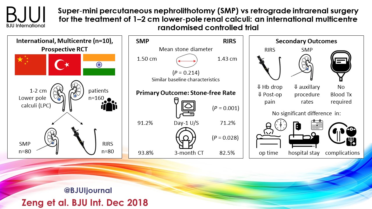
We read with interest the article by Zeng et al. [1] comparing super‐mini percutaneous nephrolithotomy (SMP) with ureteroscopy (URS) for treatment of 1–2‐cm lower pole renal calculi. In this prospective randomized controlled trial, SMP achieved significantly higher stone‐free rates (SFRs) than URS on first‐day KUB with ultrasonography (91.2% vs 71.2%) as well as on 3‐month CT (93.8% vs 82.5%). Haemoglobin drop and pain score were higher in the SMP group, although no blood transfusions were required in either group. We congratulate the authors for this well conducted multicentre study and for the comprehensive report of their results.

A few comments are worth making to aid correct interpretation of the data presented in this study. First, it remains unclear whether the superiority of SMP over URS in terms of SFR was inherent to operating techniques, or whether this might have been the result of superior skills and interest of the surgeons favouring SMP. Surgeons were (obviously) not blinded to operating technique, which could have led to a bias. No study available in the literature has yet questioned whether a surgeon might be better at one technique (SMP or URS) than another. Ultimately, results may differ if both techniques were compared between two expert centres dedicated to each technique, respectively.

Second, the study protocol allowed surgeons to leave fragments up to 2 mm at the end of URS procedures. Strikingly, ‘stone‐free’ status was defined as residual fragments ≤3 mm. This methodology may well have affected the results, as neither endoscopy, KUB, ultrasonography nor CT is precise enough to differentiate 2‐mm from 3‐mm fragments [2, 3]. Arguably, this might have contributed to a lower SFR in the URS group.

Third, the study protocol did not clearly describe indications and choices for auxiliary procedures. Consequently, four of seven SMP (57.1%) and 19 of 23 URS patients (82.6%) with ‘clinically significant’ residual fragments were offered auxiliary procedures such as SMP, shockwave lithotripsy or external physical vibration lithecbole. Remarkably, none of the patients in the URS arm was offered any second‐look intervention, while this was the case in the SMP group.

Fourth, achievements made in one country may not be transposable to others, as epidemiology of urinary stone disease, demographic characteristics, access to technologies and education differ from one country to another. This has been acknowledged by the authors, and it seems particularly important to recall the relatively low body mass index (BMI) found in this cohort (mean BMI < 25 kg/m2). Higher BMI values may arguably impact on outcomes of SMP.

We agree with the authors that both SMP and URS are safe and feasible treatment options for lower pole calculi. Importantly, expertise in percutaneous surgery is warranted for cases presenting impaired retrograde access. Nevertheless, in light of constant and rapid advances in the field of URS, it seems that superiority, if any, of percutaneous nephrolithotomy in terms of SFR is to be tackled by URS in the years to come. This is well illustrated in the present study where 1–2‐cm stones were treated by URS with a laser power range between 5 and 20 W within 52 min in 50% of all cases and within 75 min in 86.4% of all cases (calculations based on values from Table 2 [1]).

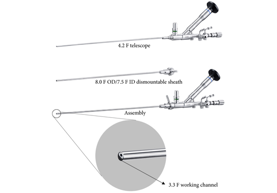
Notably, no consensus has been agreed for the definition of different sizing of percutaneous nephrolithotomy instruments [4]. In the present study, the authors refer to SMP as the use of maximal tract dilation and instrument size up to 14 F. The authors justify size reduction of instruments considering the possible reduced blood loss in favor of smaller access sheaths compared with conventional percutaneous nephrolithotomy [5]. Nevertheless, it should be recalled that whether conventional, mini, super‐mini or any other‐size percutaneous nephrolithotomy, these techniques all share the same fundamental methods of access to intrarenal cavities; therefore, their inherent potential risks and harms – particularly bleeding and iatrogenic organ injury – fundamentally remain the same. This might partly explain why solitary kidney was an exclusion criterion in this study. In contrast, URS respects the delineation of the urinary tract [6]. URS is therefore likely to maintain a superior safety profile, even if further efforts are made at reducing the size of percutaneous nephrolithotomy instruments in the years to come.

The authors’ statement that SMP is more effective than URS to treat 1–2‐cm lower pole calculi should be interpreted in the context of the above. We hope that our comments will aid the correct interpretation of the data presented in this study. We congratulate the authors for the originality of their study, and we encourage them to continue evaluating indications, efficiency and safety of SMP.

References

  1. Zeng G, Zhang T, Agrawal M et al. Super‐mini percutaneous nephrolithotomy (SMP) vs retrograde intrarenal surgery for the treatment of 1‐2 cm lower‐pole renal calculi: an international multicentre randomised controlled trial. BJU Int 2018; 122: 1034–40
  2. Kishore TA, Pedro RN, Hinck B, Monga M. Estimation of size of distal ureteral stones: noncontrast CT scan versus actual size. Urology 2008; 72: 761–4
  3. Zhu W, Liu Y, Liu L et al. Minimally invasive versus standard percutaneous nephrolithotomy: a meta‐analysis. Urolithiasis 2015; 43: 563–70
  4. Giusti G, Proietti S, Villa L et al. Current standard technique for modern flexible ureteroscopy: tips and tricks. Eur Urol 2016; 70: 188–94


Article of the month: The US opioid epidemic

Every month, the Editor-in-Chief selects an Article of the Month from the current issue of BJUI. The abstract is reproduced below and you can click on the button to read the full article, which is freely available to all readers for at least 30 days from the time of this post.

In addition to the article itself, there is an accompanying editorial written by a prominent member of the urological community. This blog is intended to provoke comment and discussion and we invite you to use the comment tools at the bottom of each post to join the conversation.

If you only have time to read one article this week, it should be this one.

The United States opioid epidemic: a review of the surgeon’s contribution to it and health policy initiatives

Katherine Theisen, Bruce Jacobs, Liam Macleod and Benjamin Davies
Department of Urology, University of Pittsburgh Medical Center, Pittsburgh, PA, USA
 
Read the full article

Abstract

Visual abstract created by Abdullatif Aydın and Rebecca Fisher

Opioid abuse and addiction is causing widespread devastation in communities across the USA and resulting in significant strain on our healthcare system. There is increasing evidence that prescribers are at least partly responsible for the opioid crisis because of overprescribing, a practice that developed from changes in policy and reimbursement structures. Surgeons, specifically, have been subject to scrutiny as ‘adequate treatment’ of post‐surgical pain is poorly defined and data suggest that many patients receive much larger opioid prescriptions than needed. The consequences of overprescribing include addiction and misuse, dispersion of opioids into the community, and possible potentiation of illicit drug/heroin use. Several solutions to this crisis are currently being enacted with variable success, including Prescription Drug Monitoring Programmes, policy‐level interventions aimed to de‐incentivize overprescribing, limiting opioid exposures through Enhanced Recovery After Surgery protocols, and the novel idea of creating surgery‐ and/or procedure‐specific prescribing guidelines. This problem is likely to require not one, but several potential solutions to reverse its trajectory. It is critical, however, that we as physicians and prescribers find a way to stop the needless overprescribing while still treating postoperative pain appropriately.


Read more Articles of the week

November 2018 – about the cover

BJUI November 2018

©istock.com/f11photo

The article of the month for November 2018 is on work carried out at the University of Pittsburgh Medical Center (UPMC), Pennsylvania, USA: The United States opioid epidemic: a review of the surgeon’s contribution to it and health policy initiatives.

The city is located at the confluence of the rivers Allegheny, Monongahela and Ohio and is known both as the “city of bridges” due to its 446 bridges and the “steel city” as it was formerly home to over 300 steel-related businesses.

The old industrial areas have been restored and redeveloped into museums, heritage centers, parks, libraries and medical centers. UPMC is now one of the biggest employers in Pennsylvania, and has been vital in treating the country’s most recent shooting victims. It has also been a pioneer in transplant surgeries with many world firsts in multiple organ transplants.


Article of the month: Effect of timing of an immediate instillation of mitomycin C after TUR in 941 patients with NMIBC

Every month, the Editor-in-Chief selects an Article of the Month from the current issue of BJUI. The abstract is reproduced below and you can click on the button to read the full article, which is freely available to all readers for at least 30 days from the time of this post.

In addition to the article itself, there is an accompanying editorial written by a prominent member of the urological community. This blog is intended to provoke comment and discussion and we invite you to use the comment tools at the bottom of each post to join the conversation.

If you only have time to read one article this week, it should be this one.

The effect of timing of an immediate instillation of mitomycin C after transurethral resection in 941 patients with non-muscle-invasive bladder cancer

Judith Bosschieter*, R. Jeroen A. van Moorselaar*, André N. Vis*, Tessa van Ginkel*, Birgit I. Lissenberg‐Witte, Goedele M.A. Beckers* and Jakko A. Nieuwenhuijzen*


Departments of *Urology and Epidemiology and Biostatistics, VU University Medical Center, Amsterdam, The Netherlands


Read the full article

Abstract

Objective

To investigate whether the timing of an immediate instillation of mitomycin C (on the day of transurethral resection of bladder tumour [TURBT] or 1 day later) has an impact on time to recurrence of non‐muscle‐invasive bladder cancer (NMIBC).

Patients and Methods

All patients with NMIBC who were enrolled in a prospective trial between 1998 and 2003, and treated with an early mitomycin C instillation (on the day of TURBT or 1 day later), were selected. Statistical analysis was performed with Kaplan–Meier curves and multivariable Cox regression.

Fig. 1 Kaplan–Meier analysis showing time to recurrence for patients treated with an immediate instillation of MMC on the day of TURBT (Day‐0 group) or 1 day after (Day‐1 group).

Results

Administering an instillation of mitomycin C on the day of TURBT or 1 day later did not show a statistically significant difference in time to recurrence in a univariable model (log‐rank P = 0.99). After correcting for the number of scheduled adjuvant instillations, no statistically significant difference could be detected either: hazard ratio 1.05 (95% confidence interval 0.81–1.35, P = 0.74).

Conclusion

These data do not support the hypothesis that a very early instillation (on the day of TURBT) of mitomycin C decreases the risk of recurrence as compared with an early instillation (1 day after TURBT).

Read more articles of the week

Editorial: Postoperative intravesical chemotherapy has an important role in reducing subsequent bladder tumours – why is it not routine?

Transurethral resection of bladder tumour (TURBT) is a frequent operation performed by urologists worldwide. Although on occasion the procedure can be quite challenging, the majority are relatively straightforward with little morbidity. In most cases, where the medical system allows, it is an outpatient procedure. Nonetheless, with the exception of small low‐grade tumours the patient is anaesthetised. It is costly, as the procedure requires medical clearance, an operating room team and equipment.

Most patients with bladder cancer have Ta or less frequently T1 tumours. Despite an initial TURBT, 30–80% of patients develop another tumour. Most are new tumours and some may be recurrences. The reasons for the high ‘recurrence’ rate are the continued impact of the carcinogen, e.g. cigarettes, incomplete resection, missed tumours, and tumour implantation on the altered urothelium. The urologist can help reduce these events by stressing the importance of limiting carcinogen exposure e.g. smoking cessation, striving to perform a complete TURBT, reviewing the entire bladder after the TURBT to avoid missing tumours (using narrow‐band imaging or fluorescent cystoscopy if available), and limiting implantation of tumour cells on the altered urothelial surface with the use of postoperative intravesical chemotherapy (POIVC).

There is a large body of evidence that POIVC reduces the chance of a subsequent tumour [1]. I became convinced that implantation occurs after animal studies demonstrated that bladder cancer cells placed into the bladder preferentially implant and grow only if the urothelial surface had been cauterised or otherwise damaged prior to exposure to the bladder cancer cells [2]. Prospective randomised trials eventually confirmed the benefit of POIVC [3]. The paper published in this issue of the BJUI by Bosschieter et al. [4] indicates that POIVC is equally effective if given the same day or the day after TURBT. Thus, if there are obstacles to instilling the medication on the day of the TURBT the drug can be administered the following day.

The evidence in favour of POIVC for bladder tumours is particularly impressive for Grade 1–2 Ta tumours. In my view, all patients with primary or ‘recurrent’ single or multiple papillary Grade 1–2 Ta tumours are the optimal candidates to receive POIVC [5]. POIVC is recommended by the European Association of Urology (EAU) and AUA/Society of Urologic Oncology (SUO) [6,7] yet, the adoption of this guideline is far from uniform. I queried my colleagues from the International Bladder Cancer group (IBCG), as they are conversant in the scientific basis for POIVC and represent several countries with different medical systems [8]. Their comments are pertinent and consistent with my understanding of the issues. Here are some of the common reasons for not following the guidelines: (i) Some urologists are not convinced that the reduction in the ‘recurrence’ rate is sufficient to use POIVC. (ii) The most common chemotherapeutic agent for POIVC in the USA is mitomycin C and it is expensive. The cost for 40 mg is ~$1000. It is approximately $500 in Europe. (iii) Hospitals have rules regarding the delivery of chemotherapy and the pharmacy and nursing departments may not make it easy to instil the drug in the postoperative setting. Some hospitals require notification a day before the surgery and the drug is wasted if the drug is not used. (iv) Urologists are concerned about extravasation and uncertainty of the tumour grade and stage. There may be other reasons but these help explain why POIVC is not routine.

On the other hand many patients with bladder cancer require frequent TURBTs. I am certain that following an uneventful TURBT or office cauterisation for Grade 1–2 Ta bladder cancer, they would choose to receive POIVC if properly informed. Urologists are proficient at judging whether a tumour fits the criteria for POIVC and if they underestimate the grade or stage the patient may still benefit. If urologists cannot instil the chemotherapy on the day of the TURBT, they can instil the drug the following day without compromising effectiveness. I believe it is our job to do what we can to help our patients and in this instance we should do our best to minimise subsequent tumour events, which includes the use of adjuvant chemotherapy.

Mark S. Soloway

Memorial Hospital Hollywood, Miami, FL, USA

References

  1. Perlis N, Zlotta AR, Beyene J, Finelli A, Fleshner NE, Kulkarni GS. Immediate post‐ transurethral resection of bladder tumor intravesical chemotherapy prevents non‐muscle invasive bladder tumor recurrence: an updated meta‐analysis on 2548 patients and quality –of‐evidence review. Eur Urol 2013; 64: 421–30
  2. Weldon TE, Soloway MS. Susceptibility of urothelium to neoplastic cellular implantation. Urology 1975; 5: 824–7
  3. Tolley DA, Hargreave TB, Smith PH et al. Effect of intravesical mitomycin C on recurrence of newly diagnosed superficial bladder cancer: interim report from the Medical Research Council Subgroup on Superficial Bladder cancer. Br Med J 1988; 296: 1259–61
  4. Bosschchieter J, von Moorselaar JA, Vis AN et al. The effect of timing of an immediate instillation of mitomycin C after transurethral resection in 941 patients with non‐muscle‐invasive bladder cancer. BJU Int 2018; 122: 571–5
  5. Klaassen Z, Soloway MS. European Association of Urology and American Urological Association/Society of Urologic Oncology guidelines on risk categories for non‐muscle‐invasive bladder cancer may lead to overtreatment for low‐grade Ta bladder tumors. Urology 2017; 105: 14–7
  6. Babjuk M, Böhle A, Burger M et al. EAU guidelines in non‐muscle invasive urothelial carcinoma of the bladder: update 2016. Eur Urol 2017; 71: 447–61
  7. Chang SS, Boorjian SA, Chou R et al. Diagnosis and treatment of non‐muscle invasive bladder cancer: AUA/SUO guideline. J Urol 2016; 196: 1021–9
  8. Brausi M, Witjes F, Lamm D et al. A review of current guidelines and best proactive recommendations for the management of nonmuscle invasive bladder cancer by the International Bladder Cancer Group. J Urol 2011; 186: 2158–67


October 2018 – about the cover

This issue’s Article of the Month is The effect of timing of an immediate instillation of mitomycin C after transurethral resection in 941 patients with non‐muscle‐invasive bladder cancer, carried out by a team from Amsterdam, The Netherlands.

The cover shows the skyline of Amsterdam, the capital of the Netherlands, although it is not the seat of government, which is The Hague. Amsterdam is well-known for its canals, its Art (particularly Rembrandt and Van Gogh) and its infamous coffee shops. In 2013 there were more bicycles than people in Amsterdam.

©istock.com/fotolupa


© 2024 BJU International. All Rights Reserved.