Tag Archive for: Article of the Week

Posts

Video: Separating the wheat from the chaff – Cognitive skills assessment during RA surgery

Cognitive skills assessment during robot-assisted surgery: separating the wheat from the chaff

Khurshid A. Guru, Ehsan T. Esfahani†, Syed J. Raza, Rohit Bhat†, Katy Wang‡,
Yana Hammond, Gregory Wilding‡, James O. Peabody§ and Ashirwad J. Chowriappa

Department of Urology, Roswell Park Cancer Institute, Buffalo, NY; †Brain Computer Interface Laboratory, Department of Mechanical & Aerospace Engineering, University at Buffalo, Buffalo, NY; ‡Department of Biostatistics, Roswell Park Cancer Institute, Buffalo, NY; and §Henry Ford Health System, Detroit, MI, USA

OBJECTIVE

To investigate the utility of cognitive assessment during robot-assisted surgery (RAS) to define skills in terms of cognitive engagement, mental workload, and mental state; while objectively differentiating between novice and expert surgeons.

SUBJECTS AND METHODS

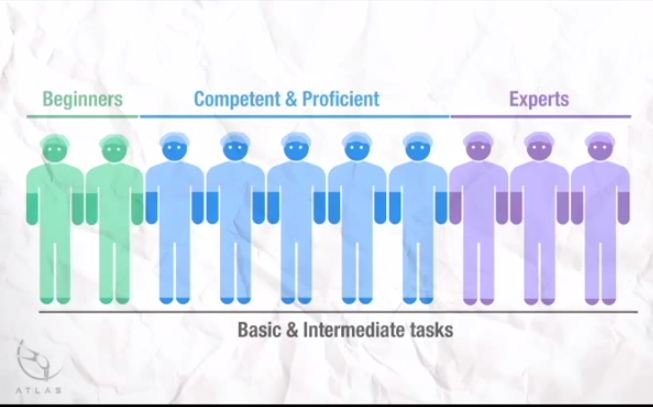
In all, 10 surgeons with varying operative experience were assigned to beginner (BG), combined competent and proficient (CPG), and expert (EG) groups based on the Dreyfus model. The participants performed tasks for basic, intermediate and advanced skills on the da Vinci Surgical System™. Participant performance was assessed using both tool-based and cognitive metrics.

RESULTS

Tool-based metrics showed significant differences between the BG vs CPG and the BG vs EG, in basic skills. While performing intermediate skills, there were significant differences only on the instrument-to-instrument collisions between the BG vs CPG (2.0 vs 0.2, P = 0.028), and the BG vs EG (2.0 vs 0.1, P = 0.018). There were no significant differences between the CPG and EG for both basic and intermediate skills. However, using cognitive metrics, there were significant differences between all groups for the basic and intermediate skills. In advanced skills, there were no significant differences between the CPG and the EG except time (1116 vs 599.6 s), using tool-based metrics. However, cognitive metrics revealed significant differences between both groups.

CONCLUSION

Cognitive assessment of surgeons may aid in defining levels of expertise performing complex surgical tasks once competence is achieved. Cognitive assessment may be used as an adjunct to the traditional methods for skill assessment during RAS.

Article of the Week: RA-RPLND – Technique and Initial Case Series of 18 Patients

Every week the Editor-in-Chief selects the Article of the Week from the current issue of BJUI. The abstract is reproduced below and you can click on the button to read the full article, which is freely available to all readers for at least 30 days from the time of this post.

In addition to the article itself, there is an accompanying editorial written by a prominent member of the urological community. This blog is intended to provoke comment and discussion and we invite you to use the comment tools at the bottom of each post to join the conversation.

Finally, the third post under the Article of the Week heading on the homepage will consist of additional material or media. This week we feature a video from Mr. Tim Dudderidge discussing his paper. 

If you only have time to read one article this week, it should be this one.

Robot-assisted retroperitoneal lymph node dissection: technique and initial case series of 18 patients

Scott M. Cheney, Paul E. Andrews, Bradley C. Leibovich* and Erik P. Castle

Mayo Clinic Arizona Department of Urology, Phoenix, AZ, and *Mayo Clinic Rochester Department of Urology, Rochester, MN, USA

Read the full article
OBJECTIVE

To evaluate outcomes of the first 18 patients treated with robot-assisted retroperitoneal lymph node dissection (RA-RPLND) for non-seminomatous germ cell tumours (NSGCT) and paratesticular rhabdomyosarcoma (RMS) at our institution.

PATIENTS AND METHODS

Between March 2008 and May 2013, 17 patients underwent RA-RPLND for NSGCT and one for paratesticular RMS. Data were collected retrospectively on patient demographics, preoperative tumour characteristics, and perioperative outcomes including open conversion rate, lymph node (LN) yield, rate of positive LNs, operative time, estimated blood loss (EBL), and length of stay (LOS). Perioperative outcomes were compared between patients receiving primary RA-RPLND vs post-chemotherapy RA-RPLND. Medium-term outcomes of tumour recurrence rate and maintenance of antegrade ejaculation were recorded.

RESULTS

RA-RPLND was completed robotically in 15 of 18 (83%) patients. LNs were positive in eight of 18 patients (44%). The mean LN yield was 22 LNs. For cases completed robotically, the mean operative time was 329 min, EBL was 103 mL, and LOS was 2.4 days. At a mean (range) follow-up of 22 (1–58) months, there were no retroperitoneal recurrences and two of 17 (12%) patients with NSGCT had pulmonary recurrences. Antegrade ejaculation was maintained in 91% of patients with a nerve-sparing approach. Patients receiving primary RA-RPLND had shorter operative times compared with those post-chemotherapy (311 vs 369 min, P = 0.03). There was no significant difference in LN yield (22 vs 18 LNs, P = 0.34), EBL (100 vs 313 mL, P = 0.13), or LOS (2.75 vs 2.2 days, P = 0.36).

CONCLUSION

This initial selected case series of RA-RPLND shows that the procedure is safe, reproducible, and feasible for stage I–IIB NSGCT and RMS in the hands of experienced robotic surgeons. Larger studies are needed to confirm the diagnostic and therapeutic utility of this technique.

Editorial: RPLND – Open Surgery’s Next Challenger Is Ready To Enter The Ring

By Tim Dudderidge

The da Vinci surgical system delivers the benefits of laparoscopic surgery with an easier and more precise human–tissue interface than conventional laparoscopic instruments. Nearly all major uro-oncological procedures are being performed robotically. In this issue of BJUI, Cheney et al. [1] present their technique and initial experience of robot-assisted retroperitoneal lymph node dissection (RA-RPLND) for patients with primary and post-chemotherapy non-seminoma germ cell tumours. Quality indicators for RA-RPLND include adequate clearance of the desired surgical field, satisfactory lymph node yield, acceptable perioperative morbidity and length of stay, as well as longer-term functional and oncological outcomes. So how well does RA-RPLND stand up to scrutiny?

The technique employed by Cheney et al., placing the robot at the head of the patient, is unfamiliar to most urologists I suspect. It appears to offer excellent access to the retroperitoneum, but still requires a re-docking when performing full bilateral dissections. Whether this technique is superior to the lateral approach that I and others have used for modified dissections requires further study [2,3]. The lymph node yield was lower than that previously reported for open RPLND and while Cheney et al. [1] observe this may be due to the use of a modified template where appropriate, the absence of any in-field recurrences at a median of 22 months is perhaps the more reliable sign that there is oncological equivalence. Concerns that a true template dissection cannot be completed with a robot-assisted laparoscopic approach are probably unjustified in my opinion. The description of surgical technique by Cheney et al., including suture ligation and division of lumbar vessels, confirms that if a surgeon is minded to do so, a complete bilateral or modified template clearance can be completed.

The absence of significant complications in this series is impressive; however, there were three out of 18 conversions to open surgery. The mean length of stay of 2.4 days is close to the 3–4 days stay I would expect after an uncomplicated open RPLND in a young fit man. However, 1–2 night stays were seen in their later cases as they gained experience. Perhaps more importantly in a group of working age men, return to full physical activity within 3 weeks is possible [2].

As highlighted by Cheney et al. [1], minimally invasive primary RPLND has been previously reported both by laparoscopic and robotic approaches. Their larger series provides an important demonstration that the robotic approach facilitates the more complex undertaking of post-chemotherapy RPLND. Furthermore they show that except for operative time, all other outcomes were similar in primary and post-chemotherapy cases.

As an enthusiast for minimally invasive therapies, I of course welcome these results and think that along with other published and presented series, they provide sufficient evidence to consider a more formal evaluation of this approach. However, how feasible is the wider introduction of RA-RPLND? Despite having experience of robotics and working in a team performing around 30 RPLNDs a year, I was only able to identify five cases during a 1-year period suitable for a robotic approach. With experience this could have been a higher proportion, but it is fair to conclude that suitable cases in typical cancer centres would be limited in number. This is particularly so for the UK and other European countries, where primary RPLND is not used. Cheney et al. [1] had similarly low numbers each year and recruited their cohort of 18 cases over 5 years.

An international multicentre registry is arguably the best way to gather more information on the safety and completeness of template dissection RPLND. Existing registries, e.g. the BAUS complex operations database, have already provided valuable insights into the results of RPLND in the UK [4] and could be combined with other international RA-RPLND databases already being compiled (Erik Castle MD personal communication). Partnership of testicular cancer surgeons without robotic experience with experienced robotic surgeons may also facilitate the development of additional centres for development of this procedure. They will also aid optimal patient selection and help avoid incomplete template dissections, which may compromise the excellent cancer control we are now used to.

There are clear potential advantages with a minimally invasive approach to RPLND, not least of which are the avoidance of a laparotomy scar, the reduction of complications and an earlier return to normal activity. Cheney et al. [1] have shown that their technique is feasible, safe and effective in the medium term and their results justify wider consideration of the procedure for further study and improvement.

Read the full article

Tim Dudderidge

University Hospital Southampton, Southampton, UK

References

1 Cheney SM, Andrews PE, Leibovich BC, Castle EP. Robot-assisted retroperitoneal lymph node dissection: technique and initial case series of 18 patients. BJU Int 2015; 115: 114–20

2 Dudderidge T, Pandian S, Nott D. Technique and outcomes for robotic assisted post-chemotherapy retroperitoneal lymph node dissection (RPLND) in Stage 2 non-seminomatous germ cell tumour (NSGCT). BJU Int 2012; 110: 97

3 Dogra PN, Singh P, Saini AK, Regmi KS, Singh BG, Nayak B. Robot assisted laparoscopic retroperitoneal lymph node dissection in testicular tumor. Urol Ann 2013; 5: 223–6

4 Hayes M, O’Brien T, Fowler S, BAUS RPLND Group. Contemporary retroperitoneal lymph node dissection (RPLND) for testis cancer in the UK – a national study. J Urol 2014; 191 (Suppl.): e89–90


Video: Robot Assisted Retroperitoneal Lymph Node Dissection

Robot-assisted retroperitoneal lymph node dissection: technique and initial case series of 18 patients

Scott M. Cheney, Paul E. Andrews, Bradley C. Leibovich* and Erik P. Castle

Mayo Clinic Arizona Department of Urology, Phoenix, AZ, and *Mayo Clinic Rochester Department of Urology, Rochester, MN, USA

Read the full article

Abstract

OBJECTIVE

To evaluate outcomes of the first 18 patients treated with robot-assisted retroperitoneal lymph node dissection (RA-RPLND) for non-seminomatous germ cell tumours (NSGCT) and paratesticular rhabdomyosarcoma (RMS) at our institution.

PATIENTS AND METHODS

Between March 2008 and May 2013, 17 patients underwent RA-RPLND for NSGCT and one for paratesticular RMS. Data were collected retrospectively on patient demographics, preoperative tumour characteristics, and perioperative outcomes including open conversion rate, lymph node (LN) yield, rate of positive LNs, operative time, estimated blood loss (EBL), and length of stay (LOS). Perioperative outcomes were compared between patients receiving primary RA-RPLND vs post-chemotherapy RA-RPLND. Medium-term outcomes of tumour recurrence rate and maintenance of antegrade ejaculation were recorded.

RESULTS

RA-RPLND was completed robotically in 15 of 18 (83%) patients. LNs were positive in eight of 18 patients (44%). The mean LN yield was 22 LNs. For cases completed robotically, the mean operative time was 329 min, EBL was 103 mL, and LOS was 2.4 days. At a mean (range) follow-up of 22 (1–58) months, there were no retroperitoneal recurrences and two of 17 (12%) patients with NSGCT had pulmonary recurrences. Antegrade ejaculation was maintained in 91% of patients with a nerve-sparing approach. Patients receiving primary RA-RPLND had shorter operative times compared with those post-chemotherapy (311 vs 369 min, P = 0.03). There was no significant difference in LN yield (22 vs 18 LNs, P = 0.34), EBL (100 vs 313 mL, P = 0.13), or LOS (2.75 vs 2.2 days, P = 0.36).

CONCLUSION

This initial selected case series of RA-RPLND shows that the procedure is safe, reproducible, and feasible for stage I–IIB NSGCT and RMS in the hands of experienced robotic surgeons. Larger studies are needed to confirm the diagnostic and therapeutic utility of this technique.

Article of the Month: Progression and treatment of incident lower urinary tract symptoms (LUTS) among men in the California Men’s Health Study

Every week the Editor-in-Chief selects the Article of the Week from the current issue of BJUI. The abstract is reproduced below and you can click on the button to read the full article, which is freely available to all readers for at least 30 days from the time of this post.

This week we feature a video from Dr. Steven Jacobsen discussing his paper. 

If you only have time to read one article this week, it should be this one.

Progression and treatment of incident lower urinary tract symptoms (LUTS) among men in the California Men’s Health Study

Lauren P. Wallner, Jeff M. Slezak*, Ronald K. Loo†, Virginia P. Quinn*, Stephen K. Van Den Eeden‡ and Steven J. Jacobsen*

Department of Medicine and Comprehensive Cancer Center, University of Michigan, Ann Arbor, MI, *Department of Research and Evaluation, Kaiser Permanente Southern California, †Department of Urology, Southern California Permanente Medical Group, Pasadena, CA, and ‡Division of Research, Kaiser Permanente Northern California, Oakland, CA, USA

Read the full article
OBJECTIVES

To characterise the progression and treatment of lower urinary tract symptoms (LUTS) among men aged 45–69 years in the California Men’s Health Study.

PATIENTS AND METHODS

A total of 39 222 men, aged 45–69 years, enrolled in the Southern California Kaiser Permanente Health Plan were surveyed in 2002–2003 and again in 2006–2007. Those men who completed both surveys who did not have a diagnosis of benign prostatic hyperplasia (BPH) and were not on medication for LUTS at baseline were included in the study (N = 19 505). Among the men with no or mild symptoms at baseline, the incidence of moderate/severe LUTS (American Urological Association Symptom Index [AUASI] score ≥8) and odds of progression to severe LUTS (AUASI score ≥20) was estimated during 4 years of follow-up.

RESULTS

Of the 9640 men who reported no/mild LUTS at baseline, 3993 (41%) reported moderate/severe symptoms at follow-up and experienced a 4-point change in AUASI score on average. Of these men, 351 (8.8%) had received a pharmacological treatment, eight (0.2%) had undergone a minimally invasive or surgical procedure and 3634 (91.0%) had no treatment recorded. Men who progressed to severe symptoms (AUASI score ≥20; n = 165) were more likely to be on medication for BPH (odds ratio [OR] 8.09, 95% confidence interval [CI] 5.77–11.35), have a BPH diagnosis (OR 4.74, 95% CI 3.40–6.61) or have seen a urologist (OR 2.49, 95% CI 1.81–3.43) when compared with men who did not progress to severe symptoms (AUASI score <20).

CONCLUSION

These data show that the majority of men who experienced progression did not have pharmacological or surgical therapy for their symptoms and, therefore, may prove to be good candidates for a self-management plan.

Video: Progression and treatment of incident lower urinary tract symptoms (LUTS) among men in the California Men’s Health Study

Progression and treatment of incident lower urinary tract symptoms (LUTS) among men in the California Men’s Health Study

Lauren P. Wallner, Jeff M. Slezak*, Ronald K. Loo†, Virginia P. Quinn*, Stephen K. Van Den Eeden‡ and Steven J. Jacobsen*

Department of Medicine and Comprehensive Cancer Center, University of Michigan, Ann Arbor, MI, *Department of Research and Evaluation, Kaiser Permanente Southern California, †Department of Urology, Southern California Permanente Medical Group, Pasadena, CA, and ‡Division of Research, Kaiser Permanente Northern California, Oakland, CA, USA

Read the full article

OBJECTIVES

To characterise the progression and treatment of lower urinary tract symptoms (LUTS) among men aged 45–69 years in the California Men’s Health Study.

PATIENTS AND METHODS

A total of 39 222 men, aged 45–69 years, enrolled in the Southern California Kaiser Permanente Health Plan were surveyed in 2002–2003 and again in 2006–2007. Those men who completed both surveys who did not have a diagnosis of benign prostatic hyperplasia (BPH) and were not on medication for LUTS at baseline were included in the study (N = 19 505). Among the men with no or mild symptoms at baseline, the incidence of moderate/severe LUTS (American Urological Association Symptom Index [AUASI] score ≥8) and odds of progression to severe LUTS (AUASI score ≥20) was estimated during 4 years of follow-up.

RESULTS

Of the 9640 men who reported no/mild LUTS at baseline, 3993 (41%) reported moderate/severe symptoms at follow-up and experienced a 4-point change in AUASI score on average. Of these men, 351 (8.8%) had received a pharmacological treatment, eight (0.2%) had undergone a minimally invasive or surgical procedure and 3634 (91.0%) had no treatment recorded. Men who progressed to severe symptoms (AUASI score ≥20; n = 165) were more likely to be on medication for BPH (odds ratio [OR] 8.09, 95% confidence interval [CI] 5.77–11.35), have a BPH diagnosis (OR 4.74, 95% CI 3.40–6.61) or have seen a urologist (OR 2.49, 95% CI 1.81–3.43) when compared with men who did not progress to severe symptoms (AUASI score <20).

CONCLUSION

These data show that the majority of men who experienced progression did not have pharmacological or surgical therapy for their symptoms and, therefore, may prove to be good candidates for a self-management plan.

Article of the Week: Spine Metastases in Prostate Cancer: Comparison of [99mTc]MDP Wholebody Bone Scintigraphy, [18F]Choline PET/CT, and [18F]NaF PET/CT

Every week the Editor-in-Chief selects the Article of the Week from the current issue of BJUI. The abstract is reproduced below and you can click on the button to read the full article, which is freely available to all readers for at least 30 days from the time of this post.

In addition to the article itself, there is an accompanying editorial written by a prominent member of the urological community. This blog is intended to provoke comment and discussion and we invite you to use the comment tools at the bottom of each post to join the conversation.

If you only have time to read one article this week, it should be this one.

Spine metastases in prostate cancer: comparison of technetium-99m-MDP whole-body bone scintigraphy, [18F]choline positron emission tomography(PET)/computed tomography (CT) and [18F]NaF PET/CT

Mads H. Poulsen, Henrik Petersen*, Poul F. Høilund-Carlsen*, Jørn S. Jakobsen, Oke Gerke*, Jens Karstoft†, Signe I. Steffansen* and Steen Walter

Research Unit of Urology, Department of Urology, and Departments of *Nuclear Medicine and †Radiology, Odense University Hospital, Odense, Denmark

Read the full article
OBJECTIVE

To compare the diagnostic accuracy of the following imaging techniques in the detection of spine metastases, using magnetic resonance imaging (MRI) as a reference: whole-body bone scintigraphy (WBS) with technetium-99m-MDP, [18F]-sodium fluoride (NaF) positron emission tomography (PET)/computed tomography (CT) and [18F]-fluoromethylcholine (FCH) PET/CT.

PATIENTS AND METHODS

The study entry criteria were biopsy-proven prostate cancer, a positive WBS consistent with bone metastases, and no history of androgen deprivation. Within 30 days of informed consent, trial scans were performed in random order. Scans were interpreted blindly for the purpose of a lesion-based analysis. The primary target variable was bone lesion (malignant/benign) and the ‘gold standard’ was MRI.

RESULTS

A total of 50 men were recruited between May 2009 and March 2012. Their mean age was 73 years, their median PSA level was 84 ng/mL, and the mean Gleason score of the tumours was 7.7. A total of 46 patients underwent all four scans, while four missed one PET/CT scan. A total of 526 bone lesions were found in the 50 men: 363 malignant and 163 non-malignant according to MRI. Sensitivity, specificity, positive and negative predictive values and accuracy were: WBS: 51, 82, 86, 43 and 61%; NaF-PET/CT: 93, 54, 82, 78 and 81%; and FCH-PET/CT: 85, 91, 95, 75 and 87%, respectively.

CONCLUSIONS

We found that FCH-PET/CT and NaF-PET/CT were superior to WBS with regard to detection of prostate cancer bone metastases within the spine. The present results call into question the use of WBS as the method of choice in patients with hormone-naïve prostate cancer.

Editorial: Bone Metastases in Prostate Cancer: Which Scan?

In this issue of BJUI, Poulsen et al. [1] present a prospective comparison of 18F-fluoride (NaF) and 18F-choline (FCH) positron emission tomography (PET)/CT with planar whole-body bone scintigraphy (WBS) using spinal MRI, including short tau inversion recovery (STIR), T1 and T2 sequences, as the reference standard in 50 hormone-naïve patients with confirmed bone metastases on WBS. They found that both PET/CT methods were significantly more sensitive and accurate than WBS and that FCH PET/CT was more specific than NaF PET/CT.

It has become increasingly recognised that planar WBS is no longer the most accurate method of assessing the skeleton for metastases and that novel imaging methods, including PET/CT, single-photon emission CT (SPECT)/CT and whole-body MRI offer advantages [2].

What is surprising in the presented results is that NaF PET/CT shows poor specificity (54%), a result that is discordant with previous literature [3, 4]. Compared with PET alone, using the CT component of hybrid PET/CT reduces false-positive interpretation of NaF uptake in benign lesions [3]. This raises the question as to whether the CT component of the PET/CT acquisition was used to full effect in the present study. The use of spinal MRI as a reference standard is also a possible limitation that is recognised by the authors, as this limits the comparison to only the spine, and MRI in itself is a method with known limitations. All patients had abnormal WBS for entry into the trial and whilst the PET methods were more sensitive on a lesion basis, a patient-based comparison was therefore not possible; however, the results imply that PET methods may identify metastatic disease in patients with normal WBS, as has been previously reported [3, 5].

Nevertheless, the authors should be congratulated in reporting valuable data from a prospective study where all imaging was performed in hormone-naïve patients, minimising confounding treatment-related effects, and within a small time window of 30 days; however, some questions remain. WBS is no longer state of the art for imaging the skeleton with radiolabelled bisphosphonates, such as 99mTc-methylene diphosphonate (MDP). Although NaF PET/CT has been shown to be superior to planar WBS augmented with SPECT [3], there have not been head-to-head comparisons with 99mTc-MDP SPECT/CT, where the potential advantages of the pharmacokinetics of NaF and the superior spatial resolution of PET compared with SPECT may not be as great. This may be particularly important given the difference in costs and availability of the two methods.

Despite the results from the present study, which show superiority of FCH PET/CT compared with NaF PET/CT with regard to specificity, taking the available literature as a whole, it remains unresolved as to what the best test for staging the skeleton in patients with high-risk prostate cancer should be at diagnosis. The different mechanisms of uptake of the PET tracers should be noted. NaF uptake reflects the local bone osteoblastic reaction to tumour within the bone marrow, whereas FCH uptake reflects metabolic activity within the tumour cells themselves. In prostate cancer, where the predominant effect is an increase in osteoblastic activity in the adjacent bone, the bone-specific tracers such as 99mTc-MDP and NaF have shown high sensitivity; however, direct imaging of tumour cell metabolism, such as increased choline kinase activity and cell membrane synthesis with FCH, may be advantageous in detecting metastases in the bone marrow before an osteoblastic reaction has occurred [6]. It is possible that both PET tracers may be required to provide optimum diagnostic accuracy and of course FCH PET/CT also provides valuable data on nodal and visceral metastatic disease. In patients with recurrent disease, better specificity has been reported with FCH [4], NaF possibly being limited by non-specific treatment-related effects such as osteoblastic flare. For similar reasons it may be that the more tumour-specific imaging methods, such as FCH PET/CT or diffusion-weighted MRI, may be better in assessing the treatment response of skeletal metastases. Questions therefore remain as to the best imaging test at different times in the management of patients with metastatic prostate cancer. 99mTc-MDP SPECT/CT deserves a full assessment, but perhaps the recent advent of PET/MRI and the potential synergies available from this hybrid technique may help resolve some of the remaining issues.

Read the full article

Gary Cook*† and Vicky Goh*‡

*Division of Imaging Sciences and Biomedical Engineering, King’s College London, † Clinical PET Centre, and ‡ Department of Radiology, Guy’s and St Thomas’ Hospitals NHS Foundation Trust, London, UK

References

1 Poulsen MH, Petersen H, Høilund-Carlsen PF et al. Spine metastases in prostate cancer: comparison of [99mTc]MDP wholebody bone scintigraphy, [18F]choline PET/CT, and [18F]NaF PET/CT. BJU Int 2014; 114: 818–23

2 Fogelman I, Blake GM, Cook GJ. The isotope bone scan: we can do better. Eur J Nucl Med Mol Imaging 2013; 40: 1139–40

3 Even-Sapir E, Metser U, Mishani E et al. The detection of bone metastases in patients with high-risk prostate cancer: 99mTc-MDP Planar bone scintigraphy, single- and multi-field-of-view SPECT, 18F-fluoride PET, and 18F-fluoride PET/CT. J Nucl Med 2006; 47: 287–974

4 Langsteger W, Balogova S, Huchet V et al. Fluorocholine (18F) and sodium fluoride (18F) PET/CT in the detection of prostate cancer: prospective comparison of diagnostic performance determined by masked reading. Q J Nucl Med Mol Imaging 2011; 55: 448–57

5 Kjölhede H, Ahlgren G, Almquist H et al. Combined 18F-fluorocholine and 18F-fluoride positron emission tomography/computed tomography imaging for staging of high-risk prostate cancer. BJU Int 2012; 110: 1501–6

6 Beheshti M, Vali R, Waldenberger P et al. Detection of bone metastases in patients with prostate cancer by 18F fluorocholine and 18F fluoride PET-CT: a comparative study. Eur J Nucl Med Mol Imaging 2008; 35: 1766–74


Editorial: Complex tumours, partial nephrectomy and functional outcomes

In the paper by Volpe et al. [1], excellent renal functional outcomes are associated with partial nephrectomy in patients with high PADUA score cancers. The study is notable because it shows that, even in patients who are typically considered candidates for radical nephrectomy, partial nephrectomy can maintain excellent estimated GFR (eGFR) and outcomes; however, because we perform nephron-sparing procedures on patients who may also be candidates for radical nephrectomy, we must consider the varied nature of some of the data on partial nephrectomy.

The literature on renal ischaemia and functional outcomes is heterogeneous and highly debated [2]. There have been several contradictory studies and changes over time in the literature based on technology, surgeon, centre, measurement and, now, correlation with parenchyma-sparing.

A study conducted by the European Organisation for the Research and Treatment of Cancer (EORTC) compared radical nephrectomy (essentially an ischaemic time of infinity) and partial nephrectomy, reporting a 10-year overall survival benefit for patients treated with radical nephrectomy [3]. Nevertheless, this oft-criticized randomised trial also showed better eGFR in partial nephrectomy. The survival benefit reported in that study is countered by population-based studies suggesting that partial nephrectomy may still be a better option when feasible [4]. Unfortunately, these population-based studies may be considered to provide a lower level of evidence than a randomised study, and are also prone to several biases, the most notable being selection of both patients and centres. Surgeons may be more likely to perform nephron-sparing in patients in lower-risk groups. There are also other questions to consider. If a patient is more likely to be referred to a larger centre for partial nephrectomy, are they not also likely to be referred for their coronary artery bypass, aortic surgery, general medical care and even emergency care? Are these patients more likely to seek out second opinions for all of their medical care? Will this affect mortality? Are they more motivated and engaged in their own overall healthcare? These are just a few of the confounding factors that could influence outcomes and are difficult to control in population-based studies. Nevertheless, I am a firm believer in partial nephrectomy, and particularly in preserving renal function, as the better choice for the treatment of both straightforward and complex lesions. It will be difficult, however, to completely negate the implications of the EORTC trial.

Does reasonable ischaemic time affect eGFR outcome? The present study by Volpe et al. [1] would suggest that reasonable ischaemic times are completely acceptable. Several contradictory studies point out the benefits and risks of a limited or minimized clamp time for partial nephrectomy. Another separate paper by White et al. [5] is consistent with other studies that show that a clamped partial nephrectomy, even for high complexity masses, results in a minimal loss of renal function, if at all. Although there is also enthusiasm for a zero ischaemia technique, it is critical to point out that this may be surgeon-, patient-, technique- and institution-dependent. Ultimately, however, we are splitting hairs over a few points of eGFR. The real issue with long-term GFR outcomes in our patients is not only the impact of a few minutes of renal ischaemia, but also control of hypertension, diabetes and their role in medical renal disease. There is an absence of urological literature that controls for patients’ glycated haemoglobin levels or measures hypertension monthly and records the response to medical therapy. These critical pieces of information confound all eGFR and comparative measurements and make it difficult to compare published outcomes. Perhaps the best medical advice we can give patients is to diet, exercise and eat healthily for better overall health. In some sense, this advice may be far more important than the decision of partial vs radical nephrectomy for a complex mass.

What are the logical conclusions of these dilemmas? Clamped partial nephrectomy is possible in complex cases, and the procedure salvages eGFR. Further refinements are also interesting academically, including papers on parenchyma-sparing. Nevertheless, if we are serious about ‘healthy kidneys’, we might take a holistic approach and encourage our patients to pursue a healthier lifestyle so they can bolster lifelong preservation of renal function and general wellness. Would the effect be more profound than a few minutes of ischaemic time? I am betting it would.

Read the full article

Sam B. Bhayani 

Division of Urological Surgery, Washington University School of Medicine and Barnes-Jewish West County Hospital, St Louis, MO, USA

References

1 Volpe A, Garrou D, Amparore D et al. Perioperative and renal functional outcomes of elective robot-assisted partial nephrectomy for renal tumors with high surgical complexity. BJU Int 2014; 114: 903–9

2 Lane BR, Russo P, Uzzo RG et al. Comparison of cold and warm ischemia during partial nephrectomy in 660 solitary kidneys reveals predominant roles of nonmodifiable factors in determining ultimate renal function. J Urol 2011; 185: 421–7

3 Van Poppel H, Da Pozzo L, Albrecht W et al. A prospective, randomized EORTC intergroup phase 3 study comparing the oncologic outcome of elective nephron-sparing surgery and radical nephrectomy for low-stage renal cell carcinoma. Eur Urol 2011; 59: 543–52

4 Sun M, Trinh Q-D, Bianchi M et al. A non-cancer related survival benefit is associated with partial nephrectomy. Eur Urol 2012; 61: 725–31

5 White MA, Georges-Pascal H, Autorino R et al. Outcomes of robotic partial nephrectomy for renal masses with nephrometry score of ≥ 7. Urology 2011; 77: 809–13


Video: Bimanual Examination Of The Retrieved Specimen And Regional Hypothermia During Robot-Assisted Radical Prostatectomy: A Novel Technique For Reducing Positive Surgical Margin And Achieving Pelvic Cooling

Bimanual examination of the retrieved specimen and regional hypothermia during robot-assisted radical prostatectomy: a novel technique for reducing positive surgical margin and achieving pelvic cooling

Wooju Jeong, Akshay Sood, Khurshid R. Ghani, Dan Pucheril, Jesse D. Sammon, Nilesh S. Gupta*, Mani Menon and James O. Peabody

Vattikuti Urology Institute and *Department of Pathology, Henry Ford Health System, Detroit, MI, USA

Read the full article
OBJECTIVE

To describe a novel method of achieving pelvic hypothermia during robot-assisted radical prostatectomy (RARP) and a modification of technique allowing immediate organ retrieval for intraoperative examination and targeted frozen-section biopsies.

PATIENTS AND METHODS

Intracorporeal cooling and extraction (ICE) consists of a modification of the standard RARP technique with the use of the GelPOINT™ (Applied Medical, Rancho Santa Margarita, CA, USA), a hand access platform, which allows for delivery of ice-slush and rapid specimen extraction without compromising pneumoperitoneum.

RESULTS

The ICE technique reproducibly achieves a temperature of 15 °C in the pelvic cavity with no obvious body temperature change. Adopting this technique during RARP, there was an absolute risk reduction by 26.6% in positive surgical margin rate in patients with pT3a disease when compared with similar patients undergoing conventional RARP (P = 0.04).

CONCLUSIONS

The ICE technique eliminates the potential handicap of decreased tactile sensation for oncological margins, especially in the high-risk patients. This technique allows the surgeon to immediately examine the surgical specimen after resection, and with the aid of frozen-section pathology determine if further resection is required. A prospective trial is underway in our centre to evaluate the effects of this novel technique on postoperative outcomes.


© 2024 BJU International. All Rights Reserved.