Tag Archive for: bone

Posts

Article of the Week: The implications of baseline bone‐health assessment at initiation of androgen‐deprivation therapy for prostate cancer

Every Week, the Editor-in-Chief selects an Article of the Week from the current issue of BJUI. The abstract is reproduced below and you can click on the button to read the full article, which is freely available to all readers for at least 30 days from the time of this post.

In addition to the article itself, there is an accompanying editorial written by a prominent member of the urological community. This blog is intended to provoke comment and discussion and we invite you to use the comment tools at the bottom of each post to join the conversation.

If you only have time to read one article this month, it should be this one.

The implications of baseline bone‐health assessment at initiation of androgen‐deprivation therapy for prostate cancer


Peter S. Kirk* , Tudor Borza*, Vah akn B. Shahinian, Megan E.V. Caram§Danil V. Makarov**, Jeremy B. Shelton††, John T. Leppert‡‡§§, Ryan M. Blake*, Jennifer A. Davis§, Brent K. Hollenbeck*, Anne Sales§¶¶ and Ted A. Skolarus *§


*Dow Division of Health Services Research, Department of Urology, Division of Nephrology, Department of Internal Medicine, Division of Hematology and Oncology, Department of Internal Medicine, University of Michigan Health System, §Veterans Affairs (VA) Health Services Research and Development, Center for Clinical Management Research, VA Ann Arbor Healthcare System, University of Michigan Medical School, Ann Arbor, MI, USA, Departments of Urology and Population Health, NYU Langone Medical Center, New York City, NY, USA, **VA New York Healthcare System, New York City, NY, USA, ††VA Greater Los Angeles Healthcare System, Los Angeles City, LA, USA, ‡‡Department of Urology, Stanford University School of Medicine, Stanford, CA, USA, §§VA Palo Alto Healthcare System, Palo Alto, CA, USA, and ¶¶Department of Learning Health Sciences, University of Michigan Medical School, Ann Arbor, MI, USA


Abstract

Objectives

To assess bone‐density testing (BDT) use amongst prostate cancer survivors receiving androgen‐deprivation therapy (ADT), and downstream implications for osteoporosis and fracture diagnoses, as well as pharmacological osteoporosis treatment in a national integrated delivery system.

Patients and methods

We identified 17 017 men with prostate cancer who received any ADT between 2005 and 2014 using the Veterans Health Administration cancer registry and administrative data. We identified claims for BDT within a 3‐year period of ADT initiation. We then used multivariable regression to examine the association between BDT use and incident osteoporosis, fracture, and use of pharmacological treatment.

Results

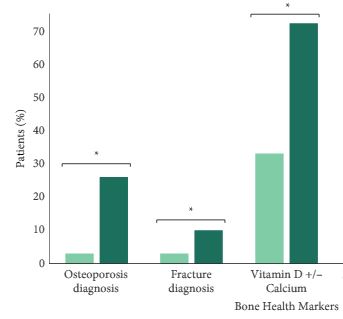
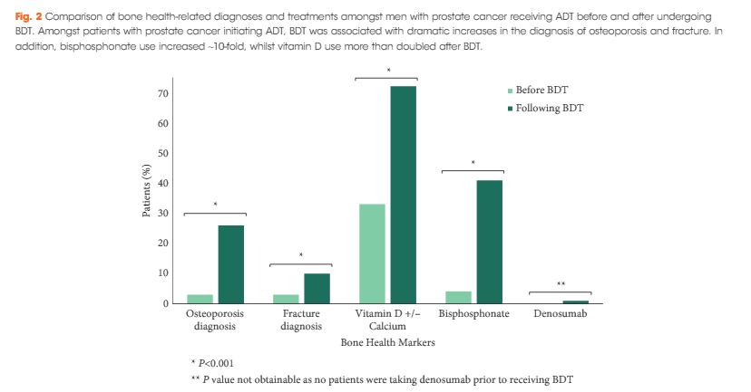
We found that a minority of patients received BDT (n = 2 502, 15%); however, the rate of testing increased to >20% by the end of the study period. Men receiving BDT were older at diagnosis and had higher‐risk prostate cancer (both P < 0.001). Osteoporosis and fracture diagnoses, use of vitamin D ± calcium, and bisphosphonates were all more common in men who received BDT. After adjustment, BDT, and to a lesser degree ≥2 years of ADT, were both independently associated with incident osteoporosis, fracture, and osteoporosis treatment.

Conclusions

BDT is rare amongst patients with prostate cancer treated with ADT in this integrated delivery system. However, BDT was associated with substantially increased treatment of osteoporosis indicating an underappreciated burden of osteoporosis amongst prostate cancer survivors initiating ADT. Optimising BDT use and osteoporosis management in this at‐risk population appears warranted.


Editorial: Low rates of bone density testing in prostate cancer survivors on androgen‐deprivation therapy: where do we go from here?

In this month’s issue of the BJU International, Kirk et al. 1 describe their findings regarding an important issue in the care of prostate cancer survivors on androgen‐deprivation therapy (ADT): the underuse of bone density testing (BDT) to screen for osteoporosis. ADT is the commonest systemic therapy in patients with prostate cancer, used in both metastatic and localised settings. Whilst it has clear survival benefits, ADT is also associated with harms including cardiovascular, cognitive, and metabolic side‐effects, as well as an increased risk of osteoporosis and fractures. These bone‐related complications are costly from a quality‐of‐life and financial perspective, especially given the critical importance of mobility in maintaining performance status and cardiovascular health during cancer treatment 23. Consequently, most clinical practice guidelines include osteoporosis screening as a recommendation for men undergoing ADT.

In their study, ‘The implications of baseline bone health assessment at initiation of androgen‐deprivation therapy for prostate cancer’, the authors describe patterns of use of BDT and diagnosis of osteoporosis amongst men treated for prostate cancer in the USA Veterans Affairs (VA) system within a 3‐year period following ADT initiation. There was a statistically significant increase in the BDT rate throughout the study period; however, overall BDT remains uncommon amongst patients with prostate cancer on ADT, used in only 15% of men in their cohort. Unsurprisingly, patients who received BDT were more likely to be diagnosed with osteoporosis, be diagnosed with a fracture, and receive treatment with vitamin D, calcium and bisphosphonates. The authors acknowledge an important limitation about the applicability of their VA study to the civilian health population. However, given that the VA and military health systems perform as well, if not better, on several important metrics in prostate cancer care 45, these results should not be ignored simply because they were obtained in the military health system.

The increase in BDT screening throughout the study may be attributable to increased awareness of guidelines published during the study period. However, the overall BDT rate remains low. This may be explained by insufficient access, lack of information technology, as well as more nebulous aspects of care such as physician culture, beliefs, and habits 6.

Studies such as this are vital to identify opportunities for improving care delivery. What are needed next are innovations to optimise the delivery of care for patients treated with ADT. Whilst improving BDT adherence may lack the cachet of next‐generation targeted therapies, this is an example of the kind of simple, measurable area where improvement in care delivery systems may yield large benefits.

There are many possible avenues for success: quality improvement collaboratives are one well‐known innovation, which may be applicable to this area: examples, such as the Michigan Urological Surgery Improvement Collaborative (MUSIC) and the AUA Quality Registry (AQUA) are success stories, but to our knowledge there are no published studies specifically attempting to improve adherence to BDT guidelines within these cohorts. Other practice‐based innovations include navigators and multidisciplinary cancer teams, either of which may yield improvements in guideline adherence. Online patient support groups can raise awareness. And although we all know how electronic reminders have frustrated countless physicians, electronic reminders about recommended tests and interventions may be an important tool. At our institution, a Prostate Cancer Foundation grant is funding the development of a mobile health app, which is targeted exclusively at men receiving ADT for prostate cancer. This app will encourage physical activity and healthy eating, which can both support bone health.

In our view, the issue of bone screening is a clear example of where innovative strategies to improve care delivery and guideline adherence may make a big difference for men living with prostate cancer. We look forward to seeing more in the years to come.


Sabrina S. Harmouch, Alexandra J. Berger, and Alexander P. Cole


Center for Surgery and Public Health, Division of Urological Surgery, Brigham and Wo mens Hospital, Harvard Medical School, Boston, MA, USA


References
  • Kirk PS, Borza T, Shahinian VB et al. The implications of baseline bone‐health assessment at initiation of androgen‐deprivation therapy for prostate cancerBJU Int2018121: 558–64


  • Cole AP, Jiang W, Lipsitz SR et al. The use of prostate specific antigen screening in purchased versus direct care settings: data from the TRICARE® military databaseJ Urol 2017198: 1295–300


  • Cullen J, Brassell SA, Chen Y et al. Racial/Ethnic patterns in prostate cancer outcomes in an active surveillance cohortProstate Cancer 20112011: 234519


Article of the week: Calcium : citrate ratio may predict severe lithogenesis

Every week the Editor-in-Chief selects the Article of the Week from the current issue of BJUI. The abstract is reproduced below and you can click on the button to read the full article, which is freely available to all readers for at least 30 days from the time of this post.

In addition to the article itself, there is an accompanying editorial written by a prominent member of the urological community. This blog is intended to provoke comment and discussion and we invite you to use the comment tools at the bottom of each post to join the conversation.

Finally, the third post under the Article of the Week heading on the homepage will consist of additional material or media. This week we feature a video of Miguel Angel Arrabal-Polo discussing his paper.

If you only have time to read one article this week, it should be this one.

Importance of citrate and the calcium : citrate ratio in patients with calcium renal lithiasis and severe lithogenesis

Miguel Angel Arrabal-Polo*, Miguel Arrabal-Martin*,  Salvador Arias-Santiago**, Juan Garrido-Gomez, Antonio PoyatosAndujar§ and Armando Zuluaga-Gomez**

Department of Urology, San Cecilio University Hospital, **Department of Medicine, Baza Hospital, Department of  Medicine, University of Granada, and Departments of Traumatology and §Biochemistry, San Cecilio University Hospital, Granada, Spain

Read the full article
OBJECTIVE

• To analyse the importance of urinary citrate and the urinary calcium : citrate  ratio in patients with calcium renal lithiasis and severe lithogenesis compared with  a control group of patients without lithiasis.

MATERIAL AND METHODS

• A cross-sectional study of 115 patients in eastern Andalusia, Spain was conducted.

• The patients were divided into two groups: Group A: 56 patients aged 25 – 60 years without calcium renal lithiasis; Group B: 59 patients aged 25 – 60 years, presenting with calcium renal lithiasis and severe lithogenesis.

• The citrate levels and the calcium : citrate ratio in the patients’ urine and the relationship of these two factors to lithiasic activity were analysed and compared.

RESULTS

• In Group B, 32.2% of the patients presented with hypocitraturia, compared with 14.3% of the patients in Group A (P = 0.02).

• The urinary citrate levels were lower in Group B than in Group A (P = 0.001) and the calcium : citrate ratio was higher in Group B than in Group A (P = 0.005).

• The results suggest that a patient urinary calcium : citrate ratio > 0.25 indicates severe lithogenesis (with a sensitivity of 89% and a specifi city of 57%).

• After linear regression analysis, we found that the urinary citrate level is an independent factor associated with the changes in bone densitometry T-score values of patients.

CONCLUSIONS

• The patients with severe lithogenesis presented with hypocitraturia, which was associated with lower bone mineral density.

• The calcium : citrate ratio, which is linearly related to the bone resorption marker β-crosslaps, could be useful in evaluating the risk of severe lithogenesis when this ratio is > 0.25.


Read Previous Articles of the Week

Editorial: The importance of citrate in patients with calcium stones and loss of bone mineral density

Stone disease and osteopaenia are both common conditions, and reduced bone mineral density (BMD) is an increasingly recognized complication in stone formers; indeed, in a previous paper in BJUI, Arrabal-Polo et al. reported that patients with recurrent stones have lower BMD compared with controls or patients with just a single episode of urolithiasis.

Although the exact pathogenesis of bone loss in stone disease is yet to be determined, the conceptually obvious relationship with hypercalciuria is well documented. In the present study, Arrabal-Polo et al. emphasise that hypocitraturia is also associated with reduced BMD. Furthermore, they found a higher calcium : citrate ratio in patients with a cumulative maximum stone diameter > 20 mm, or in those with frequent recurrences than in controls, and found that this correlated with higher levels of β-crosslaps, consistent with increased bone resorption in these patients.

We commented in our previous editorial that metabolic abnormalities should be sought in recurrent stone formers, and managed in a multi-disciplinary setting. In addition to dietary advice, options for treatment include bisphosphonates (which inhibit bone resorption, and are commonly used in osteoporosis), thiazide diuretics (which reduce calcium excretion and can increase BMD) and potassium citrate (which acts as an alkalinizing agent mitigating the bone restorative effect of acidosis). This approach is supported by recent data in medullary sponge kidneys, in which hypercalciuria and hypocitraturia were commonly detected in association with reduced BMD. Patients who were treated with potassium citrate were found to have increased urinary pH citrate levels, and an improvement in their BMD.

In the present article, Arrabal-Polo et al. suggest using a calcium : citrate ratio of 0.25 for predicting the risk of future recurrent stone formation, but this value could equally be used to predict the risk of patients having reduced BMD and the complications that may follow. Either way, their findings strengthen the argument for metabolic screening of recurrent stone formers, and for an assessment of these patients’ BMD. Patients can then be appropriately treated with a thiazide diuretic, potassium citrate, or a bisphosphonate, either singly or in combination, according to the abnormalities detected and their progress on treatment.

Daron Smith
Stone and Endourology Unit, University College Hospital, London, UK

Chris Laing
UCL Centre for Nephrology, Royal Free Hospital London, London, UK

Read the full article

Video: Commentary by Dr Arrabal-Polo on the calcium : citrate ratio.

Importance of citrate and the calcium : citrate ratio in patients with calcium renal lithiasis and severe lithogenesis

Miguel Angel Arrabal-Polo*, Miguel Arrabal-Martin*,  Salvador Arias-Santiago**, Juan Garrido-Gomez, Antonio PoyatosAndujar§ and Armando Zuluaga-Gomez**

Department of Urology, San Cecilio University Hospital, **Department of Medicine, Baza Hospital, Department of  Medicine, University of Granada, and Departments of Traumatology and §Biochemistry, San Cecilio University Hospital, Granada, Spain

Read the full article
OBJECTIVE

• To analyse the importance of urinary citrate and the urinary calcium : citrate  ratio in patients with calcium renal lithiasis and severe lithogenesis compared with a control group of patients without lithiasis.

MATERIAL AND METHODS

• A cross-sectional study of 115 patients in eastern Andalusia, Spain was conducted.

• The patients were divided into two groups: Group A: 56 patients aged 25 – 60 years without calcium renal lithiasis; Group B: 59 patients aged 25 – 60 years, presenting with calcium renal lithiasis and severe lithogenesis.

• The citrate levels and the calcium : citrate ratio in the patients’ urine and the relationship of these two factors to lithiasic activity were analysed and compared.

RESULTS

• In Group B, 32.2% of the patients presented with hypocitraturia, compared with 14.3% of the patients in Group A (P = 0.02).

• The urinary citrate levels were lower in Group B than in Group A (P = 0.001) and the calcium : citrate ratio was higher in Group B than in Group A (P = 0.005).

• The results suggest that a patient urinary calcium : citrate ratio > 0.25 indicates severe lithogenesis (with a sensitivity of 89% and a specificity of 57%).

• After linear regression analysis, we found that the urinary citrate level is an independent factor associated with the changes in bone densitometry T-score values of patients.

CONCLUSIONS

• The patients with severe lithogenesis presented with hypocitraturia, which was associated with lower bone mineral density.

• The calcium : citrate ratio, which is linearly related to the bone resorption marker β-crosslaps, could be useful in evaluating the risk of severe lithogenesis when this ratio is > 0.25.

© 2024 BJU International. All Rights Reserved.