Tag Archive for: computed tomography

Posts

Rocktober – Keep on rocking #urojc

We celebrated the two-year anniversary of the international urology journal club this month (@iurojc) with record participation. There were over 500 tweets in the 48 hour discussion of this month’s article, published in New England Journal of Medicine on September 18, 2014, Ultrasound versus Computed Tomography for Suspected Nephrolithiasis. It was a true multidisciplinary discussion with nephrologists, EM docs, and study author, radiologist Rebecca Smith-Bindman tweeting.

This was a multi-institutional prospective randomized control study evaluating bedside and radiology ultrasonography versus CT as the first test performed for patients presenting to the ED with flank or abdominal pain. Patients who initially underwent ultrasound could also receive a CT if the provider felt necessary based on clinical presentation and ultrasound findings. In terms of the primary endpoints, authors found no significant difference across the three groups in high-risk diagnoses with complications related to missed or delayed diagnoses. There was significantly less 6-month cumulative radiation exposure in patients assigned to the ultrasonography groups compared to those assigned to CT. Conclusions of this study in the form of a tweet: Get US first #noharmdone #lessradiation.

Conversation first focused on clarifying the main conclusions of this article. Notably, ED physicians were the focus of this study, who care whether the patient will be admitted or sent home. Information about size, location, etc of stones was omitted from the study since the goal was not definitive stone treatment.

Some of the limitations of the study were brought up early on. First of all, obese patients were excluded (men >129 kg and women >113 kg).

Additionally, the definition of being diagnosed with a stone only applied to individuals who reported passing a stone or having a stone surgically removed.

Much of the conversation focused on how this approach may be beneficial in recurrent stone-formers, although at least a KUB likely needed before taking a patient to the OR.

One of the main issues seemed to be the practicality of universally applying the “ultrasound first” approach. Many institutions do not have ultrasound readily available during night or weekend hours.

ER folks disagreed, and thought that point-of-care ultrasound could be easily adopted.

@soph_cash suggested urologists be the ones to perform ultrasound. Although an important skill to learn, the idea was quickly put to rest.

Author Rebecca Smith-Bindman made a brief appearance in support of the evidence in the study.

Coincidentally, the twitter-based nephrology journal club, #nephjc, discussed the same article this month. @hswapnil tweeted a useful chart comparing radiation doses (think about this next time you eat a banana) https://www.xkcd.com/radiation/

Although the conclusions among nephrologists were similar, @uretericbud said it best:

Overall, the consensus seemed to be that the paper presents good evidence for starting with ultrasound in the ED but applying this in all institutions may be difficult. Ultrasound also has limited use for urologists who are focused on stone treatment rather than catastrophic misses. Finally, some concluding thoughts from participants:

Thank you to all the tweeps over the last two years who have provided knowledge, insight, and a healthy dose of comedy to make #urojc such a huge success. Plugging an idea floated by @CanesDavid

This month’s best tweet prize was sponsored by one beautiful thing vintage furniture.

Lastly, here are the symplur analytics for the month.

Ariel Fredrick is a PGY-2 urology resident at Lahey Hospital in Burlington, Massachusetts.


sLND for Prostate Cancer Nodal Recurrence: #urojc September 2014 summary

The September 2014 edition of the International Urology Journal Club (#urojc) returned to familiar territory – prostate cancer. In particular, the discussion focused on salvage lymph node dissection following radical prostatectomy. For the second time (first in July 2014), two journal articles were selected. Both were kindly made available to open access by The Journal of Urology (@JUrology).

The first paper from the Mayo Clinic by Karnes et al., titled ‘Salvage Lymph Node Dissection (sLND) for Prostate Cancer Nodal Recurrence Detected by 11C-Choline Positron Emission Tomography/Computed Tomography (PET/CT)’, reported on a retrospective single-surgeon series of 52 men who underwent salvage lymph node dissection for nodal recurrence post radical prostatectomy. Median follow-up was 20 months. Three-year Biochemical recurrence (BCR)-free survival rate was 45.5% (PSA <0.2). Metastatic/systemic progression-free and cancer-specific survival rates were 46.9% and 92.5% respectively. They concluded that sLND may delay further progression of disease but highlighted the need for randomised controlled trials.

The second paper from German group Tilki et al., titled ‘Salvage Lymph Node Dissection for nodal recurrence of prostate cancer after Radical Prostatectomy’, also reported on a retrospective series of 58 patients who underwent sLND for nodal recurrence on PET/CT post radical prostatectomy. Median follow-up was 39 months. All but 1 patient had BCR. Five-year clinical recurrence-free and cancer-specific survival rates were 35.9% and 71% respectively.  Tilki et al. concluded that while most patients had BCR, sLND may delay ADT and clinical recurrence in selected cases.

A common sentiment shared during the discussion related to the lack of randomised evidence for sLND:

There were some serious concerns about the methodology and results from the two articles:

Discussions quickly shifted away from the two articles to the actual clinical question of sLND in oligometastatic disease and delay to ADT. Matthew Katz provided useful links to the use of stereotactic radiation therapy.

Issues surrounding sLND training and the paradigm shift in recent years were also highlighted:

Opinions were divided on the question of surgical morbidity versus the potential increase in time to ADT:

Pop culture references were in vogue this month. An article by the Mayo Clinic on the 11C-Choline PET scan sparked the linked exchange:

Some take home messages pertained to the uncertainty regarding patient selection and the role of sLND in the broader multidisciplinary arena of prostate cancer treatment:

The winner of the Best Tweet Prize is Brian Chapin (@ChapinMD) for his tweet above.  We thank the Journal of Clinical Urology for supporting this month’s prize by way of a one year electronic subscription to their journal.  We also thank the Journal of Urology for supporting this month’s discussion by way of allowing time limited open access of both articles.

Staying true to form, this month’s edition of #urojc provided a forum for lively international discussion. We look forward to next month’s installment and especially encourage trainees to make use of this excellent educational opportunity.


Isaac Thangasamy is a second year Urology Trainee currently working at the Royal Brisbane and Women’s Hospital, Brisbane, Australia. He is passionate about education and social media. Follow him on Twitter @iThangasamy


Article of the week: How useful is FDG-PET/CT in managing carcinoma invading bladder muscle?

Every week the Editor-in-Chief selects the Article of the Week from the current issue of BJUI. The abstract is reproduced below and you can click on the button to read the full article, which is freely available to all readers for at least 30 days from the time of this post.

In addition to the article itself, there is an accompanying editorial written by a prominent member of the urological community. This blog is intended to provoke comment and discussion and we invite you to use the comment tools at the bottom of each post to join the conversation.

Finally, the third post under the Article of the Week heading on the homepage will consist of additional material or media. This week we feature a video of Ms Mertens and Prof Horenblas discussing their findings.

If you only have time to read one article this week, it should be this one.

Impact of 18F-fluorodeoxyglucose (FDG)-positron-emission tomography/computed tomography (PET/CT) on management of patients with carcinoma invading bladder muscle

Laura S. Mertens, Annemarie Fioole-Bruining*, Erik Vegt, Wouter V. Vogel, Bas W. van Rhijn and Simon Horenblas

Departments of Urology, *Radiology and Nuclear Medicine, The Netherlands Cancer Institute, Antoni van Leeuwenhoek Hospital, Amsterdam, The Netherlands

Read the full article
OBJECTIVE

• To evaluate the clinical impact of 18F-fluorodeoxyglucose (FDG)-positron-emission tomography/computed tomography (PET/CT) scanning, compared with conventional staging with contrast-enhanced CT imaging (CECT).

PATIENTS AND METHODS

• The FDG-PET/CT results of 96 consecutive patients with bladder cancer were analysed. Patients included in this study underwent standard CECT imaging of the chest and abdomen/pelvis <4 weeks before FDG-PET/CT.

• Based on the original imaging reports and recorded tumour stage before and after FDG-PET/CT imaging, the preferred treatment strategies before FDG-PET/CT and after FDG-PET/CT were determined for each patient using an institutional multidisciplinary guideline. One of the following treatment strategies was chosen: (i) local curative treatment; (ii) neoadjuvant/induction chemotherapy; or (iii) palliation.

• The changes in management decisions before and after FDG-PET/CT were assessed.

RESULTS

• The median (range) interval between CECT and FDG-PET/CT was 0 (029) days.

• In 21.9% of the patients, stage on FDG-PET/CT and CECT were different. Upstaging by FDG-PET/CT was more frequent than downstaging (19.8 vs 2.1%).

• Clinical management changed for 13.5% of patients as a result of FDG-PET/CT upstaging. In eight patients, FDG-PET/CT detected second primary tumours. This led to changes of bladder cancer treatment in another four of 96 patients (4.2%).

• All the management changes were validated by tissue confirmation of the additional lesions.

CONCLUSIONS

• FDG-PET/CT provides important additional staging information, which influences the treatment of carcinoma invading bladder muscle in almost 20% of cases.

• Patient selection for neoadjuvant/induction chemotherapy was improved and futile attempts at curative treatment in patients found to have metastases were avoided.


Read Previous Articles of the Week


Editorial: Is FDG-PET/CT ready for prime time?

Fluorodeoxyglucose positron-emission tomography (FDG PET)/computed tomography (CT) in bladder cancer

In this month’s issue Mertens et al. [1] present a retrospective analysis of the clinical impact of fluorodeoxyglucose positron-emission tomography (FDG PET)/CT in 96 patients with muscle-invasive bladder cancer. Muscle invasion is present in ≈30% of patients presenting with bladder cancer and is associated with a higher incidence of nodal and metastatic disease than non-muscle-invasive tumours [2]. Accurate staging in this patient group will influence management decisions to proceed to local therapies, to instigate neoadjuvant treatment before local therapy, or to offer palliative chemotherapy where there is imaging evidence and subsequent confirmation of metastatic disease [2].

While there have been a few previous studies investigating FDG PET or FDG PET/CT for staging bladder cancer [3-7], with reported sensitivities and specificities ranging from 60 to 81% and 67 to 94% respectively, to date there are few data describing the impact on clinical management. A recent FDG PET/CT study of 57 patients with bladder cancer [3] reported that management was changed in 68% of cases after PET suggesting that FDG PET/CT has a substantial impact on the management of these patients. However, most patients in that study underwent FDG PET/CT for a suspected recurrence (72%) and the remainder for initial staging (21%) or post-treatment monitoring (chemotherapy or radiotherapy; 7%); 44% of patients had metastatic disease.

In the study reported by Mertens et al. [1], clinical data obtained in 96 patients during the patients’ clinical pathway were reviewed retrospectively. FDG PET/CT staging with standard contrast-enhanced CT was discordant in 22% of cases (21 patients), where PET/CT predominantly upstaged patients, consistent with the previous reports [3, 4]. After PET/CT, the treatment recommendations changed in 13.5% (13 patients) due to disease upstaging. In seven of the 13 patients treatment recommendations altered from local to palliative, due to the presence of metastatic disease, and in the remaining six of the 13 patients, neoadjuvant treatment was recommended in addition to planned local therapy. In another four patients management changed as a consequence of detecting other incidental primary tumours with FDG PET/CT.

However, the final clinical impact of FDG PET/CT may be less. When actual treatment changes were recorded, in only eight of these 13 patients were the recommendations implemented, due to patient co-morbidity or patient wishes in the remainder, e.g. FDG PET/CT changed actual treatment in only 8% in this study (eight of 96 patients). Including the four patients in whom incidental other primary tumours were discovered, the management impact of FDG PET/CT was 12.5%.

There is no doubt that from current published data and supported by this study by Mertens et al. [1] that FDG PET/CT improves staging in bladder cancer due to its higher sensitivity for metastatic disease. However, the actual change in management is relatively low and more prospective data will be required to confirm its clinical and cost effectiveness in terms of outcome, both in a single and multicentre setting.

Vicky Goh* and Gary Cook*
*Division of Imaging Sciences and Biomedical Engineering, King’s College London, Department of Radiology, and Clinical PET Imaging Centre, Guy’s and St Thomas’ Hospitals NHS Foundation Trust, London, UK

Read the full article

References

  1. Mertens L, Fioole-Bruining A, Vegt E, Vogel W, van Rhijn B, Horenblas S. Impact of 18F-fluorodeoxyglucose (FDG)-positron-emission tomography/computed tomography (PET/CT) on management of patients with carcinoma invading bladder muscle. BJU Int 2013; 112: 729–734
  2. Kaufman DS, Shipley WU, Feldman AS. Bladder cancer. Lancet 2009; 374: 239–249
  3. Apolo AB, Riches J, Schoder H et al. Clinical value of fluorine-18 2-fluoro-2-deoxy-d-glucose positron emission tomography/computed tomography in bladder cancer. J Clin Oncol 2010; 28: 3973–3978
  4. Kibel AS, Dehdashti F, Katz MD et al. Prospective study of [18F] Fluorodeoxyglucose positron emission tomography/computed tomography for staging of muscle-invasive bladder carcinoma. J Clin Oncol 2009; 27: 4314–4320
  5. Anjos DA, Etchebehere EC, Ramos CD, Santos AO, Albertotti C, Camargo EE. 18F-FDG PET/CT delayed images after diuretic for restaging invasive bladder cancer. J Nucl Med 2007; 48: 764–770
  6. Drieskens O, Oyen R, Van Poppel H, Vankan Y, Flamen P, Mortelmans L. FDG-PET for preoperative staging of bladder cancer. Eur J Nucl Med Mol Imaging 2005; 32: 1412–1417
  7. Kosuda S, Kison PV, Greenough R, Grossman HB, Wahl RL. Preliminary assessment of fluorine-18 fluorodeoxyglucose positron emission tomography in patients with bladder cancer. Eur J Nucl Med 1997; 24: 615–620

Video: Upstage, downstage: the spotlight on FDG-PET/CT for managing bladder cancer

Impact of 18F-fluorodeoxyglucose (FDG)-positron-emission tomography/computed tomography (PET/CT) on management of patients with carcinoma invading bladder muscle

Laura S. Mertens, Annemarie Fioole-Bruining*, Erik Vegt, Wouter V. Vogel, Bas W. van Rhijn and Simon Horenblas

Departments of Urology, *Radiology and Nuclear Medicine, The Netherlands Cancer Institute, Antoni van Leeuwenhoek Hospital, Amsterdam, The Netherlands

Read the full article
OBJECTIVE

• To evaluate the clinical impact of 18F-fluorodeoxyglucose (FDG)-positron-emission tomography/computed tomography (PET/CT) scanning, compared with conventional staging with contrast-enhanced CT imaging (CECT).

PATIENTS AND METHODS

• The FDG-PET/CT results of 96 consecutive patients with bladder cancer were analysed. Patients included in this study underwent standard CECT imaging of the chest and abdomen/pelvis <4 weeks before FDG-PET/CT.

• Based on the original imaging reports and recorded tumour stage before and after FDG-PET/CT imaging, the preferred treatment strategies before FDG-PET/CT and after FDG-PET/CT were determined for each patient using an institutional multidisciplinary guideline. One of the following treatment strategies was chosen: (i) local curative treatment; (ii) neoadjuvant/induction chemotherapy; or (iii) palliation.

• The changes in management decisions before and after FDG-PET/CT were assessed.

RESULTS

• The median (range) interval between CECT and FDG-PET/CT was 0 (029) days.

• In 21.9% of the patients, stage on FDG-PET/CT and CECT were different. Upstaging by FDG-PET/CT was more frequent than downstaging (19.8 vs 2.1%).

• Clinical management changed for 13.5% of patients as a result of FDG-PET/CT upstaging. In eight patients, FDG-PET/CT detected second primary tumours. This led to changes of bladder cancer treatment in another four of 96 patients (4.2%).

• All the management changes were validated by tissue confirmation of the additional lesions.

CONCLUSIONS

• FDG-PET/CT provides important additional staging information, which influences the treatment of carcinoma invading bladder muscle in almost 20% of cases.

• Patient selection for neoadjuvant/induction chemotherapy was improved and futile attempts at curative treatment in patients found to have metastases were avoided.

Adult-type testicular granulosa cell tumor: a case report and radiological findings of a rare testicular tumor

We report a case of an adult-type testicular granulosa cell tumor and the radiological findings of this rare tumor.

Authors: Kobayashi, Ko; Itoh, Naoki; Sakai, Shigeru; Sato, Masaaki
Corresponding Author: Kobayashi, Ko


Abstract
Among testicular neoplasms, adult-type testicular granulosa cell tumors are quite rare. We report a case of an adult-type testicular granulosa cell tumor and the radiological findings of this rare tumor. The patient was a 49-year-old man with a mass on the left testis that grew slowly for 5 years. An ultrasound scan of the left testis showed a hypoechoic and well-circumscribed mass within the normal testicular parenchyma. A computer tomographic (CT) scan of the whole abdomen revealed the mass to be mildly ring-enhanced in the left testis after contrast administration. We performed left radical orchiectomy. The pathological findings of the surgical specimen showed that the tumor was consistent with an adult-type granulosa cell tumor. The patient has no evidence of disease after 24 months of follow-up.

Introduction
Sex cord-stromal tumors account for only about 4% of testicular neoplasms [1]. Among such tumors, the adult-type testicular granulosa cell tumor is quite rare [1-3]. Therefore, radiological findings of this uncommon tumor have rarely been reported. We report a case of an adult-type testicular granulosa cell tumor including the radiological findings.

Case report
A 49-year-old man with a mass of the left testis that had grown slowly for 5 years visited our office in November 2009. We found a hard solid mass of his left testis on physical examination. An ultrasound scan of the left testis showed a hypoechoic and well-circumscribed mass within the normal testicular parenchyma (Figure 1a). A computer tomographic (CT) scan of the whole abdomen revealed the mass to be mildly ring-enhanced in the left testis after contrast-agent administration (Figure 1b). There were no enlarged lymph nodes or abnormalities in other organs in the CT findings. Tumor markers such as alpha-fetoprotein and human chorionic gonadotropin were within normal ranges. We diagnosed his scrotal mass as testicular tumor and performed left radical orchiectomy. The cut surface of the orchiectomy specimen displayed a solid, hard and white tumor occupying part of the testicular parenchyma. The size of the tumor was 2.6×2.5×2.0 cm. Microscopically, tumor cells had grooved nuclei (coffee-bean nuclei) and formed microfollicular, macrofollicular and trabecular patterns (Figure 2a). In the follicles, Call-Exner-like bodies were focally seen (Figure 2b). Immunostaining tests were positive for vimentin, CD99 and alpha-inhibin. These pathological findings showed that the tumor was consistent with an adult-type granulosa cell tumor. The patient has no evidence of disease after 24 months of follow-up.

Discussion
Granulosa cell tumors are classified under the category of sex-cord stromal tumors. Sex-cord stromal tumors account for only 4% of testicular neoplasms [1]. Granulosa cell tumors of the testis are morphologically similar to their ovarian counterparts. Two variants are distinguished, the adult and juvenile types [1-3]. Granulosa cell tumors of the ovary account for less than 5% of all malignant tumors but represent the most common malignant sex-cord stromal tumor of the ovary [4]. On the other hand, adult-type testicular granulosa cell tumors are quite rare [1-3,5].
Adult-type testicular granulosa cell tumors are mostly benign; however, malignant behavior is a possibility [1,2,5]. In a previous report, there was a case that metastasized to bone, presenting 6 years after orchiectomy [6]. Therefore we should perform long-term follow-up for our case.
In a previous case report, an ultrasound scan of a juvenile-type testicular granulosa cell tumor showed a well-defined multicystic intratesticular solid mass [7]. The present adult-type case was different in that it was hypoechoic and there was a well-circumscribed mass within the normal testicular parenchyma. Another report also showed a hypoechoic mass on the ultrasound scan of a patient with an adult-type testicular granulosa cell tumor [8]. Thus, these two variants had different findings on ultrasound scans. However, differential diagnosis of the classification of testicular neoplasms is difficult using only ultrasound scanning because approximately 80% of testicular neoplasms have a hypoechoic appearance on ultrasound scans [9].
Here we reported CT findings of a testicular tumor diagnosed as an adult-type granulosa cell tumor. To our knowledge, the current report is the third one with CT findings of the primary site of this quite rare testicular tumor. In the previous cases, there was one report of a mildly enhanced heterogenous soft tissue mass involving the testis and another with an enhanced mass in the scrotum on the CT scan [10-11]. Granulosa cell tumors of the ovary show a spectrum of imaging manifestations due to their various histologic appearances and arrangements of tumor cells [4]. Although the findings for this rare tumor on CT scans were slightly different in the three reports, it is unknown at this stage whether adult-type testicular granulosa cell tumors also present such a spectrum of imaging manifestations. We believe that accumulating knowledge from radiological findings on this rare testicular tumor will be useful for diagnosis of the disease in the future.

073fig1


Figure 1. Radiological findings.
(a) Ultrasound scan of the left testis. The black arrow indicates a hypoechoic and well-circumscribed mass within the normal testicular parenchyma.
(b) A computer tomographic scan shows a mass (white arrow) that is mildly ring-enhanced in the left testis after contrast-medium administration.

073fig2


Figure 2. Pathological findings.
(a) Black circles indicate tumor cells with grooved nuclei (coffee-bean nuclei).
(b) White circles indicate Call-Exner-like bodies.

Conflicts of interest
The authors have nothing to disclose.

References
1. Ulbright TM. Neoplasms of the testis. In: Bostwick DG, Eble JN (eds). Urologic Surgical Pathology. Mosby, St. Louis, 1997; 567-646.
2. Eble JN, Sauter G, Epstein JI, Sesterhenn IA. Pathology and Genetics of Tumors of the Urinary System and Male Genital Organs. IARCPress, Lyon, 2004.
3. Ulbright TM, Amin MB, Young RH. Tumors of the Testis, Adnexa, Spermatic Cord, and Scrotum. American Registry of Pathology, Washington, D.C.,1999.
4. Jung SE, Rha SE, Lee JM et al. CT and MRI findings of sex cord-stromal tumor of the ovary. Am J Roentgenol. 2005; 185: 207-15.
5. Hammerich KH, Hille S, Ayala GE et al. Malignant advanced granulosa cell tumor of the adult testis: case report and review of the literature. Hum Pathol. 2008; 39:701-5.
6. Suppiah A, Musa MM, Morgan DR, North AD. Adult granulosa cell tumor of the testis and bony metastasis. A report of the first case of granulosa cell tumor of the testicle metastasising to bone. Urol Int. 2005; 75:91-3.
7. Lin KH, Lin SE, Lee LM. Juvenile granulosa cell tumor of adult testis: a case report. Urology. 2008; 72: 230.e11-3.
8. Guzzo T, Gerstein M, Mydlo JH. Granulosa cell tumor of the contralateral testis in a man with a history of cryptorchism. Urol Int. 2004; 72:85-7.
9. Heiken JP. Tumors of the testis and testicular adnexa. In: Pollack HM, McClennan BL (eds). Clinical Urography. W.B. Saunders Company, Philadelphia, 2000; 1716-1742.
10. Gupta A, Mathur SK, Reddy CP, Arora B. Testicular granulosa cell tumor, adult type. Indian J Pathol Microbiol. 2008; 51: 405-6.
11. Song Z, Vaughn DJ, Bing Z. Adult type granulosa cell tumor in adult testis: report of a case and review of the literature. Rare Tumors. 2011; 3: e37.


Date added to bjui.org: 11/02/2013

DOI: 10.1002/BJUIw-2012-073-web


Primary small cell carcinoma of the ureter with hydronephrosis

We report a case of primary small cell carcinoma of the ureter with hydronephrosis.

Authors: Zhao Zhenhua (1), Wang Boyin (1), Yang Jianfeng (1), Wang Ning (2), Pan Shouhua (3)
1. Department of Radiology, Shaoxing People’s Hospital (Zhejiang University Shaoxing Hospital) Shaoxing, Zhejiang, China
2. Department of Pathology, Shaoxing People’s Hospital (Zhejiang University Shaoxing Hospital) Shaoxing, Zhejiang, China
3. Department of Urology, Shaoxing People’s Hospital (Zhejiang University Shaoxing Hospital) Shaoxing, Zhejiang, China

Corresponding Author: Yang Jianfeng Department of Radiology, Shaoxing People’s Hospital (Zhejiang University Shaoxing Hospital) 568 Zhongxing North Rd, Shaoxing, Zhejiang, China


Introduction
Small cell carcinoma (SCC) is usually found in the lungs, and its extrapulmonary counterpart is rarely encountered. Primary small cell carcinoma (PSCC) of the ureter with hydronephrosis is extremely rare. We report a 70-year-old woman who presented with left-sided flank pain. The clinical impression and diagnosis following renal ultrasound was of a calculus in the distal left ureter. Abdominal and pelvic CT indicated a mass near the distal ureter with pronounced hydronephrosis. The patient underwent left nephroureterectomy. Histological and immunohistochemical staining confirmed PSCC of the ureter. After 9 months, the patient was found to have massive metastases in the liver and lungs, lymphadenopathy in the retroperitoneum, and para-aortic region, and several implantation metastases in the bladder and left psoas major. Radiologists and clinicians should be aware of the possibility and severity of malignant PSCC of the ureter in patients with hydronephrosis.

Case report
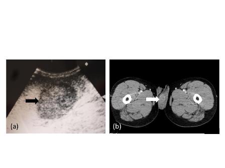
A 70-year-old woman presented with left-sided flank pain without the symptoms of bladder irritation and gross hematuria. The pain was continuous with paroxysmal irritation radiating to the hypogastrium and groin. It was accompanied by nausea but no vomiting. Physical examination was unremarkable except for sensitivity to percussion in her left renal region and tenderness over the course of the left ureter. Urinalysis indicated leukocytes (+) in her urine specimen; her other laboratory data were within normal limits. Ultrasonography revealed one 6-mm diameter calculus in the distal left ureter with hydronephrosis. The pain did not improve after several days’ treatment with anti-inflammatory agents and analgesia. The patient then underwent intravenous urography (IVU), abdominal and pelvic computed tomography (CT), and chest radiography.

The IVU showed a left hydroureter (Fig. 1A). The CT indicated pronounced left hydronephrosis and thickened perirenal tissue within the wall of the left upper ureter. Abdominal multiplanar reconstruction (MPR) demonstrated a mass near the end of the ureter and the thickened, irregular wall of the upper ureter with pronounced hydronephrosis (Fig. 1B). We did not observe any lymphadenopathy in the pelvis, retroperitoneum, or para-aortic regions. A chest radiograph was reviewed, and no abnormality was seen.

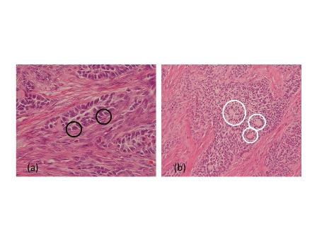
The patient underwent left nephroureterectomy. Macroscopically, a 1.6 × 1.2 × 0.5–cm grayish white lesion was identified.. Histological examination revealed that the mass was composed of small cells with hyperchromatic nuclei, round to fusiform in shape, exhibiting a high level of mitotic activity but little cytoplasm and an absence of nucleoli [Fig. 2A]. Immunohistochemical staining of the specimen was positive for chromogranin A (CgA) [Fig. 2B], CD56, synaptophysin (Syn), neuron-specific enolase, and Ki-67 80%, confirming the neuroendocrine origin of the tumor.

The abdominal and pelvic CT and chest radiography performed 9 months after the operation indicated massive metastases in the liver and lungs [Fig. 3A], lymphadenopathy involving the retroperitoneum, and para-aortic regions, and some implantation metastases in the bladder and left psoas major [Fig. 3B]. This patient died 10 months after operation due to multiple organ failure.

Discussion
Primary small cell carcinoma of the ureter is an extremely malignant and rare disease; only several cases worldwide have been reported so far [1-4]. To our knowledge, PSCC with hydronephrosis has never been reported. The cause of SCC of the ureter may be related to smoking, but it remains unclear [2]. There are two hypotheses regarding the histopathogenesis of urinary tract small cell carcinoma. One indicates that it originates from intrinsic neuroendocrine cells within the normal genitourinary tract derived from the neural crest during embryogenesis [5], whereas the other hypothesis suggests that it results from a transformation of pluripotent epithelial reserve cells in the genitourinary tract that exhibit the ability to generate any cell type [6]. According to Kim Ts [2], SCC of the ureter combined with other components, such as transitional cell carcinoma, adenocarcinoma, squamous cell carcinoma, and carcinoma sarcomatodes, supports the second hypothesis. However, in this case, there are no other components; thus, it is compatible with the first hypothesis.
Because of high mitotic activity [1, 4, 7], small cell carcinoma of the ureter usually obstructs the ureter completely. The ureter above the obstruction or renal pelvis exhibits severe dilation. To our knowledge, severe and long-standing obstruction can give rise to the renal insufficiency or renal failure; the excretion of contrast medium is then not visualized on IVU or CT after contrast administration. In this patient, we did not find these changes. We presume that this was due to the following two reasons: the pressure in the left renal pelvis decreased after the development of the hydronephrosis, and secondly that the function of the right kidney improved to compensate.

The clinical features of PSCC of the ureter are mostly hematuria and flank pain [4]. Hematuria, usually gross, is due to vascular invasion, whereas pain is secondary to hydronephrosis after obstruction of the ureter. However, in this case, the patient only presented with left-sided flank pain and radiating pain in both the hypogastrium and groin. This clinical feature was similar to the presentation of a ureteric calculus. Ultrasonography is a commonly performed examination for urogenital diseases, but its resolution is inferior to CT. We can differentiate a calculus from a mass in the ureter by CT scanning, and demonstrate the development of hydronephrosis and its relationship with surrounding tissues through MPR.

Nephroureterectomy is the primary treatment in the majority of patients with PSCC of the ureter but usually cannot achieve adequate control of the disease; cisplatin-based chemotherapy has been frequently combined with surgery, but the overall outcome is poor [1]. The lymphatics, the lung, and the liver are common metastatic sites for small cell carcinoma of the ureter, and the majority of patients with small cell carcinoma die of the disease within a year [2]. This PSCC of the ureter caused hydronephrosis, and subsequently extensive metastases involving the liver and lungs and lymphadenopathy in the retroperitoneum, and para-aortic region. A the metastasis in the psoas major muscle also occurred 9 months following the surgical procedure.

Conclusion

Primary small cell carcinoma of the ureter is rare and often misdiagnosed as a ureteric calculus or other disease. CT assisted in identifying both the lesion and subsequent metastases, but the final diagnosis depended on pathological confirmation. During operation, surgeons must be mindful of the possibility of implantation metastasis in the perirenal tissue in patients with PSCC of the ureter with hydronephrosis. Radioactive implants may be considered to help prevent metastases. A patient with an early diagnosis and comprehensive therapy may achieve a long-term relative survival
.
Conflict of interest: There are no conflicts of interest in this study.

References
1. Kozyrakis D, Papadaniil P, Stefanakis S, et al. Small cell carcinoma of the urinary tract: a case report. Cases J. 2009;2:7743.
2. Kim TS, Seong DH, Ro JY. Small cell carcinoma of the ureter with squamous cell and transitional cell carcinomatous components associated with ureteral stone.J Korean Med Sci. 2001;16:796-800.
3. Ishikawa S, Koyama T, Kumagai A, Takeuchi I, Ogawa D. A case of small cell carcinoma of the ureter with SIADH-like symptoms. Nippon Hinyokika Gakkai Zasshi. 2004;95:725-8.
4. Kho VK, Chan PH. Primary small cell carcinoma of the upper urinary tract. J Chin Med Asso. 2010;73:173-6.
5. Fetissof F, Dubois MP, Lanson Y, Jobard P. Endocrine cells in renal pelvis and ureter: an immunohistochemical analysis. J Urol. 1986;135:420-1.
6. Christopher ME, Seftel AD, Sorenson K, Resnick MI. Small cell carcinoma of the genitourinary tract: an immunohistochemical, electron microscopic and clinicopathological study. J Urol.1991; 146:382-8.
7. Sved P, Gomez P, Manoharan M, Civantos F, Soloway MS. Small cell carcinoma of the bladder. BJU Int. 2004;94:12-7.
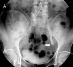
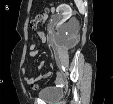
Fig1a040Fig1b040


Fig 1
The IVU shows the dilated ureter (arrow) [A]. The sagittal oblique reconstruction of the left ureter shows the lesion at the end of the ureter (arrow) and hydronephrosis (asterisk) [B]
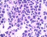
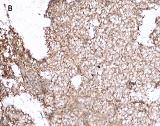
Fig2a040Fig2b040


Fig 2
Light microscopy shows small cells that exhibit hyperchromatic nuclei, round to fusiform in shape, and high mitotic activity with little cytoplasm and an absence of nucleoli (H&E, 400×) [A]. The immunohistochemical staining of the specimen is positive for CgA (100×) [B]
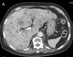
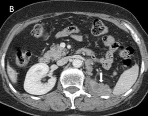
Fig3a040Fig3b040


Fig 3
Many metastases scatter over the liver [A] and one of implantation metastases are in the left psoas major (arrow) [B] 9 months postoperatively

Date added to bjui.org: 06/12/2012
DOI: 10.1002/BJUIw-2012-040-web


Shell-shaped stone in a urethral diverticulum diagnosed by 3D-CT

We report a case of a shell-shaped stone in a urethral diverticulum, which was successfully imaged by three dimensional-CT (3D-CT) filled with air.

Authors: Hisamitsu Ide, Toshiyuki China, Kojiro Nishio, Satoru Muto, Shigeo Horie. Department of Urology, Teikyo University School of Medicine
 
Corresponding Author: Shigeo Horie, M.D. Department of Urology, Teikyo University School of Medicine, Kaga 2-11-1, Itabashi, Tokyo.   Email: [email protected]


Introduction 
A 66-year-old man presented with urinary retention and scrotal hard mass. Computed tomography (CT) scan showed acute prostatitis with prostatic stones and a urethral diverticulum with stone formation. Retrograde urethrography showed little information to place the location of stones in urethra and diverticulum. We report a case of a shell-shaped stone in a urethral diverticulum, which was successfully imaged by three dimensional-CT (3D-CT) filled with air.


Introduction
 
Male urethral diverticula are rare and can be congenital or acquired [1, 2]. Some diverticula can be complicated by infection or stone formation [2]. We report a case of an acquired diverticulum with stones, accompanied by urethral stricture. We show our three dimensional-computed tomography (3D-CT) image technique, with air filling of urethra, to identify the position of diverticulum and stones.Case Report
 A 66-year-old man was referred to our hospital complaining of urinary retention and scrotal hard mass. In his past history, he received urethral injury around 40 years old. A scrotal mass was found intra-scrotally as a hard and cylindrical shape with smooth surface on physical examination. The mass was palpable around urethra and clearly delineated from the testis and epididymis, while the left testis was atrophic. Transillumination was negative. Urine analysis revealed microscopic haematuria (RBC>100/HPF) and urinary inflammation(WBC10-19/HPF). Urine culture showed Streptococcus agalactiae (5X10⁵). Physical examination and laboratory tests suggested acute prostatitis. A plain X-ray demonstrated several high-density nodules in the pelvic cavity. The patient underwent plain CT, which showed prostatitis with prostatic stones and suspected urethral diverticulum with stone formation (Figure 1).


Figure. 1 Computed tomography (CT) showing urethral stones and diverticulum with stone formation.
Computed tomography (CT) showing urethral stones and diverticulum with stone formation.


Retrograde urethrography showed little information to place the precise location of stones in urethra and diverticulum; urethral stricture was suspected in the anterior urethra. To evaluate the exact position of diverticulum and urethral stones, a 3D-CT technique employing air-filling of the urethra was performed. We observed two shell-shaped stones and several stones in a urethral diverticulum, prostatic stones, and small urethral and bladder stones (Figure 2).


Figure. 2 Three-dimensional computed tomography (3DCT) image technique filled with air in urethra. Green area; bladder, Blue area; urethra and diverticulum. Black arrow shows the shell-shaped stones in urethral diverticulum. White arrow shows prostatic stones and small urethral stones.


After treatment with antibiotic medicine to recover the acute prostatitis, he initially received cutaneous vesicostomy, endoscopic transurethral incision and cystolithotripsy. We extracted some stones endoscopically and observed the opening of diverticulum, however we could not evacuate the diverticular stones. He subsequently underwent open diverticulectomy and stone removal two months later. Two shell-shaped stones with several small stones were removed. The infrared spectroscopic analysis revealed that the stones were composed of calcium phosphate. One month after this operation, the catheter was removed and he was able to void.


Discussion  
Computed tomographic colonography is a minimally invasive technique for imaging the entire colon. In this technique, the patient’s colon is cleansed and transiently inflated with air. Subsequently, a 3D image volume is acquired of the abdomen by CT. Finally, the bowel surface images are extracted and visualized, after which the physician can examine the colonic surface virtually for abnormalities [3]. For CT urethrography or virtual urethroscopy, diluted water-soluble iodine contrast medium is usually introduced to bladder by Foley catheter [4, 5]. However, in our case, we used an air technique to diagnose provisionally urethral stones and urethral diverticulum, because the patient had urethral stricture with multiple urethral stones. There are many factors affecting the successful performance of CT uretherography. Adequate air distention, the optimal CT technique and interpretation with using the newest software by a trained reader will help ensure high accuracy for lesion detection. Air distension in the urethra for 3D-CT may improve the diagnostic accuracy and allow for low cost intervention. With continued research and development, the use of such 3D-CT imaging techniques may become appropriate for future clinical diagnostic applications to evaluate the lower urinary tract.


References
[1] Mohan V, Gupta SK, Cherian J, Tripathi VN, Sharma BB. Urethral diverticulum in male subjects: report of 5 cases. J Urol. 1980 Apr: 123:592-4
[2] Ho CH, Yu HJ, Huang KH. Scrotal mass with bladder outlet obstruction. Urology. 2008 Jul: 72:66-7
[3] Achiam MP, Bulow S, Rosenberg J. CT- and MR colonography. Scand J Surg. 2002: 91:322-7
[4] Chou CP, Huang JS, Yu CC, Pan HB, Huang FD. Urethral diverticulum: diagnosis with virtual CT urethroscopy. AJR Am J Roentgenol. 2005 Jun: 184:1889-90
[5] Chou CP, Huang JS, Wu MT, et al. CT voiding urethrography and virtual urethroscopy: preliminary study with 16-MDCT. AJR Am J Roentgenol. 2005 Jun: 184:1882-8


Date added to bjui.org: 20/04/2011 


DOI: 10.1002/BJUIw-2011-022-web


© 2024 BJU International. All Rights Reserved.