Tag Archive for: Editorial

Posts

Editorial: From Novick to the NHS – the evolution of minimally-invasive NSS

The publication in this issue of the BJUI by Veeratterapillay et al. [1] of a UK multicentre study in a community setting marks a watershed in the availability and quality of minimally invasive nephron-sparing surgery (NSS) for renal cancer. Such a turning point was predicted almost 17 years ago by Novick [2] when he wrote, ‘minimally invasive modalities of tumour resection or destruction should be reserved for highly select patients and awaits improvements in technology, standardization of technique and long-term outcomes data before they may be completely integrated options’. It appears now that robot-assisted surgery provides such a platform. The present study [1] describes the outcomes of patients treated with robot-assisted partial nephrectomy (RAPN) at four centres in Northern England, and shows very good outcomes within their first 250 cases.

The benefits of NSS have been well described. Indeed, excellent outcomes for PN were described over 20 years ago in carefully selected cases, with benefits including reduced incidence of renal insufficiency compared to radical nephrectomy, which until that time had been viewed as the ‘gold-standard’ for patients with RCC [3]. However, the popularity of PN for small renal masses appeared to decline with the advent of laparoscopy. It became apparent that a minimally invasive approach to radical nephrectomy had the advantage of improved recovery, reduced blood loss with equal cancer control to open nephrectomy [4]. Notwithstanding absolute and relative indications for PN, given the choice between an open PN and a laparoscopic radical nephrectomy, the balance for patients with an elective indication for PN was tipped in favour of a minimally invasive yet radical approach [5]. Techniques for PN were in their infancy, and even in the leading high-volume centres outcomes, including warm ischaemia time (WIT) and positive surgical margin (PSM) rate, failed to match those of open surgery [6].

Fast forward to 2017 with the increasing use of robot-assisted urological surgery carrying the advantages of three-dimensional vision, wristed movement and integrated real-time intraoperative imaging, especially beneficial for procedures such as PN where quick and accurate suturing are essential for a successful outcome. Veeratterapillay et al. [1] present a series of 250 patients from centres in the UK, in which each performs <50 RAPN procedures/year, yet the authors present favourable outcomes overall, with a PSM rate of 7.3%, major complications in 6% and trifecta in 68.4%. An impressive learning curve is seen with improving outcomes over the series, such that in the final 50 cases a trifecta (WIT <25 min, negative surgical margin and absence of complications) was achieved in 82% of cases, with a PSM rate of 2% despite increasing complex nephrometry scores, which compares favourably with larger series from internationally renowned centres [6].

So then, with the results of the present study [1], can we say that Novick’s requirements have been met, and that minimally invasive NSS is now a ‘completely integrated option’? Certainly, with the widespread adoption of robot-assisted surgery, high-quality outcomes are within the grasp of centres other than elite academic institutions. As techniques develop and experience grows robot-assisted surgery can be increasingly offered, even for resection of more complex tumours.

To ensure that minimally invasive NSS is delivered to the highest standards, it will be necessary for providers to ensure both quality assurance and quality control in their processes. The learning curve needs to be minimised with structured teaching and mentoring, and the use of adjuncts such as intraoperative ultrasonography or fluorescence should be a routine part of care.

Centres offering this technique should be mindful of the well documented volume–outcome relationship that appears to be ubiquitous among complex surgical procedures. If centres are performing less than an optimum number of cases, they may consider affiliating themselves with other such centres in networks and forming a joint clinical governance programme, as has been described for robot-assisted radical prostatectomy and which has shown demonstrable improvements in outcomes.

Finally, auditing and reporting of outcomes remains the cornerstone of quality assurance as shown by the introduction of the BAUS complex surgery audit, which is intended to drive standards of care forward. Publications such as that of Veeratterapillay et al. [1] greatly assist in documenting the progress of new techniques and emerging technologies. Increasingly, patients expect transparency from healthcare providers, and with the necessary support processes in place, such initiatives, and the data that they produce will help to further improve the delivery of complex surgery to patients from all areas of our practice.

Benjamin W. Lamb* and Daniel A. Moon*


*Division of Cance r Surgery, Peter MacCallum Cancer Centre, Epworth Healthcare, and Department of Surgery, Central Clinical School, Monash University, Melbourne, Vic., Australia


References


1 Veeratterapillay R, Addla SK, Jelley C et al. Early surgical outcomes and oncological results of robot-assisted partial nephrectomy: a multicentre study. BJU Int 2017; 120: 5505


2 Uzzo RG, Novick AC. Nephron sparing surgery for renal tumors: indications, techniques and outcomes. J Urol 2001; 166: 618


3 Polascik TJ, Pound CR, Meng MV, Partin AW, Marshall FF. Partial nephrectomy: technique, complications and pathological ndings. J Urol 1995; 154: 131218


4 Gill IS, Meraney AM, Schweizer DK et al. Laparoscopic radical nephrectomy in 100 patients. Cancer 2001; 92: 184355


5 Novick AC. Laparoscopic and partial nephrectomy. Clin Cancer Res 2004; 10: 6322S7S


Editorial: Do all patients with renal cell carcinoma need a chest computed tomography?

While all patients with RCC need chest imaging for staging evaluation, the answer to the question in the title is ‘No’, and, in fact, many patients would be adequately staged with a chest X-ray, albeit with reduced accuracy. Evidence to support this assertion is provided by Larcher et al. [1] in this issue of BJUI, who retrospectively evaluated 1946 patients with a solitary and sporadic RCC mass. While excluding patients who did not have surgery and those with visceral metastases seen on abdominal imaging, the authors observed pulmonary metastases in 6% (119 patients) of their population. In a multivariable analysis, features associated with a positive chest CT included cT1b+, cN1, systemic symptoms, anaemia, and thrombocytosis. Incorporating these features into a predictive model, the authors report a robust concordance index of 0.88, with the effect of each feature demonstrated in a nomogram. Further, the authors report that if a chest CT is only performed when the risk of a positive result is >1%, 37% of their population could have been spared a chest CT while missing a positive result in only 0.2% (four patients). Patient factors that predict for a <1% risk of a positive chest CT essentially include those with cT1aN0 RCC without systemic symptoms, anaemia, or thrombocytosis. Thus, the authors conclude that in these low-risk patients, a chest CT can be omitted, while any patient that is cT1b+, cN1, or with systemic symptoms, anaemia, or thrombocytosis warrants a dedicated chest CT at diagnosis.

The finding that patients with RCC with smaller tumours (cT1a or ≤4 cm) were unlikely to harbour pulmonary metastases is consistent with prior literature. Observations from the Memorial Sloan-Kettering Cancer Center (MSKCC) [2], and subsequently validated by our group at Mayo Clinic [3], suggested that among surgically treated patients with RCC, risk of M1 disease (at any location) at diagnosis was non-existent for tumours of <2 cm, was <1% for tumours of 2–3 cm, and was only 1–2% for tumours of 3-4 cm in size. Given that it is rare for patients with small renal masses to endorse systemic symptoms or have paraneoplastic symptoms related to the tumour, these prior observations suggest a lack of utility for chest CT for patients with small renal masses supporting the findings from Larcher et al. [1].

In patients with RCC with synchronous metastases, lung is the most common site of spread and guidelines uniformly recommend chest imaging at diagnosis. However, a ‘select-all’ strategy for chest CT in patients with renal masses leads to unnecessary findings in those with a benign primary tumour, increased use of healthcare resources, and relatively frequent findings of indeterminate lesions. In fact, contemporary observations from the MSKCC found that about half of patients with RCC undergoing surgery had indeterminate pulmonary nodules on chest CT that required either additional evaluation or subsequent chest CT to document stability [4]. Further, the presence of indeterminate pulmonary nodules was not associated with distant metastases or death from RCC after surgery unless they were >1 cm, which only represented a small portion (4%) of the entire cohort [4]. Thus, the analysis from Larcher et al. [1] in this issue of BJUI has meaningful clinical relevance; that is, patients with cT1aN0 RCC without symptoms or laboratory abnormalities do not require a chest CT for screening of their lungs.

R. Houston Thompson
Department of Urology, Mayo Clinic, Rochester, MN, USA


References


1 Larcher A, DellOglio P, Fossati N et al. When to perform preoperative chest computed tomography for renal cancer staging. BJU Int 2017; 120: 4906


2 Thompson RH, Hill JR, Babayev Y et al. Metastatic renal cell carcinoma risk according to tumor size. J Urol 2009; 182: 415


3 Umbreit EC, Shimko MS, Childs MA et al. Metastatic potential of renal mass according to original tumour size at presentation. BJU Int 2011; 109: 1904


Editorial: Immortal-Time Bias – A Crucial Yet Overlooked Confounder in Urological Research

The measurement of treatment effect through observational studies has become commonplace in the medical literature. These cohort studies provide valuable data on outcomes that can be difficult to assess in randomized controlled trials, such as long-term mortality. Accurate interpretation of observational data, however, requires accounting for potential confounders of study design, including the immortal-time bias. In this issue of BJUI, Wallis et al. [1] show how accounting for this bias can influence the measured effect of cumulative testosterone exposure on mortality. The implications of their findings extend to several other studies, whose designs may also be subject to immortal-time bias.

‘Immortal time’ refers to the portion of a follow-up period during which an outcome could not have occurred (e.g. subjects in the ‘exposure group’ cannot die before they receive the exposure); thus, potentially allowing the artificial magnification of an effect on the study outcome [2]. As the authors point out, this concept is not new. It was first identified several decades ago to highlight how a study’s finding of a survival advantage for patients undergoing heart transplant was nullified once immortal time was properly accounted for [3]. Despite its long existence in epidemiological teachings, the authors cite several studies both within and outside of the urological literature that have failed to appropriately account for this bias. Many of these studies employ binary exposure variables, but Wallis et al. delve into relatively uncharted territory by examining the effect of immortal-time bias on multi-level categorical exposures.

The relationship between testosterone replacement therapy (TRT) and mortality, the focus of the accompanying study, is apt because it is a controversial topic that weighs heavily on an accurate assessment of the therapy’s risks and benefits. The authors, using data from their own prior study, show that this delicate balance can be easily tipped when immortal-time bias is not properly accounted for. In their analysis, the overall result was the same regardless of controlling for this bias; men in the lowest tertile of TRT exposure had a higher risk of mortality, and those in the highest tertile had a lower risk of mortality; however, use of a time-fixed as opposed to the more appropriate time-varying analysis led to a substantial magnification of the effect size in each direction. While the overall result may have been the same in this example, the authors cite other instances of high-impact research whose published conclusions were shown to be completely different once accounting for immortal-time bias [4]. One can easily imagine how this type of erroneous data analysis could have deleterious consequences in the clinical setting. Healthcare providers rely on research to make decisions that have far-reaching impacts on patients’ lives. This study highlights the importance of ensuring that such analyses are carried out properly so that patients can receive the high-quality, evidence-based care they deserve.

The authors should be commended for taking the time to deconstruct and evaluate an analytical concept that is pertinent to study designs across several disciplines. Much of the research published today seeks to find answers to important clinical questions, but not nearly enough investigation is devoted to verifying that the analyses to obtain these answers are conducted properly. Urology in particular is a field that is still maturing with respect to the use of secondary data analytical techniques, such as propensity score models and instrumental variables [5]. To sustain our improvement in investigative skills alongside our fellow medical disciplines, we must pay special attention to studies that hold a magnifying glass to commonly used methodologies in the urological literature. In a similar vein, there have been increasing efforts recently to improve the process and transparency of corroborating the results of scientific studies, and these authors’ findings reinforce why these efforts are so crucial. If we expect to continue pushing forward the boundaries of medical research, it is our duty to ensure that our analytical methods are as rigorous and accurate as possible.

Sean A. Fletcher, Philipp Gild and Quoc-Dien Trinh
Division of Urological Surgery and Center for Surgery and Public Health, Harvard Medical School, Brigham and Womens Hospital, Boston, MA, USA


Read the full article


References


2 Suissa S. Immortal time bias in pharmaco-epidemiology. Am J Epidemiol 2008; 167: 4929


3 Gail MH. Does cardiac transplantation prolong life? A reassessment Ann Intern Med 1972; 76: 8157


4 van Walraven C, Davis D, Forster AJ et al. Time-dependent bias was common in survival analyses published in leading clinical journals. J Clin Epidemiol 2004; 57: 67282


5 Cole AP, Trinh QD. Secondary data analysis: techniques for comparing interventions and their limitations. Curr Opin Urol 2017; 27: 3549


Editorial: The utility of microRNAs as biomarkers in predicting progression and survival in patients with clear-cell renal cell carcinoma

RCC constitutes a diverse group of malignancies, yet the clear-cell subtype comprises ~80% of all diagnosed RCC cases [1]. The widespread use of abdominal imaging and subsequent stage migration has resulted in improved RCC 5-year cancer-specific survival. However, the overall mortality of RCC remains largely unchanged [2] and one-third of the patients have metastatic disease at the time of presentation [3]. Accordingly, the ability to precisely predict patient outcome has become an increasingly significant question in the management of these patients with RCC.

Accruing evidence suggests that changes in various biomarkers and their consequent downstream pathways affect cancer initiation and progression. Therefore, accurate prediction of the outcome and prognosis after treatment is necessary [4]. MicroRNAs (miRNAs) are small non-coding RNA molecules that can have significant functions in tumorigenesis [5]. Because of their ability in post-transcriptional regulation of gene expression, tumour-specific genetic defects in miRNA biogenesis and production correlate with development of human cancers. Thus, the differential expression of specific miRNA signatures in different tumours might become an important tool to help in directing cancer diagnosis and treatment [5].

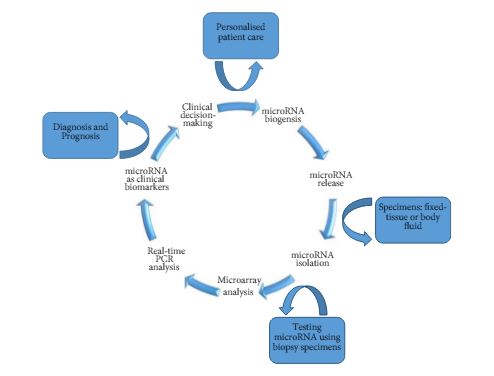
As such, Kowalik et al. [6] report on profiling miRNA to identify biomarker signatures predictive of clear-cell RCC (ccRCC) progression and survival. The authors used 202 formalin-fixed paraffin-embedded samples to isolate RNA from nephrectomy and biopsy specimens (n = 156 and n = 46, respectively) (Fig. 1).

Figure 1. Schematic diagram of miRNA-based biomarkers and potential utility in clinical decision-making approach for targeted therapy and RCC personalised treatment.

The primary analysis of their study [6] focused on the identification of miRNA signatures capable of differentiating between benign and ccRCC, as well as discerning those patients with a non-progressive ccRCC from a progressive clear-cell subtype. The secondary outcome examined the association of miRNA profiles discovered on cancer-specific survival.

In their initial microarray screening 20 differentially expressed miRNAs, comparing non-progressive with progressive tumours, were identified. The authors found four miRNA panels (10a-5p, 10b-5p, 106a-5p, and 142-5p) as a potential biomarker signature. This model was validated in nephrectomy specimens and resulted in a sensitivity of 86.7%, a specificity of 92.9%, and an area under the curve (AUC) of 0.930 for detecting ccRCC. Further analysis revealed a second signature of two biomarkers (miR-10a-5p and -223-3p) with 93.8% sensitivity, 83.3% specificity, and an AUC of 0.932 when validated for detecting progressive ccRCC. Similarly, the differential expression of these biomarkers could delineate cancer status in biopsy specimens. For correlation of miRNA expression levels with cancer-specific survival, higher expression levels of (miR-10a-5p and miR-10b-5p) and a lower expression level of (miR-223-3p) were significantly associated with survival (P< 0.001), and the median survival times were not reached.

In conclusion, the lack of precise prediction tools has led the authors to explore the potential utility of miRNAs as biomarkers to detect disease presence, biological aggressiveness, and prognosis in ccRCC. However, until future multicentre large prospective studies validate the results of the present work, the transition of miRNA from bench to bedside is emerging on the horizon and has encouraged urologists and scientists to pursue intense translational research in the field. The ability to use miRNAs as biomarkers might be promising for diagnostic and prognostic purposes. These biomarkers may exemplify different aspects of RCC pathogenesis and may potentially have important therapeutic implications to help in a clinical decision-making approach for targeted therapy and RCC personalised treatment.

Firas G. Petrosand Christopher J.D. Wallis†‡
*Department of Urology, The University of Texas MD Anderson Cancer Center, Houston, TX, USADivision of Urology, Department of Surgery, University of Toronto, Toronto, ON, Canada and Division of Urology, Department of Surgery, Sunnybrook Health Sciences Centre, Toronto, ON, Canada


Read the full article


References


1 Reuter VE, Presti JC Jr. Contemporary approach to the classication of renal epithelial tumors. Semin Oncol 2000; 27: 12437


2 Hollingsworth JM, Miller DC, Daignault S, Hollenbeck BK. Rising incidence of small renal masses: a need to reassess treatment effect. J Natl Cancer Inst 2006; 98: 13314


3 Gupta K, Miller JD , Li JZ, Russell MW, Charbonneau C. Epidemiologic and socioeconomic burden of metastatic renal cell carcinoma (mRCC): literature review. Cancer Treat Rev 2008; 34: 193205


5 Esquela-Kerscher A, Slack FJ. Oncomirs microRNAs with a role in cancer. Nat Rev Cancer 2006; 6: 25969


Editorial: Is choline-based PET imaging still relevant in recurrent prostate cancer?

The search for the ideal imaging method to detect small metastatic deposits has largely remained elusive in the field of prostate cancer. However, functional positron emission tomography (PET)/CT is now guiding us in different directions. 11C-acetate PET/CT imaging was one of the first molecular imaging probes showing promise in clinical studies, along with 11C-choline, and more recently challenged by 68Ga-prostate-specific membrane antigen (PSMA) PET/CT [1-3]. So against this background, what does this new study by Esch et al. [4] presented here offer us?

Let us rewind and focus on where molecular imaging may have an impact in prostate cancer. In primary staging, the potential to accurately identify oligometastatic disease or even widespread metastatic disease before primary gland treatment is clearly advantageous. It can allow for wider treatment fields (extended lymphadenectomy or radiation fields), directed treatment of oligometastatic disease, placement into appropriate cytoreductive trials, and in some instances consideration of the use of earlier chemo-hormonal therapy. This study did not address this important clinical scenario [4]. Nor did it focus on primary diagnosis, another ‘holy grail’ for imaging more recently dominated by MRI [5].

The clinical question addressed in this study was whether 11C-acetate PET is able to detect distant metastatic disease in men with PSA relapse after having undergone radical prostatectomy or prostate bed radiotherapy [4]. In other words, if distant metastatic disease is found then prostate bed radiation may be futile, although this assumption has not been subjected to high quality trials, but would seem logical. Also, in the era of oligometastatic disease, treatment (surgery and/or radiation) may be directed at nodal and other deposits to prolong time of systemic therapy such as androgen-deprivation therapy. So what is the ‘sweet spot’? Some would state for men having undergone surgery a PSA level of 0.2 ng/mL places the patient at risk of recurrence, although in clinical practice the level at which investigations are warranted can be as high as 1.0 ng/mL. This study found 11C-acetate PET had few positive results below a PSA level of 1.0 ng/mL. This is in contrast to a recent meta-analysis of 68Ga-PSMA PET/CT reporting positive studies at PSA levels of ≤0.2 ng/mL in 42% of patients [6].

The other group studied the detection of recurrent disease in men having undergone primary radiotherapy, although the numbers were low (six patients). We know this group of men are often undertreated for salvage treatment, and frequently placed on hormonal therapy. Again, early knowledge of locoregional or distant recurrence is required to allow best case selection and thus avoid futile salvage surgery, and also to know who may benefit from salvage lymphadenectomy and treatment of distant oligometastatic disease, if required.

The false-positive rate result for 11C-acetate PET/CT of 24% is notable and concerning. This may lead to unnecessary intervention, or even withholding prostate bed radiation that may have been of benefit. In contrast the false-positive rate for 11C-choline and 68Ga-PSMA PET/CT is far lower, although false-positive PSMA studies may occur in benign conditions and non-prostate cancer tumours.

It should be acknowledged that a strength of this study was comparative histology in a proportion of patients, allowing true sensitivity and specificity to be determined [4]. This is important information, as it guides correct decision-making, and such information is lacking for many other prostate cancer imaging probes, including 68Ga-PSMA, where these data are urgently required.

Overall, this study is important and adds to the rich milieu of available molecular imaging data on staging of possible recurrent or metastatic prostate cancer to date [4]. Current prospective trials exploring 11C-choline, 18F-FACBC (anti-1-amino-3-18F-fluorocyclobutane-1-carboxylic acid), 18F-choline and 68Ga-PSMA with MRI and clinical outcomes should provide further insight into the most appropriate molecular imaging technique for staging prostate cancer.

Nathan Lawrentschuk, BJUI USANZ Editor*,,, Julia M. Coreldand Andrew Scott,§,¶
*Department of Surgery, University of Melbourne and Olivia Newton-John Cancer Research Institute, Austin Hospital , Peter MacCallum Cancer Centre, La Trobe University, Departments of


Medicine, § Molecular Imaging and Therapy
Read the full article


References


1 Afshar-Oromieh A, Zechmann CM, Malcher A et al. Comparison of PET imaging with a (68)Ga-labelled PSMA ligand and (18)F-choline- based PET/CT for the diagnosis of recurrent prostate cancer. Eur J Nucl Med Mol Imaging 2014; 41: 1120


2 Silver DA, Pellicer I, Fair WR, Heston WD, Cordon-Cardo C. Prostate- specic membrane antigen expression in normal and malignant human tissues. Clin Cancer Res 1997; 3: 815


5 Johnson LM, Turkbey B, Figg WD, Choyke PL. Multiparametric MRI in prostate cancer management. Nat Rev Clin Oncol 2014; 11: 34653


Article of the Week: Detecting SNs in patients with BCa intra-operatively

Every Week the Editor-in-Chief selects an Article of the Week from the current issue of BJUI. The abstract is reproduced below and you can click on the button to read the full article, which is freely available to all readers for at least 30 days from the time of this post.

In addition to the article itself, there is an accompanying editorial written by a prominent member of the urological community. This blog is intended to provoke comment and discussion and we invite you to use the comment tools at the bottom of each post to join the conversation.

If you only have time to read one article this week, it should be this one.

Radio-guided sentinel lymph node detection and lymph node mapping in invasive urinary bladder cancer: a prospective clinical study

Firas Aljabery1,2,*, Ivan Shabo2,3,4, Hans Olsson2,5, Oliver Gimm2,6 and Staffan Jahnson1,2

1 Department of Urology, Region Östergötland, Linköping University Hospital, Linköping, Sweden, 2 Department of Clinical and Experimental Medicine, Faculty of Health Sciences, Linköping University, Linköping, sweden 3 Endocrine and Sarcoma Surgery Unit, Department of Molecular Medicine and Surgery, Karolinska Institute, Stockholm, Sweden 4 Department of Breast and Endocrine Surgery, Karolinska University Hospital, Solna Stockholm, Sweden 5 Department of Pathology, Region Östergötland, Linköping University Hospital, Linköping, Sweden 6 Department of Surgery, Region Östergötland, Linköping University Hospital, Linköping, Sweden

Read the full article

Abstract

Objectives

To investigate the possibility of detecting sentinel lymph nodes (SNs) in patients with urinary bladder cancer (BCa) intra-operatively and whether the histopathological status of the identified SNs reflected that of the lymphatic field.

Patients and Methods

We studied 103 patients with BCa pathological stage T1–T4 who were treated with cystectomy and pelvic lymph node (LN) dissection during 2005–2011 at the Department of Urology, Linköping University Hospital. Radioactive tracer Nanocoll 70 MBq and blue dye were injected into the bladder wall around the primary tumour before surgery. SNs were detected ex vivo during the operation with a handheld Geiger probe (Gamma Detection System; Neoprobe Corp., Dublin, OH, USA). All LNs were formalin-fixed, sectioned three times, mounted on slides and stained with haematoxylin and eosin. An experienced uropathologist evaluated the slides.

Results

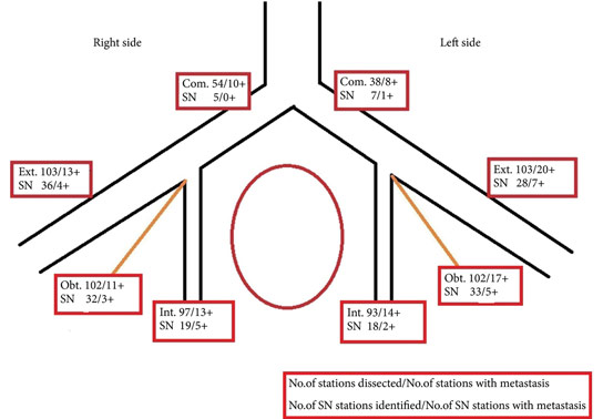
The mean age of the patients was 69 years, and 80 (77%) were male. Pathological staging was T1–12 (12%), T2–20 (19%), T3–48 (47%) and T4–23 (22%). A mean (range) number of 31 (7–68) nodes per patient were examined, totalling 3 253 nodes. LN metastases were found in 41 patients (40%). SNs were detected in 83 of the 103 patients (80%). Sensitivity and specificity for detecting metastatic disease by SN biopsy (SNB) varied between LN stations, with average values of 67% and 90%, respectively. LN metastatic density (LNMD) had a significant prognostic impact; a value of ≥8% was significantly related to shorter survival. Lymphovascular invasion (LVI) occurred in 65% of patients (n = 67) and was significantly associated with shorter cancer-specific survival (P < 0.001).

Conclusion

We conclude that SNB is not a reliable technique for peri-operative localization of LN metastases during cystectomy for BCa; however, LNMD has a significant prognostic value in BCa and may be useful in the clinical context and in BCa oncological and surgical research. LVI was also found to be a prognostic factor.

Read more articles of the week

Editorial: Positive messages for bladder cancer management in negative sentinel lymph node study

I encourage you to read the study by Aljabery et al. [1] in this edition of BJUI. Their findings are based on some very solid methodology and I think provide a robust answer to their question, which often in science means a ‘negative’ result. The principle of sentinel lymph node biopsy (SLNB) needs no introduction. It is primarily intended to detect the principal LN draining a tumour, allowing its removal and pathological determination of LN metastasis status in that individual [2].The avoidance of an unnecessary LN dissection (LND) and its associated risks is at the heart of any SLNB strategy. On the other hand, particularly for bladder cancer, there is a recognition that a higher number of LNs removed at the time of surgery confers a survival advantage to patients through more accurate staging [3]. With greater numbers of LNs removed pN0 patients are more likely to be truly N0 and pN1 patients with limited metastases have a greater chance that all disease has been completely excised. Thus, when considering SLNB in bladder cancer there is the usual conflict between maximising oncological benefit and minimising surgical harm.Aljabery et al. [1] present an excellent series of cystectomies with a 100% negative margin rate and mean LN count of 30. The 40% rate of LN involvement, is perhaps partly due to the meticulous triple sectioning of each excised LN. Their SLN technique involved four cystoscopic injections to the bladder wall surrounding the tumour and focused on the biggest lesion in multiple tumour cases. The LNs were removed in their packets and studied after removal from the patient. While this is likely to be a more precise method for determining the site of the SLN, it clearly differs from the approach one would take if trying to avoid LND in negative-SLN cases. Furthermore, examination of LNs was performed after formalin fixation. Typically when using SLN techniques frozen sections are also used to guide surgeons during surgery.The results clearly show that SLNB using radiolabelled nanocolloid does not allow accurate identification of pathologically LN-negative patients who could then avoid a complete LN dissection. Sensitivity of the technique in the detection of positive LNs ranged from 67% to 90% at the various LN stations. Overall, of patients with an identifiable SLN that was negative, 19% of patients had positive LNs elsewhere (81% negative predictive value). Effectively one in five patients who might be reassured by a negative SLN result would in fact have undetected positive LNs left behind if this technique were employed. Furthermore, this estimation does not consider errors likely to be introduced with in situ SLN identification and the use of frozen-section analysis rather than non-time-critical analysis of formalin-fixed sections.In such a dangerous disease such inaccuracy is not tolerable and so I totally agree with the authors’ [1] findings that SLNB of pelvic LNs at the time of radical cystectomy for bladder cancer is not a reliable technique for identifying LN metastasis.The positive messages from this study [1] are worth noting by those learning and undertaking cystectomy. The authors’ meticulous approach to surgery is evident from the methodology described and the accumulation of such a well-characterised series. This must be a contributing factor in achieving a 100% negative surgical margin rate and such consistently high LN yields. This should certainly be the aim of all cystectomists. The appropriate time, skill and patience should be given to this step and it should not be compromised upon, particularly when developing robot-assisted or laparoscopic cystectomy services.The findings that T-stage, N-stage and lymphovascular invasion are linked to survival are not that surprising. However, the use of LN metastatic density as a prognostic marker is interesting, as it is not usually discussed in our multidisciplinary meetings. This measure incorporates nodal tumour burden and the extent of LND. The finding of better outcomes in those with a LN metastatic density of <8% reinforces the message that even in those with LN metastases, removing greater numbers of LNs may improve prognosis. Furthermore, the finding that 30% of unilateral LN-positive tumours also had contralateral LNs settles any arguments for unilateral LN dissections.In a recent systematic review of SLNB in bladder cancer [4], the negative predictive value was found to be 92% compared to 81% in the Aljabery et al. [1] study. The authors of the systematic review suggested that SLNB is a promising technique; perhaps in view of technology advances they reviewed that might improve future outcomes of SLNB. While improvements may be possible, current evidence would not encourage me to consider SLNB using radiolabelled nanocolloid for fear of impairing cancer outcomes.

Congratulations to Aljabery et al. [1] on their work. I hope you find reading their paper as constructive as I did.

Tim Dudderidge
Department of Urology, University Hospital Southampton,
Southampton, Hampshire, UK

Read the full article

References

1 Aljabery F, Shabo I, Olson H, Gimm O, Jahnson S. Radio-guided sentinel lymph node detection and lymph node mapping in invasive urinary bladder cancer: a prospective clinical study. BJU Int 2017; 120: 329–36

2 Gould EA, Winship T, Philbin PH, Kerr HH. Observations on a “sentinel node” in cancer of the parotid. Cancer 1960; 13: 77–8

3 Koppie TM, Vickers AJ, Vora K, Dalbagni G, Bochner BH. Standardization of pelvic lymphadenectomy performed at radical cystectomy. Cancer 2006; 107: 2368–74

4 Liss M, Noguchi J, Lee H, Vera D, Kader AK. Sentinel lymph node biopsy in bladder cancer: systematic review and technology update. Indian J Urol 2015; 31: 170–5


Editorial: sLND – if yes, Robotics?

The manuscript in this issue of the BJUI by de Castro Abreu et al. [1] reports the results of the first series of patients to undergo robotic-assisted salvage lymph node dissection (sLND) for nodal recurrence of prostate cancer. Despite the absence of a high level of evidence, sLND has been gaining attention in recent years. Indeed, several series have shown promising results of such an approach, especially in terms of PSA response to surgery and delay in clinical recurrence [2-4]. However, sLND is a complex surgery and is not devoid of complications, as in up to 13.8% of patients Clavien–Dindo ≥IIIa complications occur [5]. When analysing the results of the current manuscript [1], it is impressive to read that the mean number of LNs removed was 83, ranging from 41 to 132, which is significantly higher than the reported figures of open sLND series. Moreover, despite the long median operative time (4.8 h), complication rates and postoperative course were excellent as compared to previously published series, although a direct comparison between the open and robot-assisted approach should be only addressed in prospective studies. The authors should be congratulated on the superb results obtained during the learning curve of such complex surgery, but some issues need to be discussed.

First, it is difficult to understand whether such results apply only to very expert surgeons. In other words, is it possible to translate such surgery to less experienced robotic surgeons? Second, is it necessary to extend the LND to a similar extent in all cases? Previous reports have shown that patients with retroperitoneal involvement may not benefit from sLND as much as their counterparts with only pelvic involvement [2]. The authors [1] show no significant impact of such an extended approach on complications and postoperative course, but the invasiveness of such an extended approach needs to be justified in each case. Third, the introduction of new tracer methods, such as prostate-specific membrane antigen (PSMA) positron emission tomography/CT, with higher specificity for prostate cancer may reduce the need for such extended templates, without compromising the oncological results [6]. Fourth, is the robotic approach feasible and safe in patients previously submitted to radical prostatectomy independently from the approach (open vs laparoscopic/robotic), from the extent of the previous LND, as well as from the previous administration of adjuvant/salvage radiotherapy? All these answers will need to be addressed in future studies on subgroups of patients undergoing sLND. Most importantly, until a high level of evidence is available, sLND should still be considered experimental and should be performed by highly experienced surgeons.

Read the full article
Nazareno Suardi and Francesco Montorsi
Department of Urology, Urological Research Institute, Vita Salute San Raffaele University, Milan, Italy


References

Editorial: Sexuality in men with exstrophy

It is always exciting to get new data on exstrophy from Johns Hopkins, but especially when sexual development is the subject [1]. It is the only unit with enough patients on continuous follow-up to overcome the difficulties of researching such a rare condition.

In the last 40 years, patients born with exstrophy have achieved a near normal life-expectancy. Reconstructive techniques for the bladder are now such that incontinence is rare, although often bladder emptying depends on clean intermittent self-catheterisation [2]. As with all fit young men, their minds turn frequently to sex and, occasionally, its natural consequence – pregnancy.

Current data have established that the men have a normal libido, orgasms, and erections. It is probable that the testes are normal at birth but often are damaged by recurrent infections. The penis is short, broad and has a characteristic chordee. Other erectile deformities are probably the result of corporeal damage during reconstruction in infancy. Most of these are surgically correctable. Ejaculation is poor or absent [3, 4].

Data on the men’s own satisfaction are contradictory and there are none on the partner’s opinions. Masturbation is almost universal. The incidence of erectile dysfunction is more than double that of controls (58% vs 23%) [3]. Much the commonest cause is fear of rejection by a partner because of the obvious penile anomalies. Most series show that men like to establish a good partnership before starting intercourse, although at least one group report that random and short-term relationships are common [5]. Unfortunately the published series are small and few of them address sexuality in a structured manner.

At Johns Hopkins the exstrophy database now has >1 200 patients and there is a programme for close and indefinite review. This is good for the patients and good for outcomes research. Sexual function has been investigated in 113 adult men (53% of those eligible) using a 42-question survey, which incorporated four validated instruments and additional questions related to sexuality [1].

In all, 85% had been sexually active at some time and 62% were currently in a relationship; three were homosexual and three bisexual. The divorce rate was lower than the norm in the USA! Amongst much other data, it was found that only 58% were moderately-to-very satisfied with their sex life. The mean penile perception score (PPS) was 6.2 (maximum possible 12) and most men were dissatisfied with their penile appearance to some degree. However, there was no relationship between the PPS and sexual activity or satisfaction. In all, 32 of 113 men had tried to achieve a pregnancy, of whom 72% were successful, with half of them requiring reproductive technology. Another 27% had a confirmed fertility problem.

With these new data, we can say that men born with exstrophy have a normal ambition for their sexual activity and form solid partnerships. Their overall level of satisfaction is lower than normal and the appearance of the penis is a major contributory cause. The fertility rate is significantly lower than normal. We still know nothing about the feelings of the partners.

Can anything be done to improve this situation? On the positive side, correction of the penile deformities, prompt management of urinary infections (to avoid epididymo-orchitis), and reproductive technology are helpful. It is most important not to damage the penis or its nerve supply during reconstructive surgery. At present, there are inadequate data to say whether the formation of a new phallus incorporating the native penis (similar to female–male gender reassignment) would generally be beneficial [6]. Psycho-sexual support is often recommended but the techniques used and outcomes rarely reported. However, paediatric and adolescent urologists have a vital role in discussing sexual function with their patients, encouraging ‘normality’ and providing practical help when possible.

Christopher R.J. Woodhouse


Emeritus Professor of Adolescent Urology, University College London, UK


Read the full article


References


1 Baumgartner TS, Lue KM, Sirisreetreerux P et al. Long-term sexual health outcomes in men with classic bladder exstrophy. BJU Int 2 017; 120: 422 7


2 Woodhouse CR, North A, Gearhart J. Standing the test of time: a long term outcome of reconstruction of the exstrophy bladder. World J Urol 2006; 24: 2449


3 Castagnetti M, Tocco A, Rigamonti W, Artibani W. Sexual function in men born with classic bladder exstrophy: a norm related study. J Urol 2010; 183: 111822


4 Woodhouse CR. Exstrophy and epispadias. In Adolescent Urology and Long-Term Outcomes, Oxford: Wiley Blackwell: 2015, pp 12853


5 Ben-Chaim J, Jeffs RD, Reiner WG, Gearhart JP. The outcome of patients with classic exstrophy in adult life. J Urol 1996; 155: 12512


6 Massanyi EZ, Gupta A, Goel S et al. Radial forearm free ap phalloplasty for penile inadequacy in patients with exstrophy. J Urol 2013; 190(Suppl.): 157782


Editorial: Sentinel nodes in prostate cancer– are we chasing a ghost?

Van der Poel et al. [1] report the findings of a consensus panel meeting on sentinel node (SN) biopsy at the time of radical prostatectomy. The consensus process was based on a two-round Delphi survey on the current evidence and knowledge gaps on SN detection. Then, a number of experts among those who answered the survey were invited to further discuss these issues during a consensus meeting.

The authors are to be complimented on their effort to establish standard definitions, techniques, and reporting of outcomes of SN detection. Their results push the field forward and will make it possible to compare future data between institutions, individual surgeons, and techniques of SN detection. Nevertheless, although the authors admittedly represented a group of potentially biased experts, current limitations of SN detection were fairly acknowledged.

One thorny issue remains, the definition of SN and whether a true SN exists or not. The SN concept implies that one should reliably find tumour in the first echelon of drainage when tumour is present. The SN concept further assumes that if the SN is free of tumour, then so are the next drainage stations. Does that really happen in prostate cancer? We know from previous mapping studies that primary lymphatic landing sites of the prostate are heterogeneously localised, that drainage varies from site to site of tracer injection (and thus tumour location), and that drainage varies from patient to patient [2, 3]. Thus, prostate cancer does not fit the Halstedian paradigm of a pre-defined, stepwise, uniform pathway of metastatic spread. An additional setback of SN detection is that lymph nodes that contain a large tumour burden often fail to take up the tracer [2, 4]. One might critically argue that this limitation may be dependent on the tracer used. The consensus group could not conclusively agree on which tracer technology to use. Looking forward, imaging probes that target tumour-specific molecules may improve tumour detection during pelvic lymph node dissection (PLND). The prostate-specific membrane antigen (PSMA) represents a particularly promising marker in prostate cancer imaging. However, using 68Ga-PSMA-positron emission tomography/CT for the detection of lymph node metastases before radical prostatectomy, one group failed to observe the expected improvement and reported only 33% sensitivity [5]. Future studies will determine whether these advances in prostate cancer detection will translate into more precise targeted dissection of lymph node metastases.

Altogether, the consensus group concluded that extended PLND should remain the standard of care, at least in patients with intermediate- and high-risk prostate cancer. This conclusion is laudable, but to be fair, we have no level 1 evidence for this either. However, every oncology-oriented surgeon would agree that each potential positive lymph node should be found and removed during surgery with curative intent. In case of true low-risk prostate cancer, this is probably less of an issue, but it should be mentioned that this population is at very low risk of dying from prostate cancer, even if left untreated, and the indication for radical prostatectomy should be questioned rather than that for PLND. Furthermore, pathological Gleason score is underestimated in preoperative biopsies in ~30% of all cases [6], making the decision to perform PLND or not in low-risk disease difficult.

Again, the authors should be complimented for their thorough work. We still do not know whether the SN exists, but performing surgery with image-guidance provides quality control for completeness of resection and might help detect (positive?) lymph nodes outside of the extended PLND template. Indeed, up to 35% of prostate lymphatic drainage sites may remain outside the extended anatomical template [3]. SN detection might also teach us when and where to stop dissection to decrease potential morbidity of PLND without leaving tumour behind. Thus, chasing the ghost of the SN has made and makes us better and more meticulous cancer surgeons. At the same time, it is humbling that we have been chasing a ghost for so long without bringing up level 1 evidence.

George N. Thalmann and Daniel P. Nguyen


Department of Urology, University Hospital Bern, Bern, Switzerland


Read the full article


References

1 van der Poel HG, Wit EM, Acar C et al. Sentinel node biopsy for prostate cancer: report from a consensus panel meeting. BJU Int 2017; 120: 20411


4 Weckermann D, Dorn R, Holl G, Wagner T, Harzmann R. Limitations of radioguided surgery in high-risk prostate cancer. Eur Urol 2007; 51: 154958


© 2024 BJU International. All Rights Reserved.