Tag Archive for: Editorial

Posts

Editorial: Measuring testicular asymmetry in healthy adolescent boys

The Antwerp group has provided major contributions in the field of the adolescent varicocoele before [1], leveraging their long follow‐up and school‐based screening. Here, the focus is on ultrasound measures of testis volume and the natural variation in testis size detected in healthy boys without varicocoele [2].

The cohort is a mix of secondary school evaluations and those recruited at a tertiary hospital. Hospital‐recruited subjects would be concerning for this study design, but fortunately the prevalence of medical conditions in this cohort mirrors that of other population‐based investigations (16.3% clinical varicocoele, 3.5% cryptorchidism). This reassures the reader that the results seen here are generalizable, with the caveat that it is nearly 85% Caucasian. Further favouring generalisability, we calculate the mean body mass index of the cohort at approximately the 58th percentile by Center for Disease Control and Prevention tables.

In total, 13% of screened boys had a left testis 2 mL smaller than the right, a fact made more pronounced by the younger skew of the cohort – given the known variance in ultrasound measurement it would be more likely to detect such a difference with larger volumes. With larger measures, a small linear underestimate is more likely to trigger the 2 mL volume difference as a function of geometry. The authors assert that the testicular atrophy index is normally distributed. In the narrowest sense this is unlikely to be true, as the test statistics required (e.g. Shapiro–Wilk) are not shown and are quite strict. Nevertheless, the spirit of this claim stands as without a doubt there is a ‘curve’, and readers expected to find perfect symmetry in the ultrasound‐measured gonadal size of healthy boys will be disappointed.

The authors have advanced yet another measurement of testicular asymmetry, modifying the existing testicular atrophy index, and this is difficult to support. The field is already crowded with an alphabet soup of such measures, and this new one is not algebraically equivalent to those extant [3]. It would serve us all well to agree upon a standard.

There are implications from this research on practice. The European Association of Urology (EAU) guidelines state that urologists should ‘perform surgery for […] varicocele associated with a small testis (size difference of >2 mL or 20%)’ at level of evidence 2 and grade of recommendation B [4]. In the absence of comment on persistence or longitudinal follow‐up, this is a position that both we and the authors oppose. We favour longitudinal measurements and a semen analysis, should the boys reach Tanner V status. The authors take this a step further and suggest that volume differential calculations should be used with ‘great caution’. Here we differ from the authors in opinion; difference in testis volume, especially in extremes, does appear to be associated with low total motile sperm counts, and we believe that such measures have their place [5,6].

The primary implication of this paper [2] is that differential in testis volume is common and benign. The reader should be cautioned that the latter has not been proven as the control boys have not produced semen samples or demonstrated paternity. We know only that the studied boys are presumed healthy, not fertile. There are additional limitations, largely noted by the authors. This is a cross‐sectional study, and it would be interesting to see if the volume differences are transient or persistent, as they could be present due to measurement artefact or a natural difference in growth. The growth curves by boxplot are useful, but perhaps less so than formal growth chart with percentiles (which require sophisticated techniques to generate [7]). This work also serves as a reminder that in clinical classification of adolescent development, recording Tanner stage by both genital and hair development is most rigorous.

We join the authors in cautioning against using a single volume‐based data point, such as a fixed or proportional difference in testis volumes, as a decision for surgery.


Michael P. Kurtz and David A. Diamond

Boston Children’s Hospital, Boston, MA, USA


References

  1. Bogaert G, Orye C, De Win G. Pubertal screening and treatment for varicocele do not improve chance of paternity as adult. J Urol 2013; 189: 2298–303
  2. Vaganée D, Daems F, Aerts W et al. Testicular asymmetry in healthy adolescent boys. BJU Int 2018; 122: 654–66
  3. Christman MS, Zderic SA, Kolon TF. Comparison of testicular volume differential calculations in adolescents with varicoceles. J Pediatr Urol 2014; 10: 396–8
  4. European Association of Urology. European Association of Urology Guidelines, 2015 Edition. Available at: https://uroweb.org/wp-content/uploads/EAU-Extended-Guidelines-2015-Edn.pdf. Accessed May 2018
  5. Keene DJ, Sajad Y, Rakoczy G, Cervellione RM. Testicular volume and semen parameters in patients aged 12 to 17 years with idiopathic varicocele. J Pediatr Surg 2012; 47: 383–5
  6. Kurtz MP, Zurakowski D, Rosoklija I et al. Semen parameters in adolescents with varicocele: association with testis volume differential and total testis volume. J Urol 2015; 193(Suppl.): 1843–7
  7. Department of Health and Human Services, Center for Disease Control and Prevention, National Center for Health Statistics. 2000 CDC Growth Charts for the United States: Methods and Development. Series 11, Number 246. Available at: https://www.cdc.gov/nchs/data/series/sr_11/sr11_246.pdf. Accessed May 2018

Editorial: Guidelines vs reality of practice (two sides of the same coin) and lifelong learning!

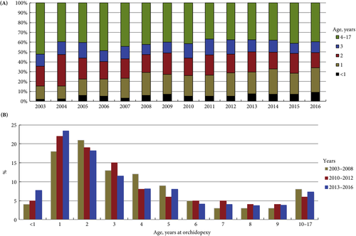
Undescended testis (UDT) is a common paediatric congenital abnormality, with an incidence of 1:100. UDT is managed by paediatricians, paediatric surgeons, paediatric and adult urologists. A consensus document was created to perform this surgery early, at ~6 months of age and definitely before 1 year of age, because of the risk of lower fertility rates and malignancy in the future [1, 2].

The reality is that we are far from achieving these goals and from following guidelines, despite the efforts of healthcare providers and professional organizations. Why is this? Is the following triad not coming together well?

  1. Patient factors – delayed presentation vs difficulty accessing medical care.
  2. Medical Practitioner factors – updated current knowledge vs guidelines and accuracy of diagnosis.
  3. Resources – healthcare costs and availability of expert medical care.

Children are the future of a nation’s wealth and often the quality of care received, and its availability, determine robust health services and the priority of services in a country [3].

In the current issue of BJUI, Boehme et al. [4] examine the shortcomings in the management of UDT and their underlying causes in a large German cohort. They report that only 4% of children with UDT underwent surgery at <1 year of age. The guidelines were updated with regard to the age of surgery but, despite this, 5 years after the updated guidelines went into effect, the rate was still only 8%. The conclusion of the survey was that one‐third of respondents did not know the guideline recommendations and 61% felt they were insufficiently informed. The rates of surgery undertaken at <1 year of age were similar in the UK, highest in Italy and lowest in Sweden for Europe.

Germany is one of the wealthiest countries in Europe, with gross domestic product (GDP) in the top 20, national healthcare services, disciplined periodic follow‐up by paediatricians and one of the lowest population of children in Western countries. Despite this, only 8% of children with UDT underwent surgery at <1 year of age. Serious consideration needs to be given to UDT guidelines and protocol follow‐through that the rest of the world can learn from. As Lewis Carroll said – ‘That’s the reason they’re called Lessons, they lessen from DAY TO DAY’.

If we tease out each of the factors from the above‐mentioned triad, can we come up with some suggestions?

Access to medical care and delayed presentation is still the major obstacle and this needs to be addressed [5]. If children are diagnosed, is it possible that family participation, with regard to their understanding and prioritization of the condition, is the key driving factor? Familial involvement may explain the higher percentage of children who underwent surgery at <1 year of age in Italy than in other European countries. Boehme et al.[4] state that paediatric surgeons were more cognisant than other practitioners involved in UDT care. Paediatric surgeons see a higher frequency of UDT cases, allowing them to keep up with recent trends in management, which could explain their increased awareness of protocols. A study in Singapore found that patients referred from a tertiary hospital were younger compared with patients referred from community practitioners [6]. Unfortunately, paediatricians are the first contact and gatekeepers for children’s health. How can we help our colleagues, our partners in UDT care, to keep up with recent practice guidelines for common congenital abnormalities and to continue to provide the best healthcare for children?

Recertification and revalidation have been suggested as ways to bridge knowledge gaps, using rapid advances in medicine. Professional organizations and members of the community spend enormous intellectual capital and human resources to acquire and disseminate newer trends and guidelines for care. Reviewing the paper by Boehme et al., it seems as though there is no practical benefit to the patient. Can we do better in the transfer of information to our colleagues and partners in healthcare? Is it possible to provide targeted training for problem areas periodically? Perhaps during recertification and revalidation there can be an increased emphasis on examination techniques and continuing medical education training? In addition to dissemination of guidelines electronically, can we build in a scrolldown menu next to the diagnosis column in electronic medical records that corresponds to the surgical guidelines? Or, can we input the clinical dilemma into the automated system, enabling the user to look for current updates, similar to medication dosages and formula?

Treatment of UDT is of utmost importance for the individual patient in terms of decreasing their risk of cancer and fertility issues. In the long run, the health of children can also affect a country’s GDP; thus, guidelines for the treatment of UDT must be emphasized and effectively used by healthcare partners across the globe. Visit the https://www.northraleighpediatrics.com/our-providers/ website to get all the details.

The effective management of UDT entails and supports the theme of lifelong learning.

Mohan S. Gundeti

Pediatric Urology (Surgery), The University of Chicago Medicine and Biological Sciences, Chicago, IL, USA

References

  1. Ritzén EM, Bergh A, Bjerknes R et al. Nordic consensus on treatment of undescended testes. Acta Paediatr 2007; 96: 638–43
  2. British Association of Pediatric Urologists. The BAPU Consensus Statement on the Management of Undescended Testis 2013. https://www.bapu.org.uk/udt-consensus-statement/
  3. Bruijnen CJ, Vogels HD, Beasley SW. Age at orchidopexy as an indicator of the quality of regional child health services. J Paediatr Child Health 2012; 48: 556–9
  4. Boehme P, Geis B, Doerner J. Shortcomings in the management of undescended testis: guideline intention vs. reality and the underlying causes – insights from the biggest German cohort. BJU Int 2018; 122: 644–53
  5. Yiee JH, Saigal CS, Lai J. Timing of orchiopexy in the United States: a quality‐of‐care indicator. Urology 2012; 80: 1121–6
  6. Nah SA, Yeo CS, How GY et al. Undescended testis: 513 patients’ characteristics, age at orchidopexy and patterns of referral. Arch Dis Child 2014; 99: 401–6


Editorial: Postoperative intravesical chemotherapy has an important role in reducing subsequent bladder tumours – why is it not routine?

Transurethral resection of bladder tumour (TURBT) is a frequent operation performed by urologists worldwide. Although on occasion the procedure can be quite challenging, the majority are relatively straightforward with little morbidity. In most cases, where the medical system allows, it is an outpatient procedure. Nonetheless, with the exception of small low‐grade tumours the patient is anaesthetised. It is costly, as the procedure requires medical clearance, an operating room team and equipment.

Most patients with bladder cancer have Ta or less frequently T1 tumours. Despite an initial TURBT, 30–80% of patients develop another tumour. Most are new tumours and some may be recurrences. The reasons for the high ‘recurrence’ rate are the continued impact of the carcinogen, e.g. cigarettes, incomplete resection, missed tumours, and tumour implantation on the altered urothelium. The urologist can help reduce these events by stressing the importance of limiting carcinogen exposure e.g. smoking cessation, striving to perform a complete TURBT, reviewing the entire bladder after the TURBT to avoid missing tumours (using narrow‐band imaging or fluorescent cystoscopy if available), and limiting implantation of tumour cells on the altered urothelial surface with the use of postoperative intravesical chemotherapy (POIVC).

There is a large body of evidence that POIVC reduces the chance of a subsequent tumour [1]. I became convinced that implantation occurs after animal studies demonstrated that bladder cancer cells placed into the bladder preferentially implant and grow only if the urothelial surface had been cauterised or otherwise damaged prior to exposure to the bladder cancer cells [2]. Prospective randomised trials eventually confirmed the benefit of POIVC [3]. The paper published in this issue of the BJUI by Bosschieter et al. [4] indicates that POIVC is equally effective if given the same day or the day after TURBT. Thus, if there are obstacles to instilling the medication on the day of the TURBT the drug can be administered the following day.

The evidence in favour of POIVC for bladder tumours is particularly impressive for Grade 1–2 Ta tumours. In my view, all patients with primary or ‘recurrent’ single or multiple papillary Grade 1–2 Ta tumours are the optimal candidates to receive POIVC [5]. POIVC is recommended by the European Association of Urology (EAU) and AUA/Society of Urologic Oncology (SUO) [6,7] yet, the adoption of this guideline is far from uniform. I queried my colleagues from the International Bladder Cancer group (IBCG), as they are conversant in the scientific basis for POIVC and represent several countries with different medical systems [8]. Their comments are pertinent and consistent with my understanding of the issues. Here are some of the common reasons for not following the guidelines: (i) Some urologists are not convinced that the reduction in the ‘recurrence’ rate is sufficient to use POIVC. (ii) The most common chemotherapeutic agent for POIVC in the USA is mitomycin C and it is expensive. The cost for 40 mg is ~$1000. It is approximately $500 in Europe. (iii) Hospitals have rules regarding the delivery of chemotherapy and the pharmacy and nursing departments may not make it easy to instil the drug in the postoperative setting. Some hospitals require notification a day before the surgery and the drug is wasted if the drug is not used. (iv) Urologists are concerned about extravasation and uncertainty of the tumour grade and stage. There may be other reasons but these help explain why POIVC is not routine.

On the other hand many patients with bladder cancer require frequent TURBTs. I am certain that following an uneventful TURBT or office cauterisation for Grade 1–2 Ta bladder cancer, they would choose to receive POIVC if properly informed. Urologists are proficient at judging whether a tumour fits the criteria for POIVC and if they underestimate the grade or stage the patient may still benefit. If urologists cannot instil the chemotherapy on the day of the TURBT, they can instil the drug the following day without compromising effectiveness. I believe it is our job to do what we can to help our patients and in this instance we should do our best to minimise subsequent tumour events, which includes the use of adjuvant chemotherapy.

Mark S. Soloway

Memorial Hospital Hollywood, Miami, FL, USA

References

  1. Perlis N, Zlotta AR, Beyene J, Finelli A, Fleshner NE, Kulkarni GS. Immediate post‐ transurethral resection of bladder tumor intravesical chemotherapy prevents non‐muscle invasive bladder tumor recurrence: an updated meta‐analysis on 2548 patients and quality –of‐evidence review. Eur Urol 2013; 64: 421–30
  2. Weldon TE, Soloway MS. Susceptibility of urothelium to neoplastic cellular implantation. Urology 1975; 5: 824–7
  3. Tolley DA, Hargreave TB, Smith PH et al. Effect of intravesical mitomycin C on recurrence of newly diagnosed superficial bladder cancer: interim report from the Medical Research Council Subgroup on Superficial Bladder cancer. Br Med J 1988; 296: 1259–61
  4. Bosschchieter J, von Moorselaar JA, Vis AN et al. The effect of timing of an immediate instillation of mitomycin C after transurethral resection in 941 patients with non‐muscle‐invasive bladder cancer. BJU Int 2018; 122: 571–5
  5. Klaassen Z, Soloway MS. European Association of Urology and American Urological Association/Society of Urologic Oncology guidelines on risk categories for non‐muscle‐invasive bladder cancer may lead to overtreatment for low‐grade Ta bladder tumors. Urology 2017; 105: 14–7
  6. Babjuk M, Böhle A, Burger M et al. EAU guidelines in non‐muscle invasive urothelial carcinoma of the bladder: update 2016. Eur Urol 2017; 71: 447–61
  7. Chang SS, Boorjian SA, Chou R et al. Diagnosis and treatment of non‐muscle invasive bladder cancer: AUA/SUO guideline. J Urol 2016; 196: 1021–9
  8. Brausi M, Witjes F, Lamm D et al. A review of current guidelines and best proactive recommendations for the management of nonmuscle invasive bladder cancer by the International Bladder Cancer Group. J Urol 2011; 186: 2158–67


Editorial: Towards an individualized approach for predicting post‐prostatectomy urinary incontinence: the role of nerve preservation and urethral stump length

Traditionally, MRI of the prostate has been mainly applied in the diagnosis and staging of prostate cancer. Kadono et al. [1] used pre‐ and postoperative pelvic MRI to assess the repositioning of the urethra 10 days and 12 months after prostatectomy, hypothesizing that these alterations could correlate with urinary incontinence and urethral function. Recent MRI measurements of anatomical structures of the pelvic floor, such as membranous urethral length and inner levator distance, were found to be independent predictors of early continence recovery at 12 months after prostatectomy [2] A meta‐analysis has also shown a strong correlation between membranous urethral length and continence recovery at 3‐, 6‐ and 12‐month follow‐up [3] Kadono et al. [1] add another metric to the pelvic floor dimensions that may help predict continence. Cranial migration of the lower end of the membranous urethra early after prostatectomy was associated with urinary incontinence and urinary sphincter function, as objectively assessed by urethral pressure profile. Interestingly, return of the membranous urethra to the more distal preoperative position after 12 months was associated with improvement in continence. In a multivariate model, urethral stump length was a strong predictor of continence outcome at 10 days as well as 12 months after prostatectomy. This observation suggests that urethral length may partly improve post-prostatectomy continence through better compression of the membranous urethra in the pelvic floor membrane rather than through transfer of the intra‐abdominal pressure onto the intra‐abdominally located urethra. If confirmed, this observation may imply that more cranial fixation of the bladder neck in a more intra‐abdominal position may not necessarily improve continence after prostatectomy, in line with data from randomized controlled studies comparing median fibrous raphe reconstruction with standard anastomosis that failed to show a benefit [4,5].

Besides anatomical location, innervation of the proximal urethra is important for post-prostatectomy continence [6]. Kadono et al. found that nerve preservation was an independent predictor of early and long‐term continence outcome, with a b value similar to that of urethral stump length at 12‐month follow‐up. To improve post-prostatectomy continence outcome, proper patient selection seems crucial. In the era of personalized medicine, MRI could be a valuable tool to assess preoperatively the risks of postoperative urinary incontinence and counsel patients accordingly. Avoiding prostatectomy in men with short preoperative membranous urethral length may be an important approach for improving outcome, in particular in light of the fact that many attempts to surgically correct anatomical alignment of the pelvic floor have not clearly improved continence outcome. If surgery is considered, nerve preservation should be performed where possible to improve continence.

Henk G. van der Poel and Nikos Grivas

Department of Urology, Netherlands Cancer Institute, Amsterdam, The Netherlands

References

  1. Kadono Y, Nohara T, Kawaguchi S et al. Investigating the mechanism underlying urinary continence recovery after radical prostatectomy: effectiveness of a longer urethral stump to prevent urinary incontinence. BJU Int 2018. 37: 463–9
  2. Grivas N, van der Roest R, Schouten D et al. Quantitative assessment of fascia preservation improves the prediction of membranous urethral length and inner levator distance on continence outcome after robot-assisted radical prostatectomy. Neurourol Urodyn 2018; 37: 417–25
  3. Mungovan SF, Sandhu JS , Akin O, Smart NA, Graham PL, Patel MI. Preoperative membranous urethral length measurement and continence recovery  following radical prostatectomy: a systematic review and meta-analysis. Eur Urol 2017; 71: 368–78
  4. Joshi N, de Blok W, van Muilekom E, van der Poel H. Impact of posterior musculofascial reconstruction on early continence after robot-assisted laparoscopic radical prostatectomy: results of a prospective parallel group trial. Eur Urol 2010; 58: 84–9
  5. Menon M, Muhletaler F, Campos M, Peabody JO. Assessment of early continence after reconstruction of the periprostatic tissues in patients undergoing computer assisted (robotic) prostatectomy: results of a 2 group parallel randomized controlled trial. J Urol 2008; 180: 1018–23
  6. van der Poel HG, de Blok W, Joshi N, van Muilekom E. Preservation of lateral prostatic fascia is associated with urine continence after robotic-assisted prostatectomy. Eur Urol 2009; 55: 892–900Dearnaley DP, Jovic G, Syndikus I et al. The. Lancet Oncol 2014; 15:464–73


Editorial: 2018 – A Spacer Odyssey

The optimisation of radical external-beam radiotherapy treatment is ultimately a compromise. The aim is to deliver a clinically effective radiation dose to the tumour target, whilst limiting the irradiation of surrounding normal tissue to minimise acute and late side effects. Modern image-guided intensity-modulated radiotherapy (IG-IMRT) allows very accurate treatment delivery, which improves this therapeutic ratio. However, there are limitations in its ability to reduce toxicity, due to the constraints of regional anatomy and the physical properties of photon beams. A variety of additional organ-specific techniques exist to further minimise the impact of radiotherapy on adjacent healthy tissue. These range in complexity from ovarian auto-transplantation in selected patients awaiting pelvic radiotherapy for gynaecological malignancies, to delivering radiotherapy for left-sided breast cancers at inspiratory breath-hold to maximise the distance of the chest wall from the left anterior descending coronary artery.

For prostate cancer radiotherapy, the normal structures that determine the optimal safely deliverable dose include the rectum (anterior rectal wall), bladder, femoral heads and penile bulb. IG-IMRT has facilitated dose-escalation, and the treatment of patients previously considered to be ineligible for a radical treatment; both with acceptable toxicity. Higher doses result in improved prostate-cancer biochemical (PSA) control rates, and metastasisfree survival, but as yet no overall survival benefit has been demonstrated in individual trials, despite 10-year follow-up [1]. This may due to the long natural history of localised prostate cancer, the impact of competing comorbidities [2], and the ever-increasing efficacy of treatments for metastatic disease on relapse.

Rectal spacers are a further refinement to the delivery of radical IG-IMRT to the prostate gland. By inserting a biodegradable substance / inflatable biodegradable balloon into the anterior perirectal space, the distance between the prostate and the anterior rectal wall can be increased by approximately 1cm. This reduces the volume of rectum lying within the high-dose radiation field, thereby reducing late bowel toxicity. A recently reported randomised Phase III trial found a reduction the 3-year incidence of ≥ Grade 1 and ≥ Grade 2 rectal toxicity (9.2% v 2.0%; p=0.28; and 5.7% v 0%; p=0.12 respectively) with the addition of a rectal spacer [3]. This was in a very select group of patients: PSA ≤20, cT1-2, International Society of Urological Pathology (ISUP) Prostate Cancer Grade 1-3 (<50% of cores involved), prostate volume <80cc, no use of androgen deprivation therapy, and the use of MRI-CT fusion to aid prostate delineation during the planning process.

In this issue of the BJUI, Chao et al. [4] report their prospective single-centre experience of using a rectal spacer device. This is not a randomised study, and no control arm exists to assist in quantifying the clinical impact of inserting a spacer. However, the patient population studied closely reflects that undergoing radical radiotherapy in most oncology departments world-wide. There were no limitations placed on prostate size (45% were 50- 100cc; 17% >100cc; maximum 187cc), PSA (10% had a PSA >20; maximum 117), or ISUP histological grade (27% were Grade 4 and 5); and 27% of patients were cT3a on imaging. They demonstrated that spacer insertion was safe across a diverse population of men with localised prostate cancer. Further, acceptable prostate-rectal wall separation and rectal dosimetry could be achieved irrespective of prostate size – which was reflected by the low rates of cumulative late rectal toxicity when assessed at a median follow-up of 14 months.

As with any new technique, additional clarification is required to determine exactly where rectal spacers fit into the radiotherapy armoury. In the study by Chao et. al. the Radiation Oncologists did not have access to CT-MRI fusion at the planning stage. As the electron densities for the prostate, rectal-spacer and rectal wall are similar, it can be challenging to determine the anatomical boundaries on CT alone. However, if growing clinical experience now permits accurate delineation by merely adjusting the window levels on the Planning CT, this could facilitate more widespread introduction of the technique, outside the specialist centres with MR-fusion capabilities.

Patient selection is also key to the introduction of this technique. Patient series have reported the safety of the spacer insertion technique across a range of prostate sizes, and prostate cancer risk groups. However, prospective randomised data is lacking on whether treatment efficacy is affected by using a spacer in high-risk / locally advanced disease – the population most likely to benefit from dose-escalated radical radiotherapy [5]. Further, in the only randomised study to date, >90% of patients in the control arm experienced no late rectal toxicity, and >94% experienced no ≥Grade 2 rectal toxicity. Based on long-term follow-up data from the RT01 study, this was an appropriate time-point at which to assess late bowel toxicity, which peaks at 12-36 months before declining [6]. Therefore, the majority of patients do not experience clinically significant late toxicity even without a spacer.

Rectal spacers have the potential to make a valuable contribution to radical IG-IMRT treatment for localised prostate cancer. However, predictive factors are required to identify which patients are likely to benefit from the technique. For many patients, with access to the latest radiotherapy planning and delivery systems, spacers may represent an additional costly procedure with limited benefit.

S.R.Hughes
Oncology Department, Guy’s & St. Thomas’ NHS Trust, London, UK

Read the full article

References

1. Dearnaley DP, Jovic G, Syndikus I, et. al. The Lancet Oncol. 2014; 15(4): 464-473

2. Lu-Yao GL, Albertsen PC, Moore DF, Lin Y, DiPaola RS, Yao SL. Fifteen-year outcomes following conservative management among men aged 65 years or older with localised prostate cancer. Eur Urol. 2015; 68(5): 805-11

3. Hamstra DA, Mariados N, Sylvester J et. al. Continued Benefit to Rectal Separation for Prostate Radiation Therapy: Final Results of a Phase III Trial. Int J Radiat Oncol Biol Phys 2017; 97(5): 976-985

4. Chao M, Ho H, Chan Y et. al. Prospective Analysis of Hydrogel Spacer for Prostate Cancer Patients Undergoing Radiotherapy. BJUI 2018

5. Kalbasi A, Li J, Berman AT. Dose-Escalated Irradiation and Overall Survival in Men with Non-metastatic Prostate Cancer. JAMA Oncol. 2015; 1(7): 897-906

6. Syndikus I, Morgan RC, Sydes MR, Graham JD, Dearnaley DP. Late Gastrointestinal Toxicity After Dose-Escalated Conformal Radiotherapy For Early Prostate Cancer:


Editorial: New robots – cost, connectivity and artificial intelligence

The amazing Da Vinci system, is about to face some market competition from other international companies with their own versions of next generation robots [1]. In order to challenge the current gold standard, these systems will need to be at least as good if not better. The alternative is to be significantly cheaper thus attracting a wider variety of institutions who could currently not afford the Da Vinci. Open consoles, 3D enhanced vision, lighter instruments and greater portability will be recurring themes in these new systems. There is even some renewed interest in automation that goes back to the days of John Wickham, who passed away just short of his 90th birthday (https://www.bjuinternational.com/bjui-blog/light-years-ahead-john-wickham-1927-2017/). The STAR robot can suture bowel better than a human hand in an animal model [2]. The water jet robot (Procept Biorobotics) takes inspiration from Wickham’s PROBOT and may prove to be a viable alternative to TURP or HOLEP but without the steep learning curve [3].

The Revo-i, a Korean robot, has completed the first clinical testing in 17 patients undergoing Retzius sparing robotic assisted radical prostatectomy (RARP). It is an example of real-life reporting where even in experienced hands, three patients underwent blood transfusion and the positive margin rate was 23% [4]. One could speculate whether the approach itself or the adoption of a new robotic system reflected the results of this paper? Either way we can expect to see more such first in man reports over coming years as new robots become available.

These new machines have the potential to reduce the cost of robotic surgery to be similar to that of laparoscopy although the initial hardware outlay may still be substantial. Cambridge Medical Robotics (CMR), UK have plans to introduce competitive cost models which cover maintenance, instruments and even assistants as a comprehensive package. This may make robotics attractive to multidisciplinary expansion, amongst high volume open and laparoscopic surgeons.

The two other aspects in the world of new robots that are causing excitement are artificial intelligence (AI) and faster digital communication. The concept of AI is not new, going back to genius of Alan Turing, who with his decoding skills had a major impact on the outcome of World War II. Machine Learning (ML) is a subset of AI, using decision-making computer algorithms to grasp and respond to specific data, keep your professional devices in good shape with IT services Morristown New Jersey. For example, a prostate recognition algorithm could make the machine learn whether a given image is that of a prostate cancer or not, thus reducing the variability in MRI readings by radiologists. The video recordings of surgeons performing RARP can now be converted through a “black box” into Automated Performance Metrics (APMs) and demonstrate paradoxical findings in that not all high volume surgeons are necessarily those with the best outcomes [5]. With Google moving into surgical robotics in collaboration with J&J, data capture and ML are likely to hold promise for the future.

The UK government amongst others has declared significant investment of > £1billion in AI, with a view to engaging with new talent and remaining a world leader in this emerging field. Led by Dame Wendy Hall (https://www.gov.uk/government/publications/growing-the-artificial-intelligence-industry-in-the-uk) this ambitious project outlines a vision of appointing new researchers from the UK and overseas in all forms of AI, while maintaining the sensitivities around data trust and ethics. However, a word of caution in that AI faces difficulty with reproducibility as a result of unpublished codes in over 90% of articles written on the subject [6].

Surgery may be further democratised in coming years with the advent of low latency ultrafast 5G connectivity. The Internet of Skills could make remote robotic surgery and mentorship easily accessible, irrespective of the location of the expert surgeon [7]. The impact of these developments on patient care will be of considerable interest to the wider surgical community.

Prokar Dasgupta FKC, Editor-in-Chief BJUI

MRC Centre for Transplantation, NIHR Biomedical Research Centre, King’s College London, UK


References

  1. Rassweiler JJ, Autorino R, Klein J et al. Future of robotic surgery in urology. BJU Int 2017; 120: 822-841. doi:10.1111/bju.13851
  2. Shademan A, Decker RS, Opfermann JD, Leonard S, Krieger A, Kim PC. Supervised autonomous robotic soft tissue surgery. Sci Transl Med 2016;8:337ra64
  3. Gilling P, Reuther R, Kahokehr A, Fraundorfer M. Aquablation – image‐guided robot‐assisted waterjet ablation of the prostate: initial clinical experience. BJU Int 2016; 117: 923-9.
  4. Chang KD, Abdel Raheem A, Choi Y D, Chung BH, Rha KH. Retzius‐sparing robot‐assisted radical prostatectomy using the Revo‐i robotic surgical system: surgical technique and results of the first human trial. BJU Int 2018; 122: 441-448
  5. Chen J, Oh PJ, Cheng N, Shah A, Montez J, Jarc A, Guo L, Gill IS, Hung AJ. Utilization of automated performance metrics to measure surgeon performance during robotic vesicourethral anastomosis and methodical development of a training tutorial. J Urol. 2018 May 21. pii: S0022-5347(18)43237-5. doi: 10.1016/j.juro.2018.05.080. [Epub ahead of print] PMID: 29792882
  6. Hutson M. Artificial intelligence faces reproducibility crisis. Science 16 Feb 2018: Vol. 359, Issue 6377, pp. 725-726
  7. Kim SS, Dohler M, Dasgupta P. The Internet of Skills: use of fifth‐generation telecommunications, haptics and artificial intelligence in robotic surgery. BJU Int 2018; 122: 356-359


Editorial: Exercise to prevent LUTS – myth and reality

The pathophysiology of LUTS is one of the most intriguing issues in urology and the conundrum remains unsolved. Their multifactorial origin imposes a differential diagnosis that is often quite straightforward but sometimes complicated and at times the enigma is cracked only ex adiuvantibus. Clinical trials and personal experience have showed us that patients rarely become asymptomatic albeit our therapeutic efforts, suggesting that part of the problem is in the ageing process. So the question comes as to whether we can halt or delay ageing. Clearly some people age without LUTS apart from a physiological decrease in urinary flow. What goes wrong in our patients that stay right in some other subjects? Most of us would like to have a pill that could fix every problem, an easy answer to swallow and remedy our troubles, but this is not always the case.

‘Lifestyle’ is one of the most frequently cited words with 1 330 000 000 on Google today, more than double the ‘hits’ for happiness (a mere 576 000 000). No doubt that behind the lifestyle mantra there is an industry that makes billions on lifestyle issues, but as there is usually ‘no smoke without fire’ there must be something to it. Who has never been advised to change his way of life? Probably none, but who actually takes up the challenge and changes their routine? Clinical trials on the therapeutic effect of lifestyle changes cannot be analysed with an intent‐to‐treat analysis because ‘there’s many a slip ‘twixt cup and lip’ and we need look at those who really undertake the challenge.

There is a growing body of evidence that a healthy lifestyle will not just help to prevent cancer and cardiovascular events or keep you ‘fit’, but will also reduce the risk of developing LUTS [12]. In this issue of the BJUI, a paper from South Korea [3] provides rather convincing evidence that sitting for ≥10 h/day will increase your risk of storage and voiding symptoms, whilst doing exercise will reduce it. But what if you are a manager and your job is to read an endless number of reports each day? You cannot read whilst walking or doing exercise; you work for a living and LUTS may be the price you pay for a wealthier life.

As a surgeon I have an obsession for fixing things and making my patients better. If my patients have a sedentary job can I suggest a change in their lifestyle (not a change of job) that can counterbalance long sitting hours? The answer from the Korean cohort seems to be negative, as multivariate analysis of a subject cohort with long sitting hours suggested an increased risk of developing LUTS notwithstanding some exercise. I never thought that sitting was that bad but ‘est modus in rebus’ as Horace put it and probably sitting for too long is bad. Actually, my watch keeps telling me to stand at regular intervals, although I think I stand for too long in theatre (it also reminds me to breathe properly but that is another story).

This is a long way to say that I would rather be told what I can do right than be told what I am doing wrong. Is my personal risk of a poor outcome reversible? I think we have enough evidence from observational studies that exercise will reduce the risk of developing LUTS, but the time has come to embark on large prospective trials of LUTS treatment with lifestyle changes including exercise.

I have a number of patients who adopted a healthier lifestyle upon retirement (more info here about how they are doing it), lost weight, lowered their arterial pressure and their glucose levels, and their LUTS improved dramatically. What I need to know is whether this is the exception or whether this is the rule. These are not easy studies but I would rather work to answer an important academic question with a difficult and long‐term trial rather than doing an easier study that will not change the way we live and the way we practise.

Andrea Tubaro and Cosimo De Nunzio
Department of Urology, SantAndrea Hospital, Rome, Italy
Read the full article

References

  1. De Nunzio C, Presicce F, Lombardo R et al. Physical activity as a risk factor for prostate cancer diagnosis: a prospective biopsy cohort analysis. BJU Int 2016117: E29–35
  2. Gacci M, Corona G, Sebastianelli A et al. Male lower urinary tract symptoms and cardiovascular events: a systematic review and meta‐analysis. Eur Urol 201670: 788–96
  3. Park HJ, Park CH, Chang Y, Ryu S. Sitting time, physical activity and the risk of lower urinary tract symptoms: a cohort studyBJU Int 2018122: 293–99

Editorial: The way towards understanding possible multiple functions of AR V7 in prostate cancer

The presence of constitutively active androgen receptors in castration therapy‐resistant prostate cancer is frequently associated with therapy resistance. This is not surprising because the transcriptional program of these receptors is not dependent on the presence of circulating androgen. In conditions of reduced expression of circulating androgen, functional activity of the receptor probably contributes to cancer progression. Investigations by Bernemann et al. [12], however, showed that some patients who present with variant ARV7, the most frequently diagnosed variant receptor, still respond to second‐generation anti‐androgen therapy. Until publication of the paper by Bernemann et al. [1], it was not completely clear whether “any findings in this area” reflected a technical error. The authors report further technical advances and similar detection of variant androgen receptors with two PCR assays, the SYBR Green and TaqMan assays. These advances in detection may open up a new area of investigation. The study reported in the current issue of BJUI may therefore shed more light on biology of truncated androgen receptors in prostate cancer [1]. According to the initial seminal publication in the field, AR‐V7‐positive patients had lower endocrine therapy response rates than those who were variant‐negative [3]. Studies aiming to detect variant androgen receptors are particularly important because of increasing interest in circulating tumour cells in prostate cancer [4]. It may be necessary to better describe subgroups of AR‐V7 which may differ in interactions with specific coactivators. Overall, relatively little is known about alterations in interactions between coactivators and the N‐terminal region of the receptor that may occur in subgroups of patients with castration therapy‐resistant prostate cancer [5]. Several important questions regarding signalling between the wild‐type and constitutively active androgen receptor have not been completely clarified, and the issue of which genes are regulated by both receptors is still a matter of discussion. The findings presented in the study by Bernemann et al. [1] may represent the next step towards individualization of therapies. If we accept that variant androgen receptors also display heterogeneity, a more differentiated classification of those receptors may guide clinical decisions in the future. Future studies should also take into consideration the fact that different variants may be expressed at different levels during and after endocrine therapy. These ratios of androgen receptor variant expression may be taken into consideration when determining the probability of success of specific anti‐androgen receptor therapy. One could also learn that application of different methodologies in variant androgen receptor diagnostics may become an established standard in monitoring castration therapy‐resistant disease. Establishment of controlled standard operating procedures in PCR diagnostics may at this stage minimize discrepancies between findings reported by different researchers and help to establish a consensus on this important topic.

Zoran Culig

Experimental Urology, Department of Urology, Medical University of Innsbruck, Anichstrasse 35, A-6020 Innsbruck, Austria

Read the full article

References

  1. Bernemann C, Steinestel J, Humberg V, Bögemann M, Schrader AJ, Lennerz JK. Performance comparison of two AR‐V7 detection methods. BJU Int 2018122: 219–26


Editorial: Prostate Artery Embolization

Andrea Tubaro, in his editorial for European Association of Urology 2006 [1], discussed the paradigm shift in the surgical management of BPH from open surgery to TURP, and postulated that more refined and less invasive techniques would further dictate the treatment pathway to reduce cost, manage more high-risk surgical cases and reduce blood loss in a population that increasingly is on antithrombotic and anticoagulant medication, to ease the management of large prostates, and to manage BPH as a day case procedure [1].

Interventional radiology has been at the forefront of minimally invasive procedures. In 1953, Seldinger [2] published his ingenious method of introducing a catheter into the vascular system after obtaining needle access and, 10 years later, Dotter recognized the potential of catheters to be used in performing intravascular surgery [3]. Superselective prostate artery embolization (PAE) was first described by DeMeritt et al. [4]. Pisco et al. [5] from Portugal and Carnevale et al. [6] from Brazil have rightly been credited with the development of the clinical service for PAE in BPH. The study by Pisco et al. in 2016, in 630 consecutive patients with moderate to severe LUTS refractory to medical therapy for at least 6 months, showed 81.9% medium-term and 76.3% long-term clinical success rates, with no urinary incontinence or sexual dysfunction reported. Carnevale et al. [6], in 2014, described a modified PAE technique that can lead to greater ischaemia and infarction of the prostate gland with the possibility of better clinical outcomes [6].

In this edition of BJUI, the UK Register of Prostate Embolization (ROPE) study [7] provides evidence for the efficacy and safety for PAE for LUTS secondary to BPH and makes an indirect comparison with TURP. What is strikingly unique and to be applauded in this registry is the collaboration between the British Society of Interventional Radiology, the BAUS and National Institute of Clinical Excellence (NICE).

A total of 305 patients across 17 UK centres were enrolled, and results were analysed over 12 months. They noted that patients who underwent PAE had a statistically significant improvement in urinary flow rate and reduction in prostate volume after the procedure. In terms of IPSS and quality-of-life improvement, there was no evidence of PAE being non-inferior to TURP. Seventy-one percent of PAE cases were performed as outpatients or day cases. By contrast, 80% of TURPs required at least one night of hospital stay and a majority two nights [7].

In April 2018, NICE revised their guidelines and have now approved PAE with certain recommendations [8].

The key to successful PAE, in our opinion, is careful patient selection. At our centre, we receive tertiary referrals of patients with very large prostates, many of whom are comorbid and elderly. We embraced the option of PAE and were delighted to be able to contribute a number of cases to the ROPE study. Our overall experience is now in excess of 200 cases and we are aware that some patients will do well, others less well. It is becoming clearer who those patients may be; those who do well tend to be those with the larger prostate with large lateral lobes and adenomatous predominant BPH, without a significant middle lobe, with big prostate vessels and with lower risk of significant renal insufficiency. The large middle lobes can ball-valve and still obstruct, and preoperative arterial CT could identify those with heavily calcified, severely diseased internal iliac arteries that may be difficult to embolize. Nonetheless, those patients who are at highest risk from surgery and those who wish to minimize the risks of sexual dysfunction or incontinence may justifiably opt for PAE as a less invasive outpatient procedure. And why should they not? For many, simply the opportunity to avoid long-term medication with a-blockers or 5-a-reductase inhibitors is the real benefit, and undergoing PAE does not exclude one from surgery afterwards.

Level 1 evidence is of course a fundamental requirement for a change in definitive practice; the ROPE study is a comparative cohort of two fundamentally different procedures. Our institute is a surgical centre for the management of massive BPH and we are convinced that PAE has a place in the management of some of our patients, but could prevention be better than cure? Ambitious it may be, but who is to say whether early PAE in symptomatic patients might reduce the progression of clinical BPH, avoiding the morbidity and cost of long-term medical treatment culminating in surgery. Perhaps the real challenge highlighted by the ROPE study is that the time has come to consider a randomized controlled trial of prostate embolization vs early non-surgical treatment of BPH (short title ‘PREVENT-BPH’), with randomization to PAE or either a-blockers and/or 5-a-reductase inhibitors or placebo. The ROPE study suggests that PAE at the least deserves a randomized controlled trial including it vs other non-invasive treatments.

Tarun Sabharwal and Rick Popert
Guy’s and St Thomas’ Hospital, London, UK

Read the full article

References

  1. Tubaro A. BPH treatment: a paradigm shift. Eur Urol 2006; 49: 939–41
  2. Seldinger SI. Catheter replacement of the needle in percutaneous arteriography; a new technique. Acta Radiol 1953; 39: 368–76
  3. Dotter CT, Judkins MP. Transluminal treatment of atherosclerotic obstructions: description of a new technique and preliminary report of its applications. Circulation 1964; 30: 654–70
  4. DeMeritt JS, Elmasri FF, Esposito MP, Rosenberg GS. Relief of benign prostatic hyperplasia-related bladder outlet obstruction after transarterial polyvinyl alcohol prostate embolization. J Vasc Interv Radiol 2000; 11: 767–70
  5. Pisco JM, Bilhim T, Pinheiro LC et al. Medium-and long-term outcome of prostate artery embolization for patients with benign prostatic hyperplasia: results in 630 patients. J Vasc Interv Radiol 2016; 27: 1115–22
  6. Carnevale FC, Moreira AM, Antunes AA. The “PErFecTED Technique”: proximal embolisation first, then embolise distal for benign prostatic hyperplasia. Cardiovasc Intervent Radiol 2014; 37: 1602–5
  7. Ray AF, Powell J, Speakman MJ et al. Efficacy and safety of prostate artery embolization for benign prostatic hyperplasia: an observational study and propensity-matched comparison with transurethral resection of the prostate (the UK-ROPE study). BJU Int 2018; 122: 270–82
  8. NICE Guidance. Prostate artery embolisation for lower urinary tract symptoms caused by benign prostatic hyperplasia. BJU Int 2018;121: 825-34


Editorial: Antibiotics and ureteroscopy: a single prophylactic dose is enough, but could we give even less?

Antibiotic resistance is internationally recognized as a threat to global health. As a consequence, there is an ongoing need to review antibiotic prescribing practice, both for treatment and prophylaxis. ‘Antibiotic stewardship’, whereby antimicrobial use, and the associated increase in bacterial resistance, is reduced, is essential worldwide [1].

In this issue of BJUI, Deng et al. [2] present the results of their systematic review and meta‐analysis of the efficacy of antibiotic prophylaxis vs no treatment in patients undergoing upper tract ureteroscopy/ureterorenoscopy. In total, 4591 patients were analysed (from 11 studies, comprising five randomized controlled trials, one prospective comparative study and five retrospective comparative studies), of whom 2700 patients received antibiotic prophylaxis and the remaining 1891 had no prophylactic antibiotics at all. To know more visit walkerstgallery .

Having excluded patients with pre‐operative urinary tract infection (UTI) or bacteriuria, they found that patients who received a single dose of pre‐operative antibiotic had a significantly lower risk of pyuria and bacteriuria than those without antibiotic, but that there was no difference in the risk of post‐operative febrile UTIs between the groups with and without the use of prophylactic antibiotic. There was also no advantage to intravenous antibiotic administration compared with oral administration in reducing febrile UTIs, nor any difference between a single dose of antibiotic drug vs a more prolonged post‐operative regime [2].

This is an important article, potentially leading many urological surgeons to change their current practice with regard to prescribing post‐operative antibiotics, and raising the question of whether antibiotic prophylaxis is needed in patients who have sterile urine pre‐operatively and no specific operative risk factors.

The next question for endourologists to answer will be ‘What is the most appropriate management of asymptomatic bacteriuria detected during pre‐operative investigations?’ Whilst current practice is to treat pre‐operative bacteriuria in patients managed in urology departments, Herr [3] has shown it is reasonable not to give prophylactic antibiotics to asymptomatic patients undergoing flexible cystoscopy, even if there is bacteriuria on pre‐procedure urine analysis. Herr evaluated >3000 outpatients undergoing flexible cystoscopies (of whom 78% had sterile urine and 22% had asymptomatic bacteriuria). The cystoscopies were performed without any antibiotic prophylaxis at all. Overall, 1.9% of patients experienced febrile UTIs, all of which resolved rapidly with oral antibiotics and without any complications (no sepsis or hospital admission). Although the rate was higher in patients with prior infected urine (UTI rate 3.7% compared with 1.4% in patients with sterile urine), Herr concluded that prophylactic antibiotics are not necessary in asymptomatic patients regardless of the presence of bacteriuria, and therefore advised that pre‐procedure urine analysis itself is not required [3].

These findings challenge the belief that pre‐operative urine analysis is essential in asymptomatic patients. Kavoussi et al. [4] studied this issue in patients undergoing insertion of an artificial urinary sphincter or inflatable penile prosthesis, of whom 41% had no pre‐operative urine culture; the authors demonstrated a low risk of 1.5% of prosthesis infection in patients receiving standard peri‐operative antibiotics. This suggests that, even in ‘high stakes’ prosthetic implantation (where the consequences of infection are considerable, requiring explanation and later re‐insertion of a new device), surgery can be performed without pre‐operative urine cultures [4].

Perhaps even more contentiously, Cai et al. [5] have questioned the need for treatment of asymptomatic bacteriuria before urological procedures when ‘standard antibiotic prophylaxis’ is given pre‐operatively. They analysed 2201 patients treated in accordance with European Association of Urology guidelines for antibiotic prophylaxis, of whom 70.1% had sterile urine and 30.4% had asymptomatic bacteriuria pre‐operatively. They reported no increased risk in patients with pre‐operative asymptomatic bacteriuria, with 10.4% of affected patients having a symptomatic post‐operative UTI and a 0.3% risk of sepsis, compared with a 8.3% UTI rate and 0.26% chance of sepsis in patients with pre‐operatively sterile urine [5].

In their article, Deng et al. [2] have shown that patients with sterile urine undergoing ureteroscopy had a similar risk of a post‐operative febrile UTI whether or not pre‐ and post‐operative antibiotics were given. This implies the need for specific high‐risk groups to be targeted for antibiotic prophylaxis, and, extending the arguments above, suggests that a more selective approach is needed for pre‐operative urine analysis in low‐risk patients.

In this regard, Grabe and Wullt [6] have commented that ‘undetected pre‐operative bacteriuria is like walking straight into a minefield’. Whilst the knowledge that one is walking into a minefield has the advantage of leading one to take a cautious approach (i.e. treating asymptomatic bacteriuria pre‐operatively), it is possible that not all of the mines in the minefield are live (i.e. certain patients with asymptomatic bacteriuria may be at lower risk of post‐operative problems than others). The real challenge is to determine which patients with asymptomatic bacteriuria need antibiotic treatment and for how long, and therefore which patients need urine analysis before which procedures in the first place. This approach, if shown to be safe, would not only reduce the cost of urine cultures and pre‐surgical eradication of asymptomatic bacteriuria, but also the wider global cost of antibiotic overuse and bacterial resistance.

Daron Smithand Bruce Macrae
*EndoLuminal EndoUrologist, Department of Urology, Westmoreland Street Hospital, and Clinical Microbiology, UCLH NHS Foundation Trust, London, UK
Read the full article

References

  1. WHO. Global action plan on antimicrobial resistance. 2015 (accessed March 23, 2018)https://apps.who.int/iris/bitstream/handle/10665/193736/9789241509763_eng.pdf?sequence=1
  2. Deng T, Liu B, Duan X et al. Antibiotic prophylaxis in ureteroscopic lithotripsy: a systematic review and meta‐analysis of comparative studies. BJU Int 2018122: 29–39
  3. Herr HW. The risk of urinary tract infection after flexible cystoscopy in patients with bladder tumor who did not receive prophylactic antibiotics. J Urol 2015193: 548–51
  4. Kavoussi NL, Viers BR, Pagilara TJ et al. Are urine cultures necessary prior to urologic prosthetic surgery? Sex Med Rev 20186: 157–61
  5. Cai T, Verze P, Palmieri A et al. Is preoperative assessment and treatment of asymptomatic bacteriuria necessary for reducing the risk of postoperative symptomatic urinary tract infections after urologic surgical procedures? Urology 201799: 100–5
  6. Grabe M, Wullt B. Re: Is preoperative assessment and treatment of asymptomatic bacteriuria necessary for reducing the risk of postoperative symptomatic urinary tract infection after urologic surgical procedures? Eur Urol 2017; 73: 476-477
© 2024 BJU International. All Rights Reserved.