Tag Archive for: #kcsm

Posts

Video: Stereotactic ablative body radiotherapy for inoperable primary kidney cancer

Stereotactic ablative body radiotherapy for inoperable primary kidney cancer

Abstract

Objective

To assess the feasibility and safety of stereotactic ablative body radiotherapy (SABR) for renal cell carcinoma (RCC) in patients unsuitable for surgery. Secondary objectives were to assess oncological and functional outcomes.

Materials and Methods

This was a prospective interventional clinical trial with institutional ethics board approval. Inoperable patients were enrolled, after multidisciplinary consensus, for intervention with informed consent. Tumour response was defined using Response Evaluation Criteria In Solid Tumors v1.1. Toxicities were recorded using Common Terminology Criteria for Adverse Events v4.0. Time-to-event outcomes were described using the Kaplan–Meier method, and associations of baseline variables with tumour shrinkage was assessed using linear regression. Patients received either single fraction of 26 Gy or three fractions of 14 Gy, dependent on tumour size.

Results

Of 37 patients (median age 78 years), 62% had T1b, 35% had T1a and 3% had T2a disease. One patient presented with bilateral primaries. Histology was confirmed in 92%. In total, 33 patients and 34 kidneys received all prescribed SABR fractions (89% feasibility). The median follow-up was 24 months. Treatment-related grade 1–2 toxicities occurred in 26 patients (78%) and grade 3 toxicity in one patient (3%). No grade 4–5 toxicities were recorded and six patients (18%) reported no toxicity. Freedom from local progression, distant progression and overall survival rates at 2 years were 100%, 89% and 92%, respectively. The mean baseline glomerular filtration rate was 55 mL/min, which decreased to 44 mL/min at 1 and 2 years (P < 0.001). Neutrophil:lymphocyte ratio correlated to % change in tumour size at 1 year, r2 = 0.45 (P < 0.001).

Conclusion

The study results show that SABR for primary RCC was feasible and well tolerated. We observed encouraging cancer control, functional preservation and early survival outcomes in an inoperable cohort. Baseline neutrophil:lymphocyte ratio may be predictive of immune-mediated response and warrants further investigation.

Editorial: Stereotactic radiotherapy for primary renal cell carcinoma: time for larger-scale prospective studies

A number of important trends in kidney cancer diagnosis have emerged in recent decades, including the increasing detection of renal tumours in older patients with more comorbidities. In the UK in 2012–2014, 50% of new cases were diagnosed in people aged 70 years and over. Whilst many of these lesions are incidental small renal masses suitable for active surveillance, the dilemma of how to manage the higher-risk lesion (rapid growth kinetics, larger size, symptomatic lesion) is increasingly encountered. Surgical management may pose an unacceptable risk of morbidity, mortality or dialysis, yet these patients may live long enough to experience the consequences of disease progression. Thermal ablation is an option for small cortical tumours (≤3 cm), but there are limitations for larger or centrally located tumours.

In this issue of BJUI, Siva and colleagues [1] report promising early efficacy and toxicity data using stereotactic ablative body radiotherapy (SABR) for the treatment of primary RCC in this difficult cohort. SABR is a non-invasive treatment that delivers very high doses of radiation over one to five outpatient sessions. It uses advanced motion management, radiation planning and image guidance techniques to ensure delivery of an ablative dose with millimetre precision. Survival benefits with stereotactic radiosurgery have been demonstrated in patients with solitary brain metastases [2], and SABR is now an accepted standard of care for patients with medically inoperable early-stage lung cancer [3]. Randomized phase III trials are currently under way, testing SABR against standard of care in primary prostate (clinicaltrials.gov ID NCT01584258) and liver cancer (NCT01730937) and in the oligometastatic setting (NCT02759783). Historically considered radio-resistant, both pre-clinical and clinical data now support the sensitivity of RCC to high-dose per fraction radiotherapy, as used in SABR [4].

The study by Siva and colleagues is one of the largest, early-phase, prospective studies of SABR for primary RCC to date, accruing 37 patients with cT1a–cT2a RCC not suitable for other therapies. Importantly, this was not a cohort of incidentally detected small renal masses. The majority (65%) of tumours were >4 cm (median 4.8 cm), were growing on surveillance or symptomatic, and were biopsy-proven. The inclusion of enlarging T1a tumours is not unreasonable. A recent analysis of patients with localized T1a kidney cancer from the Surveillance, Epidemiology and End Results (SEER) Medicare data reported an excess of kidney cancer deaths for non-surgically managed patients aged >75 years, highlighting how difficult it can be to find the right balance between active and expectant management in this group [5]. Indeed 11% of patients in the present study by Siva et al. developed distant metastases by 2 years.

In the present study, tumours <5 cm received a single 26-Gy fraction of SABR, whilst tumours >5 cm received 42 Gy over three fractions. Whilst acknowledging a number of uncertainties in modelling, this should equate to an equivalent biological dose in excess of 100 Gy. The authors found that delivering this SABR regimen was feasible and well tolerated with one grade 3 toxicity (transient fatigue) and no grade 4–5 toxicities. Most patients sustained only transient minor side effects (78%) or no treatment-related side effects (18%). The mean baseline estimated GFR was 55 mL/min, which decreased to 44 mL/min at 1 year, and was maintained for those with 2 years follow-up.

Similarly, short-term efficacy appears promising. With a median follow-up of 24 months, freedom from local progression at 2 years was 100%, with one patient subsequently progressing locally with concurrent distant metastases 28 months after treatment. Local progression was defined using Response Evaluation Criteria In Solid Tumors (RECIST) v1.1. This is a pragmatic definition and takes into account the challenges in interpreting standard imaging after SABR and the difficulties in obtaining and interpreting repeat biopsies in this cohort. Similarly to the study by Sun et al. [6] it appears that stable or partial radiological responses will predominate in the early years after SABR and, unlike thermal ablation, changes in enhancement patterns can be very slow to evolve.

Longer-term follow-up is required to confirm these promising tumour control and nephron preservation rates, in addition to evaluating longer-term late effects. To that end, the present study has provided a platform for the authors to launch an international phase II clinical trial under the auspices of the TransTasman Radiation Oncology Group (TROG 15.03 FASTRACK, clinicaltrials.gov ID NCT02613819). Larger-scale prospective studies are essential to confirm the efficacy and safety of this non-invasive, nephron-sparing, ablative technique and provide further information to help refine patient selection and develop better biomarkers of response.

David I. Pryor *† and Simon Wood†‡


*Department of Radiation Oncology, Princess Alexandra Hospital, Wooloongabba, School of Medicine, University of Queensland, Brisbane, and Department of Urology, Princess Alexandra Hospital, Wooloongabba, Qld, Australia


References


1 Siva S. Stereotactic ablative body radiotherapy for inoperable primary kidney cancer: a prospective clinical trial. BJU Int 2017; 120: 62330


2 Andrews DW, Scott CB , Sperduto PW et al. Whole brain radiation therapy with or without stereotactic radiosurgery boost for patients with  one to three brain metastases: phase III results of the RTOG 9508 randomised trial. Lancet (London, England) 2004; 363: 166572


4 De Meerleer G, Khoo V, Escudier B et al. Radiotherapy for renal-cell carcinoma. Lancet Oncol 2014; 15: e1707


6 Sun MR, Brook A, Powell MF et al. Effect of stereotactic body radiotherapy on the growth kinetics and enhancement pattern of primary renal tumors. AJR Am J Roentgenol 2016; 206: 54453


Article of the Week: Stereotactic ablative body radiotherapy for inoperable primary kidney cancer: a prospective clinical trial

Every week the Editor-in-Chief selects an Article of the Week from the current issue of BJUI. The abstract is reproduced below and you can click on the button to read the full article, which is freely available to all readers for at least 30 days from the time of this post.

In addition to the article itself, there is an accompanying editorial written by a prominent member of the urological community. This blog is intended to provoke comment and discussion and we invite you to use the comment tools at the bottom of each post to join the conversation.

If you only have time to read one article this week, it should be this one.

Stereotactic ablative body radiotherapy for inoperable primary kidney cancer: a prospective clinical trial


Shankar Siva*,, Daniel Pham*, Tomas Kron*,, Mathias Bressel*, Jacqueline Lam*, Teng Han Tan*, Brent Chesson*, Mark Shaw*, Sarat Chander*, Suki Gill*,Nicholas R. Brook§, Nathan Lawrentschuk*,, Declan G. Murphy*,† and Farshad Foroudi*,


*Peter MacCallum Cancer Centre, Melbourne, Vic., Australia, † Sir Peter MacCallum Department of Oncology, University of Melbourne, Melbourne, Vic. Australia, Sir Charles Gairdner Hospital, Nedlands Perth, WA, Australia, §Royal Adelaide Hospital, Adelaide, SA, Australia, and Olivia Newton John Cancer Centre, Heidelberg, Vic., Australia


Abstract

Objective

To assess the feasibility and safety of stereotactic ablative body radiotherapy (SABR) for renal cell carcinoma (RCC) in patients unsuitable for surgery. Secondary objectives were to assess oncological and functional outcomes.

Materials and Methods

This was a prospective interventional clinical trial with institutional ethics board approval. Inoperable patients were enrolled, after multidisciplinary consensus, for intervention with informed consent. Tumour response was defined using Response Evaluation Criteria In Solid Tumors v1.1. Toxicities were recorded using Common Terminology Criteria for Adverse Events v4.0. Time-to-event outcomes were described using the Kaplan–Meier method, and associations of baseline variables with tumour shrinkage was assessed using linear regression. Patients received either single fraction of 26 Gy or three fractions of 14 Gy, dependent on tumour size.

Results

Of 37 patients (median age 78 years), 62% had T1b, 35% had T1a and 3% had T2a disease. One patient presented with bilateral primaries. Histology was confirmed in 92%. In total, 33 patients and 34 kidneys received all prescribed SABR fractions (89% feasibility). The median follow-up was 24 months. Treatment-related grade 1–2 toxicities occurred in 26 patients (78%) and grade 3 toxicity in one patient (3%). No grade 4–5 toxicities were recorded and six patients (18%) reported no toxicity. Freedom from local progression, distant progression and overall survival rates at 2 years were 100%, 89% and 92%, respectively. The mean baseline glomerular filtration rate was 55 mL/min, which decreased to 44 mL/min at 1 and 2 years (P < 0.001). Neutrophil:lymphocyte ratio correlated to % change in tumour size at 1 year, r2 = 0.45 (P < 0.001).

Conclusion

The study results show that SABR for primary RCC was feasible and well tolerated. We observed encouraging cancer control, functional preservation and early survival outcomes in an inoperable cohort. Baseline neutrophil:lymphocyte ratio may be predictive of immune-mediated response and warrants further investigation.

Read more articles of the week

Article of the Week: Silicone renal models and complex tumour resections prior to RALPN

Every week the Editor-in-Chief selects an Article of the Week from the current issue of BJUI. The abstract is reproduced below and you can click on the button to read the full article, which is freely available to all readers for at least 30 days from the time of this post.

In addition to the article itself, there is an accompanying editorial written by a prominent member of the urological community. This blog is intended to provoke comment and discussion and we invite you to use the comment tools at the bottom of each post to join the conversation.

Finally, the third post under the Article of the Week heading on the homepage will consist of additional material or media. This week we feature a video discussing the paper.

If you only have time to read one article this week, it should be this one.

Utility of patient-specific silicone renal models for planning and rehearsal of complex tumour resections prior to robot-assisted laparoscopic partial nephrectomy

Friedrich-Carl von Rundstedt*,, Jason M. Scovell*, Smriti Agrawal, Jacques Zaneveld§ and Richard E. Link*,,**


*Scott Department of Urology, Baylor College of Medicine, Houston, TX, USA, Department of Urology, Jena University Hospital, Friedrich-Schiller University, Jena, Germany, Department of Molecular and Human Genetics, Baylor College of Medicine, §Lazarus 3D LLC, Dan L. Duncan Comprehensive Cancer Center, Baylor College of Medicine, and **Center for Reproductive Medicine, Baylor College of Medicine, Houston, TX, USA


Read the full article

How to Cite this Article

von Rundstedt, F.-C., Scovell, J. M., Agrawal, S., Zaneveld, J. and Link, R. E. (2017), Utility of patient-specific silicone renal models for planning and rehearsal of complex tumour resections prior to robot-assisted laparoscopic partial nephrectomy. BJU International, 119: 598–604. doi: 10.1111/bju.13712

Abstract

Objective

To describe our experience using patient-specific tissue-like kidney models created with advanced three-dimensional (3D)-printing technology for preoperative planning and surgical rehearsal prior to robot-assisted laparoscopic partial nephrectomy (RALPN).

Patients and Methods

A feasibility study of 10 patients with solid renal masses who underwent RALPN after preoperative rehearsal using 3D-print kidney models. A single surgeon performed all surgical rehearsals and procedures. Using standard preoperative imaging and 3D reconstruction, we generated pre-surgical models using a silicone-based material. All surgical rehearsals were performed using the da Vinci® robotic system (Intuitive Surgical Inc., Sunnyvale, CA, USA) before the actual procedure. To determine construct validity, we compared resection times between the model and actual tumour in a patient-specific manner. Using 3D laser scanning in the operating room, we quantified and compared the shape and tumour volume resected for each model and patient tumour.

aotw-apr-results-5

Results

We generated patient-specific models for 10 patients with complex tumour anatomy. R.E.N.A.L. nephrometry scores were between 7 and 11, with a mean maximal tumour diameter of 40.6 mm. The mean resection times between model and patient (6:58 vs 8:22 min, P = 0.162) and tumour volumes between the computer model, excised model, and excised tumour (38.88 vs 38.50 vs 41.79 mm3, P = 0.98) were not significantly different.

Conclusions

We have developed a patient-specific pre-surgical simulation protocol for RALPN. We demonstrated construct validity and provided accurate representation of enucleation time and resected tissue volume. This simulation platform can assist in surgical decision-making, provide preoperative rehearsals, and improve surgical training.

Read more articles of the week

Editorial: Touching the future – 3D printing facilitates preoperative planning, realistic simulation and enhanced precision in RALPN

Practice taking that match-winning penalty knowing which way the keeper will dive, or take that last putt knowing the lie of the green; it would be very handy wouldn’t it?

Virtual reality (VR), augmented reality (AR), computer-generated images (CGI), and stereotactic overlay, have all been documented as adjuncts in enhancing operative patient care through planning, simulation and increased precision. But what if you could actually handle the specimen and practice operating on a model to refine operative technique before the definitive procedure? With three-dimensional (3D) printing this has now become a reality.

The work of von Rundstedt et al. [1] has the potential to transform surgical planning, operative accuracy, and training, with the development of a life-like kidney and tumour model. Their paper validates a patient-specific simulation protocol to assist in surgical decision-making through operative rehearsal. They assessed the benefits of 10 patient-specific 3D renal models for preoperative planning using tissue-like silicone, and performed model tumour excision with the robot before actual robot-assisted laparoscopic partial nephrectomy (RALPN). Nephrometry scores for tumours ranged from 7 to 11, with a relatively large mean maximal tumour diameter of 40.6 mm. In validating the model the investigators compared resection times between the model and patient (6.61 vs 7.93 min, P = 0.16) and tumour volumes between computer model, excised model, and excised tumour (38.88 vs 38.50 vs 41.79 mm3, P = 0.98), showing no significant differences.

The key principles in nephron-sparing surgery are adequate oncological excision, whilst preserving maximal renal parenchyma, with minimal ischaemia time, and avoiding complications. RALPN is challenging for complex tumours, with an extended learning curve, due partly to limitations in accurate surgical planning and surgical technique. Key anatomical considerations for planning including kidney orientation, tumour position and depth, and locality of adjacent anatomical structures (vessels, collecting system), are difficult to appreciate on conventional two-dimensional axial imaging platforms; with variance in imaging and model-planned approaches clearly noted in previous studies [2].

The development of nephrometry scores have been designed to predict surgical complexity along with various simulation and modelling reconstructions to aid excisional techniques. Several other surgical specialties (orthopaedics, maxillofacial and craniofacial surgery, neurosurgery, plastic surgery [3]) have used 3D-printing technology for organ/lesion modelling or to produce accurate imaging-based prostheses. In the era of minimally invasive surgery and personalised medicine, 3D printing can be a powerful tool for uro-oncologists to better understand individual tumour characteristics and anatomical variations.

Currently there is limited published data on 3D renal tumour printing. This paper [1] represents the first model and validation of its kind. Previous studies of 3D-printed kidneys with renal masses have been reported but limited to depictions of anatomical visualisation of arteries, collecting system and the tumour itself [4, 5]. This is the first time that a model not only provides a 3D representation of the tumour anatomy but also allows high-fidelity simulated excision. Construct validity of these reproductions has been assessed and demonstrate a striking similarity in tumour volume, morphology, and resection time, the main limitations of previous studies [4].

Preoperative planning has never achieved this accuracy before. Operative rehearsal significantly altered the ultimate approach to tumour excision in several cases as noted by the authors. In addition, 3D printing represents a breakthrough in surgical training as it offers a great opportunity, especially in facilities were wet laboratories are not available.

With the small sample assessed and the subjective nature of the surgical technique modification between model and tumour excision, early generalisation may not be appropriate. Other limitations include the models inability to replicate viable orthotopic anatomy such as adjacent organs, dissection planes, perinephric fat thickness/adherence, and bleeding; while also excluding the renorrhaphy component of the procedure. Additionally, production costs and 3D printer access may be an initial deterrent to widespread use of this technique; however, it does address the lack of tactile feeling in AR or VR, avoids the specialised facilities required by animal or cadaveric models, can readily be accurately reproduced, and most importantly provide an accurate anatomical representation of the individual patient.

This is an important and interesting paper as it presents and validates a novel model with extirpative technique in a prospective manner. It provides a life-like model useful for patient education, procedural practice with realistic simulation, an accurate training platform, and is the easiest to access given current technology. Clinical trials are needed to confirm how 3D modelling is ultimately useful in: i) improving patient education, ii) enhancing surgical training, and iii) conferring superior clinical outcomes. Evolution of 3D printers and shrinking production costs will eventually contribute to the widespread usage of this technology.

Further development will provide functional models that replicate not only macroscopic structures but elements such as the collecting system, segmental vessels, and bleeding parenchyma. However, it may be that high-fidelity VR simulators or CGI that can generate patient-specific graphics or even provide an intraoperative stereotactic 3D overlay to guide tumour excision may eventually supersede 3D modelling. Urology has entered the 3D printing era. This study [1] shows that 3D printing is both a feasible and useful technique that may enhance current practice, while providing an improved training platform. The future is here today.

Read the full article

How to Cite this Article

von Rundstedt, F.-C., Scovell, J. M., Agrawal, S., Zaneveld, J. and Link, R. E. (2017), Utility of patient-specific silicone renal models for planning and rehearsal of complex tumour resections prior to robot-assisted laparoscopic partial nephrectomy. BJU International, 119: 598–604. doi: 10.1111/bju.13712

Nicolo de Luyk, Benjamin Namdarian* and Benjamin Challacombe*
*Department of Urology, Guys and St Thomas Hospit als NHS Foundation Trust and Kings College, London, UK and Department of Urology, University Hospital of VeronaVerona, Italy


References


2 Wake N , Rude T, Kang SK et al. 3D printed renal cancer models derived from MRI data: application in pre-surgical planning. Abdom Radiol (NY) 2017; [Epub ahead of print]. doi: 10.1007/s00261-016-1022-2


3 LiJ, Chen M, Fan X, Zhou H. Recent advances in bioprinting techniques: approaches, applications and future prospects. J Transl Med 2016; 14: 271. doi:10.1186/s12967-016-1028-0


4 Silberstein JL, Maddox MM, Dorsey P, Feibus A, Thomas R, Lee BRPhysical models of renal malignancies using standard cross-sectional imaging and 3- dimensional printers: a pilot study. Urology 2014; 84: 26872


5 Bernhard JC, Isotani S, Matsugasumi T et al. Personalized 3D printed model of kidney and tumor anatomy: a useful tool for patient education. World J Urol 2016; 34: 33745


Video: Silicone renal models and complex tumour resections prior to RALPN

Utility of patient-specific silicone renal models for planning and rehearsal of complex tumour resections prior to robot-assisted laparoscopic partial nephrectomy

Read the full article

Abstract

Objective

To describe our experience using patient-specific tissue-like kidney models created with advanced three-dimensional (3D)-printing technology for preoperative planning and surgical rehearsal prior to robot-assisted laparoscopic partial nephrectomy (RALPN).

Patients and Methods

A feasibility study of 10 patients with solid renal masses who underwent RALPN after preoperative rehearsal using 3D-print kidney models. A single surgeon performed all surgical rehearsals and procedures. Using standard preoperative imaging and 3D reconstruction, we generated pre-surgical models using a silicone-based material. All surgical rehearsals were performed using the da Vinci® robotic system (Intuitive Surgical Inc., Sunnyvale, CA, USA) before the actual procedure. To determine construct validity, we compared resection times between the model and actual tumour in a patient-specific manner. Using 3D laser scanning in the operating room, we quantified and compared the shape and tumour volume resected for each model and patient tumour.

Results

We generated patient-specific models for 10 patients with complex tumour anatomy. R.E.N.A.L. nephrometry scores were between 7 and 11, with a mean maximal tumour diameter of 40.6 mm. The mean resection times between model and patient (6:58 vs 8:22 min, P = 0.162) and tumour volumes between the computer model, excised model, and excised tumour (38.88 vs 38.50 vs 41.79 mm3, P = 0.98) were not significantly different.

Conclusions

We have developed a patient-specific pre-surgical simulation protocol for RALPN. We demonstrated construct validity and provided accurate representation of enucleation time and resected tissue volume. This simulation platform can assist in surgical decision-making, provide preoperative rehearsals, and improve surgical training.

Article of the Week: Safety, reliability and accuracy of small renal tumour biopsies: results from a multi-institution registry

Every week the Editor-in-Chief selects an Article of the Week from the current issue of BJUI. The abstract is reproduced below and you can click on the button to read the full article, which is freely available to all readers for at least 30 days from the time of this post.

In addition to the article itself, there is an accompanying editorial written by a prominent member of the urological community. This blog is intended to provoke comment and discussion and we invite you to use the comment tools at the bottom of each post to join the conversation.

If you only have time to read one article this week, it should be this one.

Safety, reliability and accuracy of small renal tumour biopsies: results from a multi-institution registry

Patrick O. Richard*,, Michael A. S. Jewett*, Simon Tanguay, Olli Saarela§, Zhihui Amy Liu§, Frederic Pouliot, Anil Kapoor**, Ricardo Rendon†† and Antonio Finelli*


*Departments of Surgery and Surgical Oncology, Princess Margaret Cancer Centre, University Health Network and the University of Toronto, Toronto, ON, Canada, Centre Hospitalier Universitaire de Sherbrooke, Universite de Sherbrooke, Sherbrooke, QC, Canada, Department of Surgery, Division of Urology, McGill University Health Center, McGill University, Montreal§Dalla Lana School of Public Health, University of Toronto, Universite Laval, Centre de Recherche du Centre Hospitalier Universitaire de Quebec, Quebec **Department of Surgery, Division of Urology, McMaster University, Hamilton and ††QEII Health Sciences Centre, Department of Urology, Dalhousie University, Halifax, NS, Canada


Read the full article

Abstract

Objective

To validate, in a multi-institution review, the safety, accuracy and reliability of renal tumour biopsy (RTB) and its role in decreasing unnecessary treatment.

Materials and Methods

We conducted a multi-institution retrospective study of patients who underwent RTB to characterize a small renal mass (SRM) between 2011 and May 2015. Patients were identified using the prospectively maintained Canadian Kidney Cancer information system. Diagnostic and concordance rates were presented using proportions, whereas factors associated with a diagnostic RTB were identified using a logistic regression model.

scasca

Results

Of the 373 biopsied SRMs, the initial biopsy was diagnostic in 87% of cases. Of the 47 non-diagnostic biopsies, 15 had a repeat biopsy of which, 80% were diagnostic. When both were combined, therefore, a diagnosis was obtained in 91% of SRMs. Of these, 18% were benign. Size was the only factor found to be associated with achieving a diagnostic biopsy. RTB histology and nuclear grade (high or low) were found to be highly concordant with surgical pathology (86 and 81%, respectively). Of the discordant tumours (n = 16), all were upgraded from low to high grade on surgical pathology. Adverse events were rare (<1% of cases).

Conclusion

The present multi-institution study confirms that RTB of SRMs is safe, accurate and reliable across institutions, while decreasing unnecessary treatment. Given our findings, RTBs may be a helpful tool with which to triage SRMs and guide appropriate management.

Read more articles of the week

Article of the Week: PADUA and R.E.N.A.L. nephrometry scores correlate with perioperative outcomes of RAPN: analysis of the Vattikuti GQI-RUS database

Every week the Editor-in-Chief selects an Article of the Week from the current issue of BJUI. The abstract is reproduced below and you can click on the button to read the full article, which is freely available to all readers for at least 30 days from the time of this post.

In addition to the article itself, there is an accompanying editorial written by a prominent member of the urological community. This blog is intended to provoke comment and discussion and we invite you to use the comment tools at the bottom of each post to join the conversation.

If you only have time to read one article this week, it should be this one.

PADUA and R.E.N.A.L. nephrometry scores correlate with perioperative outcomes of robot-assisted partial nephrectomy: analysis of the Vattikuti Global Quality Initiative in Robotic Urologic Surgery (GQI-RUS) database


Riccardo Schiavina*, Giacomo Novara,, Marco Borghesi*, Vincenzo Ficarra§Rajesh Ahlawat, Daniel A. Moon**, Francesco Porpiglia††,BenjaminJ.Challacombe‡‡Prokar Dasgupta‡‡, Eugenio Brunocilla*, Gaetano La Manna§§, Alessandro Volpe¶¶Hema Verma***, Giuseppe Martorana* and Alexandre Mottrie,†††


*Department of Urology, University of Bologna, Bologna,† Department of Surgery, Oncology, and Gastroenterology – Urology Clinic, University of Padua, Padua, Italy, OLV Vattikuti Robotic Surgery Institute, Aalst, Belgium, §Department of
Experimental and Clinical Medical Sciences, University of Udine, Udine, Italy, Division of Urology and Renal Transplantation, Medanta Kidney and Urology Institute, Medanta-The Medicity, Gurgaon, India, **Department of Surgery, Peter MacCallum Cancer Centre, University of Melbourne, Melbourne, Vic., Australia, ††San Luigi Gonzaga Hospital, University of Turin, Orbassano, Italy, ‡‡Department of Urology, Guys and St Thomas NHS Foundation Trust and National Institute for Health Research (NIHR) Biomedical Research Centre, Kings College London, London, UK, §§Department Nephrology and Experimental, Diagnostic and Specialty Medicine, University of Bologna, Bologna, ¶¶University of Eastern Piedmont, Novara, Italy, ***Department of Radiology, Guys and St Thomas NHS Foundation Trust and National Institute for Health Research (NIHR) Biomedical Research Centre, Kings College London, London, UK, and †††Department of Urology, Onze-Lieve-Vrouw Hospital, Aalst, Belgium


Read the full article

Abstract

Objectives

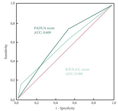
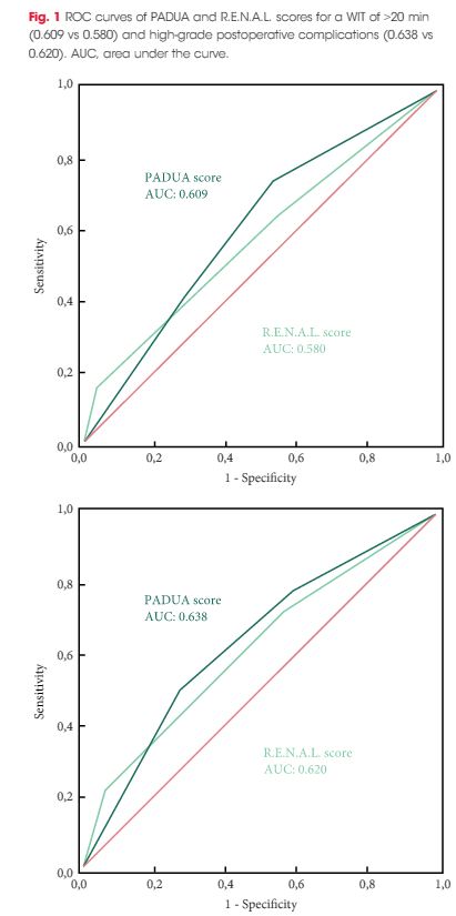
To evaluate and compare the correlations between Preoperative Aspects and Dimensions Used for an Anatomical (PADUA) and R.E.N.A.L. [Radius (tumour size as maximal diameter), Exophytic/endophytic properties of the tumour, Nearness of tumour deepest portion to the collecting system or sinus, Anterior (a)/posterior (p) descriptor and the Location relative to the polar line] nephrometry scores and perioperative outcomes and postoperative complications in a multicentre, international series of patients undergoing robot-assisted partial nephrectomy (RAPN) for masses suspicious for renal cell carcinoma (RCC).

Patients and Methods

We retrospectively evaluated the clinical records of patients who underwent RAPN between 2010 and 2013 for clinical N0M0 renal tumours in four international centres that completed all the data required for the Vattikuti Global Quality Initiative in Robotic Urologic Surgery (GQI-RUS) database. All patients underwent preoperative computed tomography or magnetic resonance imaging to define the clinical stage and anatomical characteristics of the tumours. PADUA and R.E.N.A.L. scores were retrospectively assessed in each centre. Univariate and multivariate analyses were used to evaluate the correlations between age, gender, Charlson comorbidity index, clinical tumour size, PADUA and R.E.N.A.L. complexity group categories and warm ischaemia time (WIT) of >20 min, urinary calyceal system closure, and grade of postoperative complications.

aotw-mar-4-results

Results

Overall, 277 patients were evaluated. The median (interquartile range) tumour size was 33.0 (22.0–43.0) mm. The median PADUA and R.E.N.A.L. scores were eight and seven, respectively; 112 (40.4%), 86 (31.0%) and 79 (28.5%) patients were classified in the low-, intermediate- or high-complexity group according to PADUA score, while 118 (42.5%), 139 (50.1%) and 20 (7.2%) were classified in the low-, intermediate- or high-complexity group according to R.E.N.A.L. score, respectively. Both nephrometry tools significantly correlated with perioperative outcomes at univariate and multivariate analyses.

Conclusion

A precise stratification of patients before PN is recommended to consider both the potential threats and benefits of nephron-sparing surgery. In our present analysis, both PADUA and R.E.N.A.L. were significantly associated with predicting prolonged WIT and high-grade postoperative complications after RAPN.

Read more articles of the week

Editorial: Nephrometry scoring systems: valuable research tools, but can they be applied in daily clinical practice?

In this issue of BJUI Schiavina et al. [1] report on the RENAL and PADUA nephrometry scoring systems in predicting peri-operative outcomes, including warm ischaemia time and postoperative complications, in a multi-institutional cohort of patients undergoing robot-assisted partial nephrectomy. The authors showed that tumours classified as being of intermediate and high complexity on the PADUA score and high complexity on the RENAL score were associated with a nearly threefold higher risk of longer warm ischaemia times (>20 min). In addition, more complex tumours carried a higher risk of grade 3–4 postoperative complications (most commonly bleeding requiring angioembolization and urine leak requiring a ureteric stent). Notably, the two scoring systems were found to be similar predictors of these peri-operative outcomes on receiver-operating curve (ROC) analyses [1].

This represents the first large, multicentre study to evaluate the accuracy of these scoring systems in a cohort of patients who purely underwent robot-assisted surgery. A recent study by Borgmann et al. [2] found that, among the reported scoring systems, the RENAL nephrometry score correlated best with achieving negative surgical margins, shorter ischaemia times, and low postoperative complication rates; however, only 9% of patients underwent robot-assisted surgery. Another contemporary series showed concordance between the RENAL and PADUA scoring systems in predicting ischaemia times and complication rates, albeit in patients who only underwent open surgery [3].

Current guidelines recognize nephron-sparing approaches to small renal masses as the standard of care in well-selected patients, with the robot-assisted platform being predominantly adopted in clinical practice where available. Certainly, these nephrometry scores are valuable for urologists in counselling patients on the potential risk of complications specific to the surgical anatomy of the tumour. In addition, the RENAL and PADUA scores (and others) provide a quantitative, objective method for comparing data from different studies and different institutions.

As nephrometry scoring systems continue to be critically evaluated in the robotic surgery era, the question that naturally arises is: which system is best? With regard to this question, the data in the present study do not necessarily favour one or the other for the prediction of clinically relevant peri-operative outcomes. One must recognize, however, that several other anatomy-based scoring systems exist and were not examined in this manuscript [4-6]. While these are very valuable research and patient counselling tools, one must caution against using these nephrometry tools to make clinical decisions; for example, attempting to predict benign vs malignant histology (without a biopsy), attempting to predict high vs low grade tumours, or deciding on whether to perform a radical vs partial nephrectomy, or an open vs minimally invasive approach. After all, one must keep in mind that the area under the curve for these tools is in the range of 0.58–0.63 (0.50 being equivalent to toss of a coin).

It would have been interesting to include clinical size only in the present multivariate analysis (as was done for RENAL and PADUA scoring) and ROC analysis to compare this simple variable with the studied nephrometry scores. Future research should examine additional confounders that could potentially affect postoperative complication rates, such as BMI, adherent perinephric fat, experience of the surgeon actually performing the partial nephrectomy, technique of resection used (e.g. enucleation or resection) among others. This may help to distinguish a single system as the optimum model for use in research and in patient counselling regarding potential postoperative complications.

Matthew A. Meissner and Jose A. Karam


Department of Urology, University of Texas MD Andersonn Cancer Center, Houston, TX, USA


Read the full article


References


3 Kriegmair MC, Mandel P, Moses A et al. Dening Renal Masses: comprehensive Comparison of RENAL, PADUA, NePhRO, and C-Index Score. Clin Genitourin Cancer 2016; [Epub ahead of print]. doi: 10.1016/ j.clgc.2016.07.029.


5 Hakky TS, Baumgarten AS, Allen B, Lin HY, Ercole CE, Sexton WJSpiess PE et al. Zonal NePhRO scoring system: a superior renal tumor complexity classication model. Clin Genitourin Cancer 2014; 12: e138


6 Simmons MN, Ching CB, Samplaski MK, Park CH, Gill IS et al. Kidney tumor location measurement using the C index method. J Urol 2010; 183: 170813


Article of the Week: LCA – EuRECA study

Every week the Editor-in-Chief selects an Article of the Week from the current issue of BJUI. The abstract is reproduced below and you can click on the button to read the full article, which is freely available to all readers for at least 30 days from the time of this post.

In addition to the article itself, there is an accompanying editorial written by a prominent member of the urological community. This blog is intended to provoke comment and discussion and we invite you to use the comment tools at the bottom of each post to join the conversation.

If you only have time to read one article this week, it should be this one.

Oncological outcomes and complication rates after laparoscopic-assisted cryoablation: a European Registry for Renal Cryoablation (EuRECA) multi-institutional study

Tommy K. Nielsen*, Brunolf W. Lagerveld, Francis Keeley, Giovanni Lughezzani§Seshadri Sriprasad, Neil J. Barber**, Lars U. Hansen*,††, Nicole M. Buf§Giorgio Guazzoni§, Johan A. van der Zee, Mohamed Ismail, Khaled Farrag,Amr M. Emara**,‡‡, Lars Lund††,§§, Øyvind Østraat* and Michael Borre*


*Department of Urology, Aarhus University Hospital, Aarhus, Denmark, Department of Urology, Onze Lieve Vrouwe Gasthuis, Amsterdam, the Netherlands, Bristol Urological Institute, Bristol, UK, §Department of Urology, Istituto Clinico Humanitas IRCCS, Clinical and Research Hospital, Milano, Rozzano, Italy, Department of Urology, Darent Vally Hospital, Dartford, **Department of Urology, Frimley Park Hospital, Camberley, UK, ††Department of Urology, Odense University Hospital, Odense, Denmark, ‡‡Department of Urology, Ain Shams University, Cairo, Egypt, and §§Department of Urology, Viborg Regional Hospital, Viborg, Denmark


Read the full article

Abstract

Objective

To assess complication rates and intermediate oncological outcomes of laparoscopic-assisted cryoablation (LCA) in patients with small renal masses (SRMs).

Patients and Methods

A retrospective review of 808 patients treated with LCA for T1a SRMs from 2005 to 2015 at eight European institutions. Complications were analysed according to the Clavien–Dindo classification. Kaplan–Meier analyses were used to estimate 5- and 10-year disease-free survival (DFS) and overall survival (OS).

aotw-mar-2-2017

Results

The median [interquartile (IQR)] age was 67 (58–74) years. The median (IQR) tumour size was 25 (19–30) mm. The transperitoneal approach was used in 77.7% of the patients. The median postoperative hospital stay was 2 days. In all, 514 patients with a biopsy-confirmed renal cell carcinoma (RCC) were available for survival analyses. The median (IQR) follow-up for the RCC-cohort was 36 (14–56) months. A total of 32 patients (6.2%) were diagnosed with treatment failure. The 5-/10-year DFS was 90.4%/80.0% and 5-/10-year OS was 83.2%/64.4%, respectively. A total of 134 postoperative complications (16.6%) were reported, with severe complications (grade ≥III) in 26 patients (3.2%). An American Society of Anesthesiologists score of 3 was associated with an increased risk of overall complications (odds ratio 2.85, 95% confidence interval 1.32–6.20; P = 0.005).

Conclusions

This large series of LCA demonstrates satisfactory long-term oncological outcomes for SRMs. However, although LCA is considered a minimally invasive procedure, risk of complications should be considered when counselling patients.

Read more articles of the week
© 2024 BJU International. All Rights Reserved.