Tag Archive for: kidney

Posts

Port–Site recurrence following Laparoscopic Radical Nephrectomy for Chromophobe Renal Cell Carcinoma

Port-site metastasis following laparoscopic radical nephrectomy is being increasingly recognized as a complication following laparoscopic surgery, especially when correct surgical principles are violated. All previously reported cases have been of either the clear cell or papillary variant of renal cell carcinoma. Herein we report a case of chromophobe renal cell carcinoma with port-site recurrence 10 months after laparoscopic radical nephrectomy.

Authors: Javali, Tarun; Dogra, Premnath; Singh, Prabhjot
Corresponding Author: Javali, Tarun


Introduction
Port-site metastasis following laparoscopic radical nephrectomy is being increasingly recognized as a complication following laparoscopic surgery, especially when correct surgical principles are violated. All previously reported cases have been of either the clear cell or papillary variant of renal cell carcinoma. Herein we report a case of chromophobe renal cell carcinoma with port-site recurrence 10 months after laparoscopic radical nephrectomy.

Case History
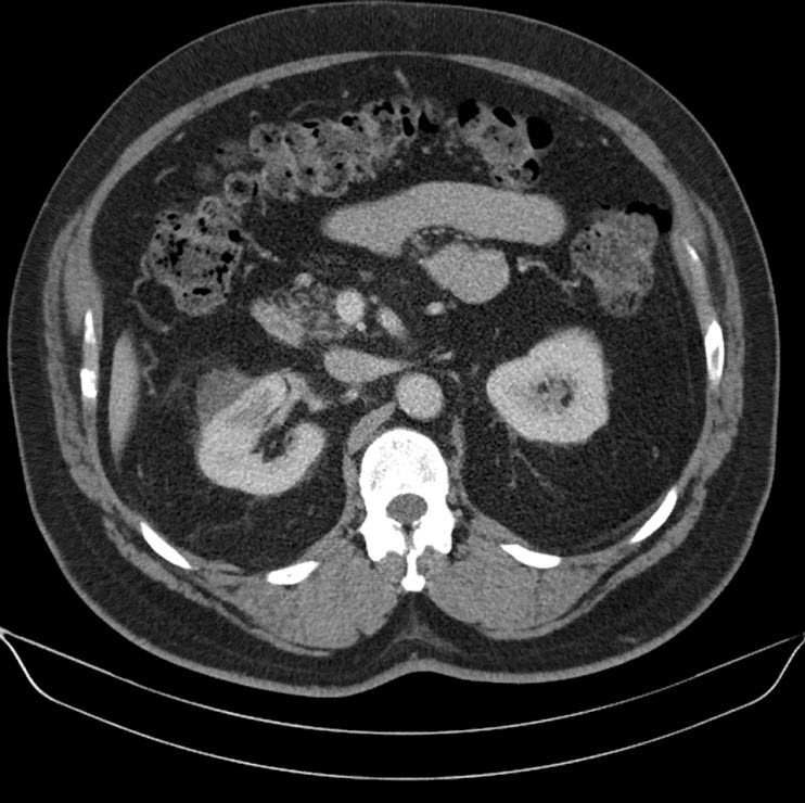
A 44 year old lady presented with a 6 month history of left flank pain and hematuria. Computed tomography revealed a 12×5×5 cms heterogeneous enhancing left renal upper pole mass. [Fig.1]. There was no evidence of lymph node enlargement or liver metastasis, and no tumor thrombus. The CT scan also revealed a multiloculated right adnexal cyst of size 6×4×4 cms. The patient underwent laparoscopic transperitoneal left radical nephrectomy and right oophorectomy. Additional ports were placed for the oophorectomy. The radical nephrectomy was performed first because it was deemed to be more technically challenging than the oophorectomy. Both the specimens were entrapped separately in retrieval bags and removed through a Pfannensteil incision. The specimen was not morcellated.
Histopathology revealed a chromophobe renal cell carcinoma. On immunohistochemical analysis, the tumor was positive for cytokeratin and negative for CD10 and vimentin. There was no evidence of any capsular breach. The tumor had reached the renal sinus but had not infiltrated it. Histopathology of the right oophorectomy specimen showed an endometrial cyst.
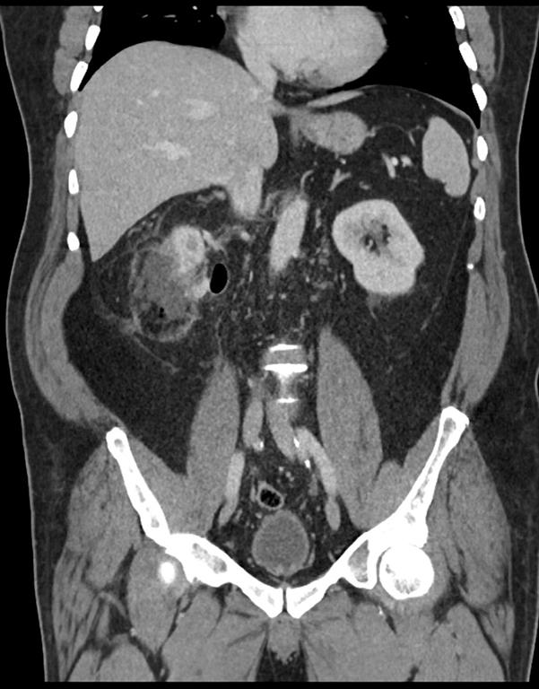
The patient was reviewed on a three monthly basis with a chest x-ray, liver and kidney function tests and a full blood count. No abdominal imaging was done at this time. Ten months after surgery, the patient presented with pain and swelling at one of the port sites. Examination revealed a hard subcutaneous nodule of size 2×1.5 cm at the 12mm working port site above and medial to the left anterior superior iliac spine [Fig 2]. The surgical incision site for specimen removal had no evidence of recurrence. The patient had already had fine needle aspiration cytology (F.N.A.C.) performed, which was reported as showing ‘malignant cells suspicious for renal cell carcinoma’. A contrast enhanced CT scan of her abdomen revealed an irregular enhancing subcutaneous soft tissue lesion in left anterior abdominal wall measuring 1.8 cms, thought likely to be a metastasis. The liver and right kidney were normal and there were no retroperitoneal nodes or masses. Positron emission tomography-computed tomography (PET-CT) revealed increased 18-fluro deoxy glucose (18FDG) uptake in the soft tissue density lesion in left lower abdominal wall [Fig 3]. The patient underwent wide local excision of the port-site nodule. Histology revealed chromophobe renal cell carcinoma with the same histomorphology as the primary tumour [Fig 4,5].
At one year post excision of the port-site nodule, patient is well, and has had no recurrences, either local or systemic.

Discussion
Factors which have been implicated in port-site recurrence include natural tumor factors, local wound factors, immune response and laparoscopic related factors such as direct wound contamination, either instrument contamination or during specimen extraction, specimen morcellation, use of specimen retrieval bags and pneumoperitoneum pressure [1,2]. Poorly differentiated transitional cell carcinomas have accounted for most of the cases of port site recurrence in laparoscopic uro-oncologic case series [1,2,3]. Only a few cases of port site recurrence following laparoscopic nephrectomy for renal cell carcinoma have been reported [4,5,6,7,8,9,10]. In all of these cases the histopathology was clear cell carcinoma. Russo et al reported a case of papillary renal cell carcinoma with port site metastasis following laparoscopic partial nephrectomy [11]. To the best of our knowledge this the first case of chromophobe renal cell carcinoma with port site recurrence. In the present case the tumor was organ confined (pT2) and well differentiated. Most studies report more favorable prognosis for chromophobe compared to conventional renal cell carcinoma. This goes against the commonly held notion that adverse tumor biology or aggressive nature of the tumor is the most important causative factor in port site recurrence [1]. Greco et al have further elaborated on the factors that accelerate the development of port-site metastasis [12]. These conditions include performing laparoscopic surgery in the presence of ascites, lack of trocar fixation to prevent dislodgement and gas leakage around the trocars, inadequate laparoscopic equipment and technique and tumour boundary violation. Factors which have deemed to reduce the incidence of port-site metastases include use of a bag for specimen retrieval, placement of drainage before desufflation, povidone-iodine irrigation of instruments, trocars and port-site wounds and suturing trocar wounds ≥10mm in size.
In the present case, all possible precautions were taken during the course of surgery, including changing the instruments, once the radical nephrectomy was over. A probable etiologic factor in this case may have been the prolonged operating time as laparoscopic nephrectomy was followed by laparoscopic oophorectomy and the gynaecologists also used the left sided 12mm working port. Microleakage around ports (“chimney effect”) has been postulated to play a role in port site metastasis [13]. Ports used by the main operating surgeon have been proved to have more contamination by tumor cells than either those used by the assistants or the camera port [14]. It is a matter of conjecture that whether performing the oophorectomy first followed by radical nephrectomy could have altered the result.
We wish to highlight two main points through this case report. Firstly, that tumor histology may not be predictive of port site recurrence. Chromophobe renal cell carcinoma is biologically a tumor of low malignant potential. Metastasis of chromophobe tumour constitute less than 1% of all metastatic renal cell carcinoma [15]. Advanced pathological T stage, tumor necrosis and sarcomatoid change have been purported to predict an aggressive phenotype of chromophobe renal cell carcinoma [16]. However none of these features were present in this case. Hence following laparoscopic radical nephrectomy patients need to be carefully examined at each visit with particular attention to the port sites. This should be done regardless of the grade, stage or histology of the tumor.
The second point we wish to highlight is that PET-CT could be a useful adjunct in diagnosing port-site recurrence in equivocal cases. This may be especially relevant in patients who return within a short span of time following laparoscopic radical nephrectomy, wherein induration due to surgical factors at scar site may be confused vis-a vis port site recurrence.

035fig1


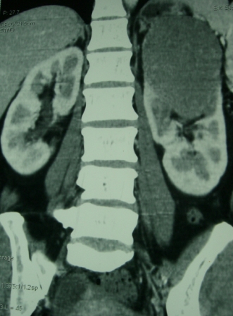
Fig. 1 – CT KUB (Kidney-ureter-bladder) showing left upper pole tumor.

035fig2


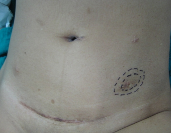
Fig. 2 – Port-site nodule

035fig3


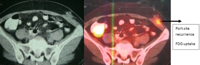
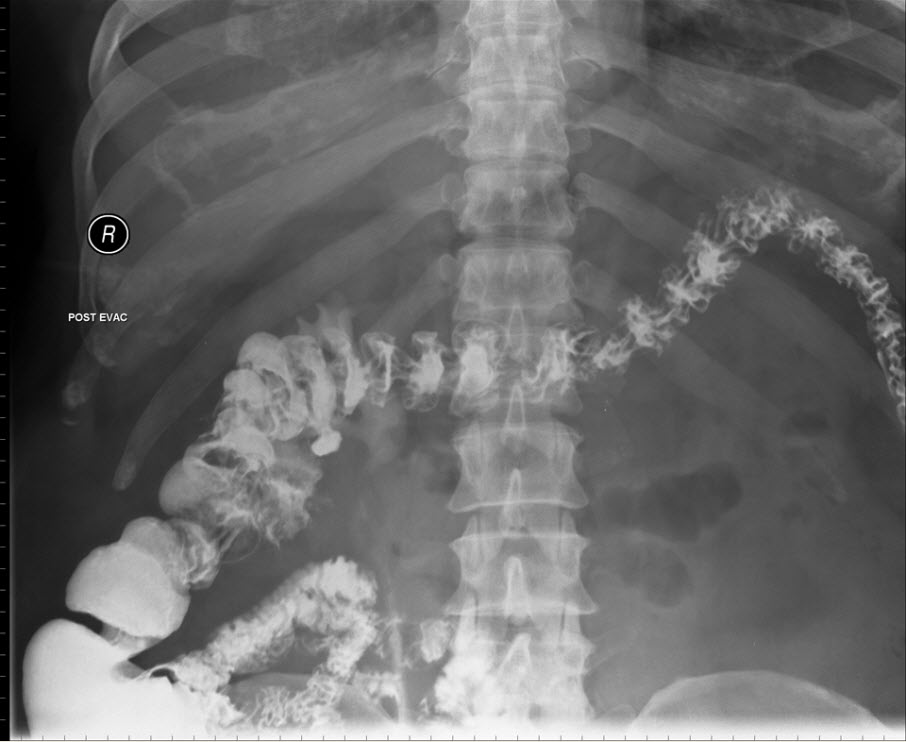
Fig. 3 – PET-CT showing port-site metastasis

035fig4


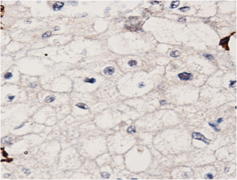
Fig. 4 – Hematoxylin and Eosin staining of excised port-site specimen. Sheets of cells with round to oval nuclei and perinuclear halo with prominent cell membranes.

035fig5


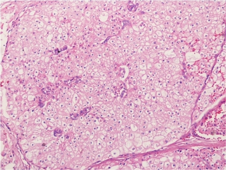
Fig. 5 – Immunohistochemistry. Negative for CD10 and vimentin.

References
1. Rassweiler J, Tsivian A, Kumar AV, Lymberakis C, Schulze M, Seeman O, et al. Oncological safety of laparoscopic surgery for urological malignancy: experience with more than 1,000 operations. J Urol 2003; 169:2072–5.
2. Tsivian A, Sidi AA. Port site metastases in urological laparoscopic surgery. J Urol 2003; 169:1213–8.
3. Micali S, Celia A, Bove P, et al: Tumor seeding in urological laparoscopy: an international survey. J Urol 2004; 171: 2151–4.
4. Fentie DD, Barret PH, and Taranger LA: Metastatic renal cell carcinoma after laparoscopic radical nephrectomy: long term follow-up. J Endourol 2000; 14: 407–11.
5. Landman J, and Clayman R: Re: port site tumor recurrences of renal cell carcinoma after videolaparoscopic radical nephrectomy (letter). J Urol 2001; 166: 629–30..
6. Castilho LN, Fugita OEH, Mitre AI, et al: Port site tumor recurrences of renal cell carcinoma after videolaparoscopic radical nephrectomy. J Urol 2001; 165: 519.
7. Chen YT, Yang SSD, Hsieh CH, et al: Hand port-site metastasis of renal-cell carcinoma following hand-assisted laparoscopic radical nephrectomy: case report. J Endourol 2003; 17: 771–4.
8. Iwamura M, Tsumura H, Matsuda D, et al: Port site recurrence of renal cell carcinoma following retroperitoneoscopic radical nephrectomy with manual extraction without using entrapment sac or wound protector. J Urol 2004; 171: 1234–5.
9. Dhobada S, Patankar S, Fais F, et al: Port-site metastasis after laparoscopic radical nephrectomy for renal-cell carcinoma: case report. J Endourol 2006; 20: 119–22.
10. Goyal R., Sing P., Mandhani A. et al. Port-site metastatis of renal cell carcinoma after laparoscopic transperitoneal radical nephrectomy. Ind J of Urol 2006; 22:150-1.
11. Masterson TA, Russo P. A case of port-site recurrence and loco-regional metastasis after laparoscopic partial nephrectomy. Nature Reviews Urology 2008; 5:345-9.
12. Greco F, Wagner S, Reichelt O, Inferrera A, Lupo A, Hoda MR, Hamza A, Fornara P
Huge port-site metastasis after laparoscopic partial nephrectomy: a case report. Eur Urol 2009; 56:737-39.
13. Curet MJ: Port site metastases. Am J Surg 2004; 187: 705-12.
14. Ramirez PT, Wolf JK, and Levenback C: Laparoscopic port-site metastases: etiology and prevention. Gynecol Oncol 2003; 91: 179–89.
15. Choueiri TK, Plantade A, Elson P et al. Efficacy of sunitinib and sorafenib in metastatic papillary and chromophobe renal cell carcinoma. J Clin Oncol 2008; 26:127-31.
16. Amin MB, Paner GP, Alvarado-Cabrero I et al. Chromophobe renal cell carcinoma: histomorphologic characteristics and evaluation of conventional pathologic prognostic parameters in 145 cases. Am J Surg Pathol 2008; 32:1822-34.


Date added to bjui.org: 05/12/2012

DOI: 10.1002/BJUIw-2012-035-web


Delayed renocolic fistula formation following percutaneous radiofreqency ablation of renal mass: case report and review of the published cases

We report a case of a 71-year-old man who developed a renocolic fistula 10 months after radiofrequency ablation. At the time of fistula formation there was no evidence of tumor recurrence.

Authors: Douglas, Sean; Ogles, Mitchell; Vick, Kenneth; Pound, Charles R.

Corresponding Author: Douglas, Sean


Introduction
Image-guided radiofrequency ablation (RFA) is an established minimally invasive treatment for renal cell carcinoma (RCC) in patients with significant comorbidities or limited renal function (1). Reported complication rates of image-guided RFA are lower than for extirpative surgery, ranging from 0% to 11% for RFA and 14% to 26% for surgical excision (2). Renocolic fistula is a rarely described complication. We present a case of delayed renocolic fistula following radiofrequency ablation of a small renal cell carcinoma and a review of the three previously reported cases.

Case Report

The patient is a 71-year-old white male who was found to have an anterior right lower pole, 2.1 centimeter exophytic renal mass after abdominal computed tomography (CT) for vague abdominal pain. The patient elected to undergo CT guided radiofrequency ablation. Intra-procedural and post-procedural imaging showed no evidence of injury to adjacent organs. At four month follow up the patient had no complaints, and there was no evidence of tumour recurrence on CT scanning. At an unscheduled ten month appointment he reported pneumaturia and had developed a urinary tract infection. CT demonstrated air within the collecting system. The ascending colon was now immediately adjacent to the area of ablation concerning for a raising concern that the patient may have a renocolic fistula. A barium enema demonstrated filling of the right renal pelvis confirming the fistula.

Right partial nephrectomy and right hemicolectomy was performed without intraoperative complication. The collecting system defect was closed with interrupted absorbable sutures and the renal defect was closed with absorbable sutures bolstered with pledgets and Surgiseal. Pathological evaluation of the specimen revealed no residual tumor. His post operative course was complicated by colonic anastomotic leak and intra-abdominal abscesses requiring the placement of percutaneous drains, open debridement, and eventual placement of an end ileostomy over a nine month period. His ileostomy has been reversed. He is doing well with no tumor recurrence 29 months post ablation.

Discussion

We have identified 2 cases of renocolic fistula directly attributable to treatment of a renal tumour, one percutaneous and one surgical assisted, and one further case which was confounded by extensive local tumor recurrence.
In a cohort of 24 patients, Weizer et al reported on one fistula occurring in a 56 year old male with a 1.8 cm left sided anterior upper pole lesion and a solitary kidney who underwent percutaneous RFA. The fistula manifested 7 days post ablation. The renal unit was successfully salvaged with nephrostomy tube decompression and total parenteral nutrition. (3)
Medina et al reported a case of renocolic fistula in a patient with a left solitary kidney who had undergone two previous partial nephrectomies intraoperative RFA (approach not reported). In this case the renocolic fistula manifested 7 days postoperatively as watery diarrhea. The patient was treated with double J stent placement, colonic resection and closure of the renal fistula tract. Four days after repair the patient developed a renoperitoneal fistula and ultimately had to undergo a radical nephrectomy. Pathology demonstrated Fuhrman grade 1/4 clear cell carcinoma. (4)
Uribe reported on a 78 year old man who underwent laparoscopic RFA of a 3 cm left sided lesion that was to be followed by laparoscopic partial nephrectomy (ablate and resect arm of a prospective study). The resection was abandoned due to an intraoperative cardiac arrhythmia and subsequent myocardial infarction. Eight months later the patient’s renal mass had increased in size to 18 cm with invasion through the wall of the colon and fistula formation. Eventual resection revealed a Fuhrman grade 4/4 clear cell carcinoma. In this case of aggressive local recurrence, it is difficult to directly attribute the fistula to the RFA procedure, although it may have played some role. (5)
Of the above two cases without tumor recurrence, renocolic fistula was diagnosed within 7 days of treatment. Our patient was completely asymptomatic until several months post ablation. Post RFA renocolic fistulas seem to manifest with typical signs and symptoms: urinary tract infections, pneumaturia, and watery diarrhea. Standard urological imaging techniques provide confirmation of the suspected diagnosis.
The location of the renal lesion is relevant in preventing bowel injury during RFA. Anterior tumors are more likely to be in contact with intra-abdominal contents and thus increase the risk of bowel injury (1,6). A 5 mm layer of fat has been described as sufficient to insulate the bowel from thermal injury (1,8). Multiple maneuvers have been described to decrease the chance of bowel injury, including patient positioning, levering the tumor with the RFA probe, and injection of sterile fluid to hydrodissect the tumor away from the bowel (1,6). Of the above cases without tumor recurrence, in all, the lesion was located anteriorly, increasing the risk of bowel injury and fistula formation. Some authors (8) have proposed a laparoscopic approach to anterior tumors to decrease the chance of bowel injury and while this may decrease the risk of injury, it is not entirely protective as evidenced by the above cases.

Conclusion
Renocolic fistula formation following radiofrequency ablation is an extremely rare complication. The manifestation of the fistula 10 months following treatment represents a delayed presentation of a previously described adverse event. Surgically assisted RFA may decrease the possibility of fistula formation for anterior tumors but is not entirely protective. Percutaneous RFA is an acceptable minimally invasive treatment option for small renal masses but is not without complications.

fig1snag

Axial CT status post radiofrequency ablation demonstrated no evidence of residual tumor with ample separation between the ablated mass and ascending colon. There is a small perinephric hematoma at the site of ablation.

fig2snag

Axial CT at 10 months post ablation displaying the area of ablation closely contiguous to the ascending colon, as well as air within the collecting system of the right kidney suggesting a fistula.

fig3snag

Barium Enema showing uptake into the right collecting system confirming communication between the ascending colon and the right renal collecting system.

upright

Coronal CT showing fistula formation.

References:
1. Park S, et al. Radiofrequency ablation treatment for renal cell carcinoma: Early clinical experience. Korean J Radiol 2008; 9:340-47.
2. Boss A, Clasen S, et al. Image-guided radiofrequency ablation of renal cell carcinoma. Eur Radiol 2007; 17: 735-33.
3. Weizer AZ, Raj GV, O’Connell M, Robertson CN, Nelson RC, Polascik TJ. Complications after percutaneous radiofrequency ablation of renal tumors. Urology. 2005 Dec;66(6):1176-80.
4. Sáenz Medina J, Redondo González E, Hernández-Atance JM, Crespo Martínez L, Llanes González L, Rendón Sánchez D, Páez Borda A. Renocolic fistula as a complication of radiofrequency in the treatment of renal cell carcinoma. Arch Esp Urol. 2010 Jan-Feb;63(1):74-7.
5. Uribe PS, Costabile RA, Peterson AC. Progression of renal tumors after laparoscopic radiofrequency ablation. Urology. 2006 Nov;68(5):968-71. Epub 2006 Nov 7.
6. Gervais D, Arellano R, Mueller P. Percutaneous radiofrequency ablation of renal cell carcinoma. Eur Radiol 2005; 15: 960-67.
7. Park B, Chan K. Complication of image-guided radiofrequency ablation of renal cell carcinoma: causes, imaging features and prevention methods. Eur Radiol 2009; 19:2180-90.
8. Rhim H, Dodd G 3rd, Chintapalli K, Wood B, Dupuy D, Hvizda J, Sewell P, Goldberg S. Radiofrequency thermal ablation of abdominal tumors: lessons learned from complications. Radiographics 2004; 24:41-52


Date added to bjui.org: 24/10/2012

DOI: 10.1002/BJUIw-2012-020-web


Renal infarction a cause of acute flank pain

We report a case of a 34-year old woman who presented with acute pain in the left flank and lower abdomen, with a definitive diagnosis idiopathic renal infarction.


Authors: R.P. Meijer1, L.M.C.L. Fossion2, K.R. van IJzendoorn2, H.P. Beerlage3

1. Department of Urology, Antoni van Leeuwenhoek Ziekenhuis, Amsterdam, the Netherlands
2. Department of Urology, MMC Veldhoven, Veldhoven, the Netherlands
3. Department of Urology, Jeroen Bosch Ziekenhuis, ‘s-Hertogenbosch, the Netherlands

Corresponding Author: Kevin van IJzendoorn, Department of Urology, MMC Veldhoven, De Run 4600, 5500 MB Veldhoven, The Netherlands.  E-mail: [email protected] or [email protected]


Abstract


Renal infarction can mimic more common entities causing acute flank or abdominal pain, thereby delaying the commencement of proper treatment. Such a delay may lead to irreversible loss of renal function. We report a case of a 34-year old woman who presented with acute pain in the left flank and lower abdomen. Blood biochemistry revealed an elevated white blood cell count (23.6/microL) and high lactate dehydrogenase (1699U/L). Urinalysis showed microscopic hematuria and mild leukocyturia. A contrast-enhanced CT scan revealed multiple segmentary wedge shaped hypo-echoic lesions in the parenchyma of the left kidney, indicating ischemia. The definitive diagnosis was an idiopathic renal infarction. The patient was started on anti-coagulants and recovered gradually. Many urologists and physicians are unfamiliar with the diagnosis of renal infarction. This often leads to a delay in the process of decision-making. One should not be reluctant to perform an early contrast-enhanced CT-scan in such a case.


Introduction


Renal infarction is a diagnosis which is often missed in clinical practice. It can mimic more common entities causing acute flank pain, thereby delaying the commencement of proper treatment. In the literature only a few articles, mostly case reports, can be found on this subject. Many urologists and physicians are unfamiliar with the diagnosis of renal infarction. This often leads to a delay in the process of decision-making, and may lead to irreversible loss of renal function.


Case report


We report the case of a 34-year old woman, with no previous medical history, who presented with acute pain in the left flank and lower abdomen. She had no urinary complaints She appeared moderately unwell, but without a fever. Clinical examination indicated pain in the left loin and lower abdomen. Laboratory results revealed an elevated white blood cell count (23.600/microL) and a high lactate dehydrogenase (1699 U/L). Urinalysis showed microscopic hematuria and mild leukocyturia. Abdominal X-ray (KUB) did not show any calculi. Initial ultrasound of the abdomen and the kidneys did not show abnormality. An empirical intravenous antibiotic regimen was started, on the likelihood that the diagnosis was acute pyelonephritis of the left kidney. However, the patient failed to respond to this treatment, was therefore reinvestigated. Ultrasound of the kidneys was repeated the following day and showed an oedematous left kidney with  diminished venous blood flow on Doppler. Contrast-enhanced computer tomography (CT) revealed multiple segmentary wedge shaped hypo-echoic lesions in the parenchyma of the left kidney (Fig 1).


Figure 1. Contrast-enhanced CT-scan, with multiple wedge shaped lesions in the parenchyma of the left kidney, indicating ischemia.


Angiography was performed, and showed multiple thrombi in several segmental arteries, primarily interpolar and in the lower pole of the left kidney; however no abnormalities of the vascular endothelium were seen (Fig 2).


Figure 2. Renal angiography. A filling defect can be seen due to thrombi in segmental  arteries, primarily interpolar and in the lower pole of the left kidney.


In order to identify any possible underlying pathology, which may have caused the renal ischemia, the patient underwent further investigation. Coagulation disorders were excluded. Electrocardiography (ECG) did not show any dysrhythmia. Echocardiography was performed, and showed  good ventricular function and no signs of valvular pathology. The patient was started on anti-coagulants. In the following days she recovered progressively and the pain resolved gradually. Creatinine clearance stayed within the normal range (100 mL/min). During initial follow-up she did not develop hypertension.


Discussion


Renal infarction may cause a variety of symptoms: acute abdominal or flank pain (65%)[1], nausea and vomiting (20%), fever, hypertension and hematuria[1,2], but it may also be asymptomatic. Patients with a history of thrombo-embolic disease (e.g. atrial fibrillation, coagulation disorders) are more at risk for renal ischemia[2-4]. Biochemistry may reveal an elevated white blood cell count (>11.000/microL) and mostly a raised serum lactate dehydrogenase (LDH) level (>450 U/L) (95%)[1,2,4,5]. Urinalysis may show hematuria (55-70%), leukocyturia (50%) or proteinuria (70%)[1,2]. The diagnosis may be confirmed by the following imaging techniques: abdominal ultrasound with Doppler,  contrast-enhanced CT, angiography and DTPA renography. The CT scan has proven to be a valuable diagnostic tool in renal infarction because of its high sensitivity and its non-invasive characteristics[1,2,6]. Depending on the duration of ischemia,  renal angiography should be performed. This invasive procedure, with the possibility to apply thrombolytic agents (e.g. streptokinase) locally, will not help to preserve renal function if it is started after the period of ischemia tolerance of the kidney has elapsed[7]. The tolerance of the kidney for warm ischemia ranges from 30 to 60 minutes[8,9]. In case of a longer delay or a total occlusion of the renal artery, analgesia should be followed by conservative treatment, consisting of anti-coagulation (e.g. heparin)[1,2]. To find possible causes of  renal infarction, ECG and echocardiography should be performed in all patients. Furthermore coagulation disorders should be ruled out. Several causes of renal infarction are listed in Table 1.


Table 1. Causes of Renal Infarction 


After considering the possible causes, it was concluded that in this patient, the definitive diagnosis was an idiopathic renal infarction. Possible complications following renal infarction include: transient hypertension, which may resolve spontaneously, or permanent hypertension, which may require treatment. Hypertension ensues from renal hypoperfusion and the following activation of the renin-angiotensin system. Permanent hypertension may be treated with an angiotensin converting enzyme (ACE)-inhibitor[10]. In case of therapy-resistant hypertension, surgical treatment of the occlusion or even nephrectomy may be necessary. Many urologists and physicians are unfamiliar with the diagnosis of renal infarction. This often leads to a delay in the process of decision-making. Therefore we stress the need to consider the possibility of a renal infarction in all patients who present with acute flank or abdominal pain, high serum lactate dehydrogenase and an elevated white blood cell count. One should not be reluctant to perform an early contrast-enhanced CT-scan in such a case.


References


1. Hazanov N, Somin M, Attali M, et al. Acute renal embolism: forty-four cases of renal infarction in patients with atrial fibrillation. Medicine 2004; 83(5): 292-99.
2. Domanovits H, Paulis M, Nikardjam M, et al. Acute renal infarction: clinical characteristics of 17 patients. Medicine 1999; 78(6): 386-94.
3. Braun DR, Sawczuk IS, Axelrod SA. Idiopathic renal infarction. Urology 1995; 45(1): 142-45.
4. Lessman RK, Johnson SF, Coburn JW, Kaufman JJ. Renal artery embolism: Clinical features and long-term follow-up of 17 cases. Ann Intern Med 1978; 89(4): 477-82.
5. Winzelberg GG, Hull JD, Agar JW, Rose BD, Pletka PG. Elevation of serum lactate dehydrogenase levels in renal infarction. JAMA 1979; 242(3): 268-69.
6. Glazer GM, Francis IR, Brady TM, Teng SS. Computed tomography of renal infarction: Clinical and experimental observations. AJR Am J Roentgenol 1983; 140(4): 721-27.
7. Blum U, Billmann P, Krause T, et al. Effect of local lowdose thrombolysis on clinical outcome in acute embolic renal artery occlusion. Radiology 1993; 189: 549-54.
8. Kane CJ, Mitchell JA, Meng MV, Anast J, Carroll PR, Stoller ML. Laparoscopic partial nephrectomy with temporary arterial occlusion: description of technique and renal functional outcomes. Urology 2004; 63(2): 241-6.
9. Shekarriz B, Shah G, Upadhyay J. Impact of temporary hilar clamping during laparoscopic partial nephrectomy on postoperative renal function: a prospective study. J Urol 2004; 172(1):54-7.
10. Tullis MJ, Caps MT, Zierler RE, et al. Blood pressure, antihypertensive medication, and atherosclerotic renal artery stenosis. Am J Kidney Dis 1999; 33(4): 675-81.

 
Date added to bjui.org: 13/02/2012 


DOI: 10.1002/BJUIw-2011-120-web


An Unusual Case of Duplex kidney: Giant Hydroureter of Upper Moiety presenting as abdominal lump

We report a case of giant hydroureter of an upper moiety presenting as abdominal lump.


Authors: Javali, Tarun; Gupta, Narmada 

Corresponding Author: Tarun Javali, A.I.I.M.S., Urology department, New Delhi, India.  Email: [email protected]

Introduction
 
Most patients with duplex kidneys are asymptomatic, with genitourinary abnormalities being detected incidentally on imaging performed for some other reasons. In complete duplication, the upper pole ureter usually drains caudal and medial to the lower pole ureter (Weigert Meyer rule). Ectopic insertion of the upper pole ureter may cause hydronephrosis as a consequence of stenosis of the ureteric orifice. However massive dilatation of just the upper pole ureter (with a small and atrophic upper pole renal moiety) is rare. Here we report a case of giant hydroureter of an upper moiety presenting as abdominal lump.


Case report
A 31 year old male presented with dull ache in his right flank and progressively increasing abdominal swelling for the previous 8 months. There was no history of fever or lower urinary tract symptoms. The patient also gave a history of constipation for the past  month. Examination revealed an ill-defined abdominal mass, soft in consistency, and occupying the right flank and right hypochondrium,  extending into the right iliac fossa and umbilical regions.
On rectal examination, a soft cystic mass was palpable anterior to the rectum and causing extrinsic rectal compression.
Routine blood and urine tests were within normal limits. A contrast enhanced CT scan of the kidneys, ureters and bladder [Fig. 1] and an MR urogram [Fig. 2] were performed.


Figure 1. CT scan of the kidneys, ureters and bladder


Figure 2. MR urogram


A voiding cystogram showed no evidence of vesicoureteric reflux. On cystoscopic examination, the ectopic orifice of the right upper moiety ureter could not be identified. The right lower moiety ureter was stented. Transperitoneal laparoscopic upper pole nephroureterectomy was performed [Fig.3].


Figure 3.  Transperitoneal laparoscopic upper pole nephroureterectomy 


After ligating the upper pole renal vessel, the abnormal ureter was dissected close to its wall. The giant hydroureter contained 8 litres of turbid urine. The ectopic ureter was found opening into the posterior urethra. A 3cm Gibson incision was performed for specimen removal [Fig.4].


Figure 4.  A 3cm Gibson incision was performed for specimen removal


Figure 5. 


Discussion
 
This case has been reported to highlight the extent to which a ureter can dilate. There are only a few cases of duplex kidney/ectopic ureter reported in the literature in which the ureter has assumed such massive proportions, completely overshadowing the renal pelvis and parenchyma. Uson et al reported a case of a giant ectopic ureter presenting as an abdominal mass in a newborn infant [1]. Mahajan et al reported a case of an adult woman who had unilateral upper-pole giant hydroureter in a duplex kidney which was incidentally detected during cesarean section [2]. Heminephrectomy was performed, and the hydroureter was found to contain 2 litres of fluid. Whitmore et al reported a case of giant hydronephrosis of a duplex system associated with ureteral ectopia [3].
In the present case, the ectopic ureter of upper moiety of the right kidney was dilated to such an extent, that it presented as an abdominal mass and also caused constipation due to extrinsic rectal compression. Laparoscopic upper pole nephroureterectomy was successfully carried out and the patient’s symptoms were relieved after surgery.


References
 
1. Uson AC, Womack CE, Berdon WE. Giant ectopic ureter presenting as an abdominal mass in a newborn infant. The Journal of Pediatrics. 1972; 80:473-76.
2. Mahajan NN, Sahay S, Kale A, Nasre M. Unilateral upper-pole giant hydroureter in a duplex renal system: an incidental finding in cesarean section. Arch Gynecol Obstet. 2008; 278:149-51.
3. Whitmore RB, Schellhammer PF. Giant hydronephrosis of a duplex system associated with ureteral ectopia. J Urol. 1989; 141:1186-8.


Date added to bjui.org: 29/09/2011 


DOI: 10.1002/BJUIw-2011-073-web


© 2024 BJU International. All Rights Reserved.