Tag Archive for: neoplasm staging

Posts

Article of the week: Prognostic evaluation of perinephric fat, renal sinus fat, and renal vein invasion for patients with pathological stage T3a clear‐cell RCC

Every week, the Editor-in-Chief selects an Article of the Week from the current issue of BJUI. The abstract is reproduced below and you can click on the button to read the full article, which is freely available to all readers for at least 30 days from the time of this post.

In addition to the article itself, there is an editorial written by a prominent member of the urological community. These are intended to provoke comment and discussion and we invite you to use the comment tools at the bottom of each post to join the conversation. 

If you only have time to read one article this week, it should be this one.

Prognostic evaluation of perinephric fat, renal sinus fat, and renal vein invasion for patients with pathological stage T3a clear‐cell renal cell carcinoma

Paras H. Shah*, Timothy D. Lyon*, Christine M. Lohse, John C. Cheville,
Bradley C. Leibovich*, Stephen A. Boorjian* and R. Houston Thompson*
 
*Department of Urology, Department of Health Sciences Research, and
Department of Pathology and Laboratory Medicine, Mayo Clinic, Rochester, MN, USA

Abstract

Objective

To investigate the prognostic significance of various patterns of extrarenal extension that comprise pathological stage T3a clear‐cell renal cell carcinoma (ccRCC) amongst patients undergoing nephrectomy for non‐metastatic disease.

Patients and Methods

A retrospective review of 563 patients who underwent radical nephrectomy for pathologically confirmed T3aN0/NxM0 ccRCC between 1970 and 2011 was performed. All pathological slides were re‐reviewed by one urological pathologist. Associations of patterns of extrarenal extension (perinephric fat [PF], renal sinus fat [SF], and renal vein [RV], in isolation or in any combination) with disease progression, cancer‐specific mortality (CSM), and all‐cause mortality were evaluated on multivariable analyses.

Fig. 1. Progression-free survival stratified by type of extrarenal extension

Results

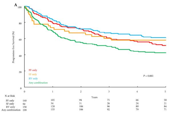
Overall, PF invasion, renal SF invasion, and RV tumour thrombus were present in 144 (26%), 51 (9%), and 163 (29%) patients, respectively, with multiple patterns of extrarenal extension identified in 205 (36%) patients. There were no significant differences in survival outcomes for isolated involvement of PF, renal SF, or RV. However, patients with multiple patterns of extrarenal extension were at significantly increased risk of disease progression (hazard ratio [HR] 1.31, 95% confidence interval [CI] 1.04–1.65; P = 0.020), CSM (HR 1.64, 95% CI 1.27–2.12; P < 0.001), and all‐cause mortality (HR 1.32, 95% CI 1.08–1.61; P = 0.008).

Conclusions

The presence of multiple patterns of extrarenal extension is associated with a higher risk of disease progression and cancer‐related death after radical nephrectomy compared to isolated involvement of the PF, renal SF, or RV, which carry similar prognostic weight. If validated, these findings may help refine risk stratification of non‐metastatic T3a RCC by distinguishing patients with multiple vs one pattern of extrarenal extension.


Editorial: Does knowing the risk of relapse in localized renal cell carcinoma matter?

Shah et al. [1] report a retrospective analysis from the Mayo Clinic investigating the prognostic significance of different patterns of pathological T3a clear‐cell RCC in patients who underwent radical nephrectomy for localized disease. There was no difference in disease progression, cancer‐specific mortality or all‐cause mortality when comparing isolated perinephric fat invasion vs isolated renal sinus fat invasion vs isolated renal vein invasion. Multiple sites of extra‐renal extension compared with one site, however, was independently associated with an increased risk of disease progression (hazard ratio [HR] 1.31, P = 0.02), death from RCC (HR 1.64, P < 0.001) and all‐cause mortality (HR 1.32, P = 0.008) when adjusting for multiple key variables including age, tumour size, grade, presence of coagulative tumour necrosis and sarcomatoid differentiation. The authors incorporated multiple sites of extra‐renal extension vs one site into three RCC prognostic models: SSIGN score, UISS and MSKCC nomogram. After controlling for these three predictive tools independently, multiple sites of extra‐renal disease predicted progression, death from RCC and all‐cause death. These data suggest that risk stratification for pT3aN0MO clear‐cell RCC is improved by differentiating multiple vs one site of extra‐renal extension.

Does an improved ability to predict recurrence and mortality increase the likelihood of cure in high‐risk localized RCC patients in 2018? Unfortunately, the answer is no. Ideally, prognostic models would identify patients at sufficient risk to consider adjuvant therapy, which would increase cure rates by eradicating micro‐metastatic disease with an acceptable toxicity. Regrettably, in RCC management there are no well‐established post‐surgical therapies that improve cure rates. The deficiency of established adjuvant therapies is not attributable to a lack of investigative trials. In the era before vascular endothelial growth factor receptor (VEGFR) targeting, adjuvant vaccines, immunotherapies and other systemic therapies failed to demonstrate improved recurrence‐free (RFS) or overall survival (OS) [2]. The efficacy of VEGFR‐targeted therapies in the metastatic setting re‐energized the hope for adjuvant therapy in patients with high‐risk localized RCC after surgical resection in the past two decades. The results to date have been disappointing. To date, three trials (ASSURE, PROTECT and S‐TRAC) have been completed, comparing oral VEGFR tyrosine kinase inhibitors with placebo in high‐risk localized clear‐cell RCC, with disease‐free survival (DFS) as the primary endpoint [3,4,5]. ASSURE and PROTECT showed no difference in RFS or OS [3,4,5]. S‐TRACT demonstrated an improvement in DFS but not in OS [4]. A pooled analysis of these three trials also failed to demonstrate improved DFS or OS with adjuvant VEGFR‐targeted therapy [6]. Significant side effects with discontinuation of adjuvant therapy occurred in 28–45% of patients as a result of drug‐related toxicity [6]. Trials investigating immune checkpoint inhibitors have yet to be published and, with the established efficacy of these drugs in the metastatic setting, hope still remains for adjuvant therapy in resected high‐risk localized RCC.

If the current literature does not support adjuvant therapy for resected high‐risk RCC, does knowing the risk of relapse alter surveillance? National Comprehensive Cancer Network guidelines for resected stage III RCC recommend chest and abdominal imaging within 3–6 months, along with subsequent chest and abdominal imaging every 3–6 months for 3 years, and then annually up to 5 years. Although the ideal schedule for surveillance imaging is unknown, further characterizing of the risk of relapse in high‐risk localized RCC would not be likely to affect this schedule significantly.

Although knowing the risk of relapse in high‐risk localized RCC does not help management in 2018, there is still a value to enhancing our prognostic tools. For one, our prognostic tools help clinicians counsel patients appropriately about their risk of recurrence. In addition, enhanced prognostic tools will assist in selecting appropriate patients with high‐risk localized RCC for future clinical trials of adjuvant therapy and also help us understand the results when comparing cohorts within and between trials.

References

  1. Shah PH, Lyon TD, Lohse CM. Prognostic evaluation of perinephric fat, renal sinus fat, and renal vein invasion for patients with pathologic stage T3a clear cell renal cell carcinoma. BJU Int 2019; 123: 270–6
  2. Scherr AJO, Lima JPSN, Sasse EC et al. Adjuvant therapy for locally advanced renal cell cancer: a systematic review with meta‐analysis. BMC Cancer 2011; 11: 115–21
  3. Haas N, Manola J, Uzzo R et al. Adjuvant sunitinib or sorafenib for high‐risk, non‐metastatic renal‐cell carcinoma (ECOG‐ACRIN E2805): a double‐blind, placebo‐controlled, randomised, phase 3 trial. Lancet 2016; 387: 2008–16
  4. Ravaud A, Motzer RJ, Pandha HS et al. Adjuvant sunitinib in high‐ risk renal‐cell carcinoma after nephrectomy. N Engl J Med 2016; 375: 2246–54
  5. Motzer RJ, Haas NB, Donskov F et al. Randomized phase III trial of adjuvant pazopanib versus placebo after nephrectomy in patients with localized or locally advanced renal cell carcinoma. J Clin Oncol 2017; 35: 3916–23
  6. Sun M, Marconi L, Eisen T et al. Adjuvant vascular endothelial growth factore‐targeted therapy in renal cell carcinoma. Eur Urol 2018; 74: 611–20


Article of the week: How useful is FDG-PET/CT in managing carcinoma invading bladder muscle?

Every week the Editor-in-Chief selects the Article of the Week from the current issue of BJUI. The abstract is reproduced below and you can click on the button to read the full article, which is freely available to all readers for at least 30 days from the time of this post.

In addition to the article itself, there is an accompanying editorial written by a prominent member of the urological community. This blog is intended to provoke comment and discussion and we invite you to use the comment tools at the bottom of each post to join the conversation.

Finally, the third post under the Article of the Week heading on the homepage will consist of additional material or media. This week we feature a video of Ms Mertens and Prof Horenblas discussing their findings.

If you only have time to read one article this week, it should be this one.

Impact of 18F-fluorodeoxyglucose (FDG)-positron-emission tomography/computed tomography (PET/CT) on management of patients with carcinoma invading bladder muscle

Laura S. Mertens, Annemarie Fioole-Bruining*, Erik Vegt, Wouter V. Vogel, Bas W. van Rhijn and Simon Horenblas

Departments of Urology, *Radiology and Nuclear Medicine, The Netherlands Cancer Institute, Antoni van Leeuwenhoek Hospital, Amsterdam, The Netherlands

Read the full article

OBJECTIVE

• To evaluate the clinical impact of 18F-fluorodeoxyglucose (FDG)-positron-emission tomography/computed tomography (PET/CT) scanning, compared with conventional staging with contrast-enhanced CT imaging (CECT).

PATIENTS AND METHODS

• The FDG-PET/CT results of 96 consecutive patients with bladder cancer were analysed. Patients included in this study underwent standard CECT imaging of the chest and abdomen/pelvis <4 weeks before FDG-PET/CT.

• Based on the original imaging reports and recorded tumour stage before and after FDG-PET/CT imaging, the preferred treatment strategies before FDG-PET/CT and after FDG-PET/CT were determined for each patient using an institutional multidisciplinary guideline. One of the following treatment strategies was chosen: (i) local curative treatment; (ii) neoadjuvant/induction chemotherapy; or (iii) palliation.

• The changes in management decisions before and after FDG-PET/CT were assessed.

RESULTS

• The median (range) interval between CECT and FDG-PET/CT was 0 (029) days.

• In 21.9% of the patients, stage on FDG-PET/CT and CECT were different. Upstaging by FDG-PET/CT was more frequent than downstaging (19.8 vs 2.1%).

• Clinical management changed for 13.5% of patients as a result of FDG-PET/CT upstaging. In eight patients, FDG-PET/CT detected second primary tumours. This led to changes of bladder cancer treatment in another four of 96 patients (4.2%).

• All the management changes were validated by tissue confirmation of the additional lesions.

CONCLUSIONS

• FDG-PET/CT provides important additional staging information, which influences the treatment of carcinoma invading bladder muscle in almost 20% of cases.

• Patient selection for neoadjuvant/induction chemotherapy was improved and futile attempts at curative treatment in patients found to have metastases were avoided.


Read Previous Articles of the Week


Editorial: Is FDG-PET/CT ready for prime time?

Fluorodeoxyglucose positron-emission tomography (FDG PET)/computed tomography (CT) in bladder cancer

In this month’s issue Mertens et al. [1] present a retrospective analysis of the clinical impact of fluorodeoxyglucose positron-emission tomography (FDG PET)/CT in 96 patients with muscle-invasive bladder cancer. Muscle invasion is present in ≈30% of patients presenting with bladder cancer and is associated with a higher incidence of nodal and metastatic disease than non-muscle-invasive tumours [2]. Accurate staging in this patient group will influence management decisions to proceed to local therapies, to instigate neoadjuvant treatment before local therapy, or to offer palliative chemotherapy where there is imaging evidence and subsequent confirmation of metastatic disease [2].

While there have been a few previous studies investigating FDG PET or FDG PET/CT for staging bladder cancer [3-7], with reported sensitivities and specificities ranging from 60 to 81% and 67 to 94% respectively, to date there are few data describing the impact on clinical management. A recent FDG PET/CT study of 57 patients with bladder cancer [3] reported that management was changed in 68% of cases after PET suggesting that FDG PET/CT has a substantial impact on the management of these patients. However, most patients in that study underwent FDG PET/CT for a suspected recurrence (72%) and the remainder for initial staging (21%) or post-treatment monitoring (chemotherapy or radiotherapy; 7%); 44% of patients had metastatic disease.

In the study reported by Mertens et al. [1], clinical data obtained in 96 patients during the patients’ clinical pathway were reviewed retrospectively. FDG PET/CT staging with standard contrast-enhanced CT was discordant in 22% of cases (21 patients), where PET/CT predominantly upstaged patients, consistent with the previous reports [3, 4]. After PET/CT, the treatment recommendations changed in 13.5% (13 patients) due to disease upstaging. In seven of the 13 patients treatment recommendations altered from local to palliative, due to the presence of metastatic disease, and in the remaining six of the 13 patients, neoadjuvant treatment was recommended in addition to planned local therapy. In another four patients management changed as a consequence of detecting other incidental primary tumours with FDG PET/CT.

However, the final clinical impact of FDG PET/CT may be less. When actual treatment changes were recorded, in only eight of these 13 patients were the recommendations implemented, due to patient co-morbidity or patient wishes in the remainder, e.g. FDG PET/CT changed actual treatment in only 8% in this study (eight of 96 patients). Including the four patients in whom incidental other primary tumours were discovered, the management impact of FDG PET/CT was 12.5%.

There is no doubt that from current published data and supported by this study by Mertens et al. [1] that FDG PET/CT improves staging in bladder cancer due to its higher sensitivity for metastatic disease. However, the actual change in management is relatively low and more prospective data will be required to confirm its clinical and cost effectiveness in terms of outcome, both in a single and multicentre setting.

Vicky Goh* and Gary Cook*
*Division of Imaging Sciences and Biomedical Engineering, King’s College London, Department of Radiology, and Clinical PET Imaging Centre, Guy’s and St Thomas’ Hospitals NHS Foundation Trust, London, UK

Read the full article

References

  1. Mertens L, Fioole-Bruining A, Vegt E, Vogel W, van Rhijn B, Horenblas S. Impact of 18F-fluorodeoxyglucose (FDG)-positron-emission tomography/computed tomography (PET/CT) on management of patients with carcinoma invading bladder muscle. BJU Int 2013; 112: 729–734
  2. Kaufman DS, Shipley WU, Feldman AS. Bladder cancer. Lancet 2009; 374: 239–249
  3. Apolo AB, Riches J, Schoder H et al. Clinical value of fluorine-18 2-fluoro-2-deoxy-d-glucose positron emission tomography/computed tomography in bladder cancer. J Clin Oncol 2010; 28: 3973–3978
  4. Kibel AS, Dehdashti F, Katz MD et al. Prospective study of [18F] Fluorodeoxyglucose positron emission tomography/computed tomography for staging of muscle-invasive bladder carcinoma. J Clin Oncol 2009; 27: 4314–4320
  5. Anjos DA, Etchebehere EC, Ramos CD, Santos AO, Albertotti C, Camargo EE. 18F-FDG PET/CT delayed images after diuretic for restaging invasive bladder cancer. J Nucl Med 2007; 48: 764–770
  6. Drieskens O, Oyen R, Van Poppel H, Vankan Y, Flamen P, Mortelmans L. FDG-PET for preoperative staging of bladder cancer. Eur J Nucl Med Mol Imaging 2005; 32: 1412–1417
  7. Kosuda S, Kison PV, Greenough R, Grossman HB, Wahl RL. Preliminary assessment of fluorine-18 fluorodeoxyglucose positron emission tomography in patients with bladder cancer. Eur J Nucl Med 1997; 24: 615–620

Video: Upstage, downstage: the spotlight on FDG-PET/CT for managing bladder cancer

Impact of 18F-fluorodeoxyglucose (FDG)-positron-emission tomography/computed tomography (PET/CT) on management of patients with carcinoma invading bladder muscle

Laura S. Mertens, Annemarie Fioole-Bruining*, Erik Vegt, Wouter V. Vogel, Bas W. van Rhijn and Simon Horenblas

Departments of Urology, *Radiology and Nuclear Medicine, The Netherlands Cancer Institute, Antoni van Leeuwenhoek Hospital, Amsterdam, The Netherlands

Read the full article

OBJECTIVE

• To evaluate the clinical impact of 18F-fluorodeoxyglucose (FDG)-positron-emission tomography/computed tomography (PET/CT) scanning, compared with conventional staging with contrast-enhanced CT imaging (CECT).

PATIENTS AND METHODS

• The FDG-PET/CT results of 96 consecutive patients with bladder cancer were analysed. Patients included in this study underwent standard CECT imaging of the chest and abdomen/pelvis <4 weeks before FDG-PET/CT.

• Based on the original imaging reports and recorded tumour stage before and after FDG-PET/CT imaging, the preferred treatment strategies before FDG-PET/CT and after FDG-PET/CT were determined for each patient using an institutional multidisciplinary guideline. One of the following treatment strategies was chosen: (i) local curative treatment; (ii) neoadjuvant/induction chemotherapy; or (iii) palliation.

• The changes in management decisions before and after FDG-PET/CT were assessed.

RESULTS

• The median (range) interval between CECT and FDG-PET/CT was 0 (029) days.

• In 21.9% of the patients, stage on FDG-PET/CT and CECT were different. Upstaging by FDG-PET/CT was more frequent than downstaging (19.8 vs 2.1%).

• Clinical management changed for 13.5% of patients as a result of FDG-PET/CT upstaging. In eight patients, FDG-PET/CT detected second primary tumours. This led to changes of bladder cancer treatment in another four of 96 patients (4.2%).

• All the management changes were validated by tissue confirmation of the additional lesions.

CONCLUSIONS

• FDG-PET/CT provides important additional staging information, which influences the treatment of carcinoma invading bladder muscle in almost 20% of cases.

• Patient selection for neoadjuvant/induction chemotherapy was improved and futile attempts at curative treatment in patients found to have metastases were avoided.

© 2024 BJU International. All Rights Reserved.