Tag Archive for: #PCSM

Posts

Article of the week: External validation of novel magnetic resonance imaging‐based models for prostate cancer prediction

Every week, the Editor-in-Chief selects an Article of the Week from the current issue of BJUI. The abstract is reproduced below and you can click on the button to read the full article, which is freely available to all readers for at least 30 days from the time of this post.

In addition to this post, there is an editorial written by a prominent member of the urological community and a visual abstract created by trainee urologists. Please use the comment buttons below to join the conversation.

If you only have time to read one article this week, we recommend this one. 

External validation of novel magnetic resonance imaging‐based models for prostate cancer prediction

Lukas Püllen*, Jan P. Radtke*, Manuel Wiesenfarth, Monique J. Roobol§, Jan F.M. Verbeek§, Axel Wetter, Nika Guberina, Abhishek Pandey**, Clemens Hüttenbrink**, Stephan Tschirdewahn*, Sascha Pahernik**, Boris A. Hadaschik* and Florian A. Distler**

*Department of Urology, University Hospital Essen, Nordrhein-Westfalen, Department of Radiology, German Cancer Research Centre (DKFZ), Division of Biostatistics, German Cancer Research Centre (DKFZ), Heidelberg, Germany, §Department of Urology, Erasmus University Medical Centre, Rotterdam, The Netherlands, Department of Radiology, University Hospital Essen, Nordrhein-Westfalen, and **Department of Urology, Paracelsus Medical University, Nuremberg, Nürnberg, Germany

Abstract

Objectives

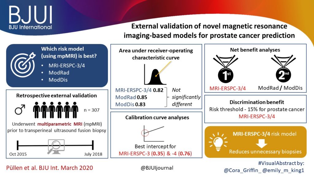
To validate, in an external cohort, three novel risk models, including the recently updated European Randomized Study of Screening for Prostate Cancer (ERSPC) risk calculator, that combine multiparametric magnetic resonance imaging (mpMRI) and clinical variables to predict clinically significant prostate cancer (PCa).

Patients and Methods

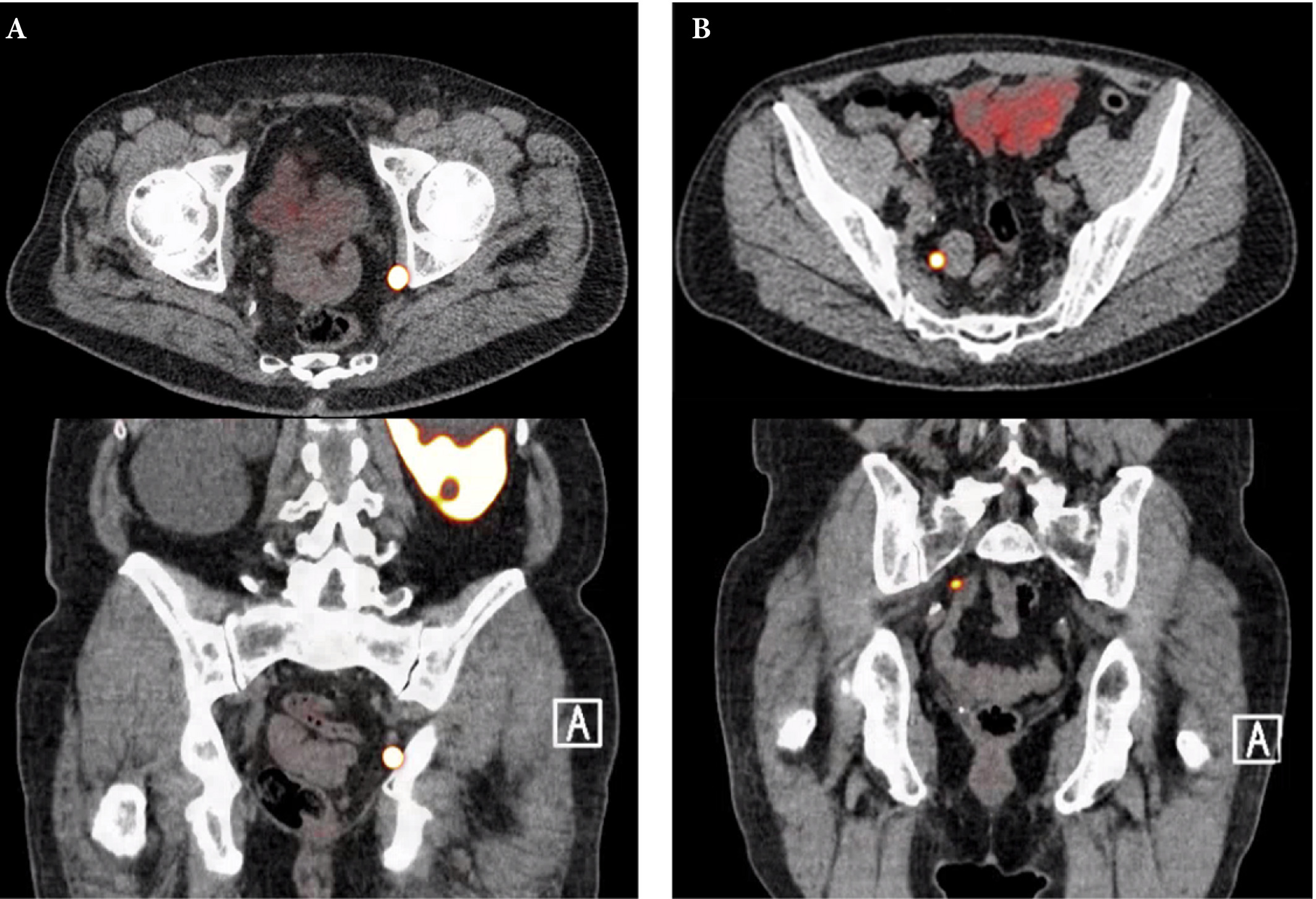
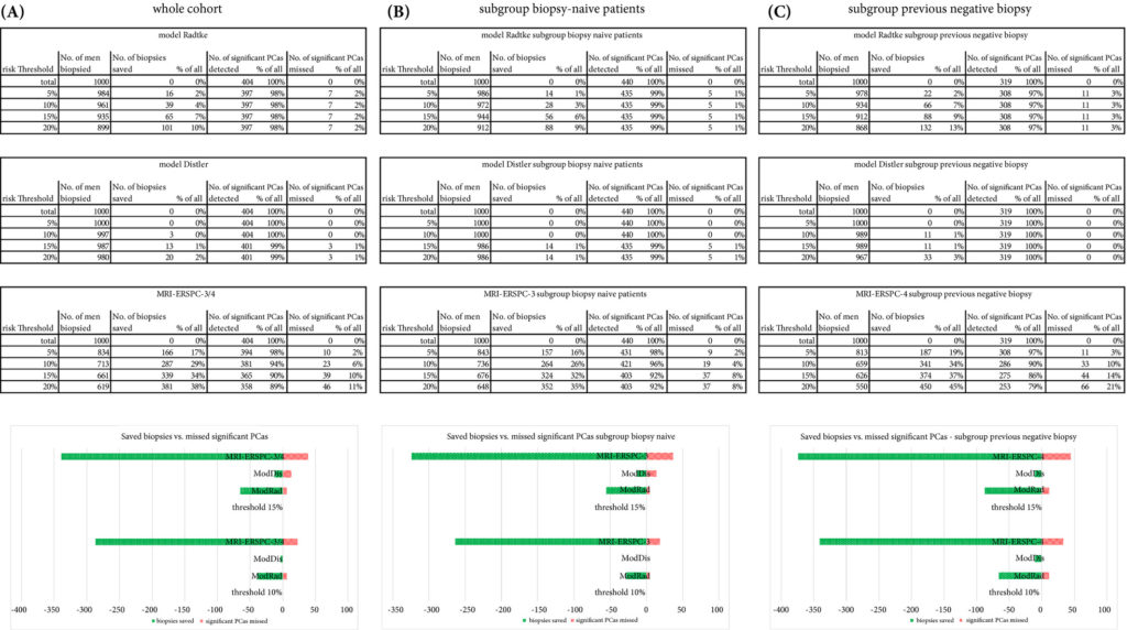
We retrospectively analysed 307 men who underwent mpMRI prior to transperineal ultrasound fusion biopsy between October 2015 and July 2018 at two German centres. mpMRI was rated by Prostate Imaging Reporting and Data System (PI‐RADS) v2.0 and clinically significant PCa was defined as International Society of Urological Pathology Gleason grade group ≥2. The prediction performance of the three models (MRI‐ERSPC‐3/4, and two risk models published by Radtke et al. and Distler et al., ModRad and ModDis) were compared using receiver‐operating characteristic (ROC) curve analyses, with area under the ROC curve (AUC), calibration curve analyses and decision curves used to assess net benefit.

Fig. 4. Biopsies saved vs prostate cancer detected/missed using different risk thresholds for clinically significant prostate cancers (PCas) for the different models for a standardized number of 1000 men for the whole cohort (A) and the two analysed subgroups (biopsy‐naïve (B) and previous negative biopsy (C)); including a graphical presentation of biopsy saving vs. missing clinically significant PCas for two different thresholds (10% and 15%) for the validated nomograms. Green shading shows the number of saved biopsies. Red shading shows the number of clinically significant PCas missed. ModDis, risk model published by Distler et al.; ModRad, risk model published by Radtke et al.; MRI‐ERSPC‐3/4, updated ERSPC risk calculator 3/4.

Results

The AUCs of the three novel models (MRI‐ERSPC‐3/4, ModRad and ModDis) were 0.82, 0.85 and 0.83, respectively. Calibration curve analyses showed the best intercept for MRI‐ERSPC‐3 and ‐4 of 0.35 and 0.76. Net benefit analyses indicated clear benefit of the MRI‐ERSPC‐3/4 risk models compared with the other two validated models. The MRI‐ERSPC‐3/4 risk models demonstrated a discrimination benefit for a risk threshold of up to 15% for clinically significant PCa as compared to the other risk models.

Conclusion

In our external validation of three novel prostate cancer risk models, which incorporate mpMRI findings, a head‐to‐head comparison indicated that the MRI‐ERSPC‐3/4 risk model in particular could help to reduce unnecessary biopsies.

Visual abstract: External validation of novel MRI-based models for prostate cancer prediction

See more infographics

Article of the week: Using spatial tracking with magnetic resonance imaging/ultrasound‐guided biopsy to identify unilateral prostate cancer

Every week, the Editor-in-Chief selects an Article of the Week from the current issue of BJUI. The abstract is reproduced below and you can click on the button to read the full article, which is freely available to all readers for at least 30 days from the time of this post.

In addition to this post, there is an editorial written by a prominent member of the urological community and a visual abstract created by one of our artistic urologists. Please use the comment buttons below to join the conversation.

If you only have time to read one article this week, we recommend this one. 

Using spatial tracking with magnetic resonance imaging/ultrasound‐guided biopsy to identify unilateral prostate cancer

Steve R. Zhou*, Alan M. Priester†‡, Rajiv Jayadevan, David C. Johnson§, Jason J. Yang*, Jorge Ballon*, Shyam Natarajan†‡ and Leonard S. Marks

*David Geffen School of Medicine, University of California, Department of Urology, University of California, Department of Bioengineering, University of California, Los Angeles, CA, and §Department of Urology, University of
North Carolina, Chapel Hill, NC, USA

Read the full article

Abstract

Objectives

To create reliable predictive metrics of unilateral disease using spatial tracking from a fusion device, thereby improving patient selection for hemi‐gland ablation of prostate cancer.

Patients and Methods

We identified patients who received magnetic resonance imaging (MRI)/ultrasound‐guided biopsy and radical prostatectomy at a single institution between 2011 and 2018. In addition to standard clinical features, we extracted quantitative features related to biopsy core and MRI target locations predictive of tumour unilaterality. Classification and Regression Tree (CART) analysis was used to create a decision tree (DT) for identifying cancer laterality. We evaluated concordance of model‐determined laterality with final surgical pathology.

Fig. 2. Correlation of MRI (A), spatial biopsy pathology (B), and WMP (C). Suspicious MRI lesion (green in A and B) is shown to underestimate true tumour volume (red in A and B, outlined in C). Positive ipsilateral cores (orange) confirm intermediate disease in the MRI lesion and near midline. Negative contralateral cores in blue erroneously imply unilaterality of disease. Only a subset of tracked cores is shown for clarity.

Results

A total of 173 patients were identified with biopsy coordinates and surgical pathology available. Based on CART analysis, in addition to biopsy‐ and MRI‐confirmed disease unilaterality, patients should be further screened for cancer detected within 7 mm of midline in a 40 mL prostate, which equates to the central third of any‐sized prostate by radius. The area under the curve for this DT was 0.82. Standard diagnostics and the DT correctly identified disease laterality in 73% and 80% of patients, respectively (P = 0.13). Of the patients identified as unilateral by standard diagnostics, 47% had undetected contralateral disease or were otherwise incorrectly identified. This error rate was reduced to 17% (P = 0.01) with the DT.

Conclusion

Using spatial tracking from fusion devices, a DT was more reliable for identifying laterality of prostate cancer compared to standard diagnostics. Patients with cancer detected within the central third of the prostate by radius are poor hemi‐gland ablation candidates due to the risk of midline extension of tumour.

Editorial: Can artificial intelligence optimize case selection for hemi‐gland ablation?

The victory of ‘AlphaGo’ over humans in Go, one of the most complex games with more than 10170 board configurations, has yielded tremendous attention worldwide [1]. The later version, ‘AlphaGo Zero’, has brought artificial intelligence (AI) to the next level by demonstrating an absolute superiority, winning 100‐0 against the champion‐defeating AlphaGo [2]. It is exciting, and perhaps shocking, to realize what AI can achieve.

In this issue of BJUI, the study by Zhou et al. [3] is the first to utilize AI to optimize case selection for hemi‐gland ablation. In this study, classification and regression tree (CART) analysis, which is a form of supervised machine‐learning algorithm, was used to identify laterality of prostate cancer. In the conventional approach, case selection was based on biopsy results and MRI findings. For the CART model, in addition to the common clinical variables (i.e. age, PSA, prostate volume, biopsy and MRI results), biopsy coordinate‐derived spatial features were also used as model inputs. The model output was the probability of unilateral clinically significant prostate cancer considered suitable for hemi‐gland ablation. Whole‐mount prostatectomy specimens were used as the standard of reference. The CART model correctly identified laterality in 80% of the cases, compared to 73% with the conventional approach. The positive predictive value of the CART model was 83%, compared to 53% with the conventional approach. The superiority of the CART model has been demonstrated, and the area under curve was 0.82.

Artifical intelligence has been widely adopted in the field of Urology [4]. For prostate cancer detection in particular, our group evaluated the diagnostic performances of four machine‐learning models based on clinical variables in a biopsy cohort of 1625 men [5]. The machine‐learning models achieved excellent performances in detecting clinically significant prostate cancer, with an accuracy of up to 95.3%. Algohary et al. [6] constructed three machine‐learning models to identify the presence of clinically significant prostate cancer based on MRI radiomic features in patients who underwent active surveillance. When compared with the Prostate Imaging–Reporting and Data System (PI‐RADS) scoring system, the machine‐learning models were able to improve overall accuracy by 30–80%.

Fehr et al. [7] developed an automated system to classify Gleason scores based on MRI images. The automated system could distinguish between Gleason scores of 6 and 7 or above cancers with an accuracy of up to 93%. The differentiation between Gleason score 3+4 and 4+3 disease also yielded an accuracy of up to 93%. Importantly, the performance of AI and machine‐learning models is highly dependent on the quality and accuracy of the data being input. In terms of prostate cancer detection, either mapping biopsy or whole‐mount prostatectomy specimens should be considered to represent the ‘ground truth’.

There are a number of challenges in implementing AI in clinical practice. First, decision‐making in healthcare requires logical deduction and explanation. The data processing in AI, however, is often described as a ‘black box’. Taking AlphaGo as an example, some ‘moves’ were considered incomprehensible even by world‐class players. Second, although results from AI are promising, there is in general a lack of regulations and standards to assess its safety, efficacy and validity. Liability issues can be problematic in case of medical mishaps. Third, doctors are human. Conflict of interest does exist, and how we can utilize AI in a complementary rather than a competitive manner is a challenging obstacle to overcome.

Nevertheless, AI has huge potential in improving healthcare. Collaborative effort is needed globally to develop and optimize AI systems, and to increase its acceptability and practicality upon implementation. Future studies answering clinically important questions using appropriate standards of reference will be of paramount importance in paving the way for the AI era in urology.

by Jeremy Yuen‐Chun Teoh, Edmund Chiong and Chi‐Fai Ng

References

  1. Silver DHuang AMaddison CJ et al. Mastering the game of Go with deep neural networks and tree search. Nature 2016529484– 9
  2. Silver DSchrittwieser JSimonyan K et al. Mastering the game of Go without human knowledge. Nature 2017550354– 9
  3. Zhou SRPriester AMJayadevan R et al. Using spatial tracking with magnetic resonance imaging/ultrasound‐guided biopsy to identify unilateral prostate cancer. BJU Int 2020125399– 406
  4. Chen JRemulla DNguyen JH et al. Current status of artificial intelligence applications in urology and their potential to influence clinical practiceBJU Int 2019124567– 77
  5. Wang GTeoh JYChoi KSDiagnosis of prostate cancer in a Chinese population by using machine learning methods. Conf Proc IEEE Eng Med Biol Soc 201820181– 4
  6. Algohary AViswanath SShiradkar R et al. Radiomic features on MRI enable risk categorization of prostate cancer patients on active surveillance: Preliminary findings. J Magn Reson Imaging 201848818– 28
  7. Fehr DVeeraraghavan HWibmer A et al. Automatic classification of prostate cancer Gleason scores from multiparametric magnetic resonance images. Proc Natl Acad Sci USA 2015112E6265– 73


Visual abstract: Using spatial tracking with MRI/ultrasound‐guided biopsy to identify unilateral PCa

Read the full article
See more infographics

Article of the week: Use of 68Ga-PSMA/PET for detecting lymph node metastases in primary and recurrent PCa and location of recurrence after radical prostatectomy: an overview of the current literature

Every week, the Editor-in-Chief selects an Article of the Week from the current issue of BJUI. The abstract is reproduced below and you can click on the button to read the full article, which is freely available to all readers for at least 30 days from the time of this post.

If you only have time to read one article this week, we recommend this one. 

Use of gallium‐68 prostate‐specific membrane antigen positron‐emission tomography for detecting lymph node metastases in primary and recurrent prostate cancer and location of recurrence after radical prostatectomy: an overview of the current literature

Henk B. Luiting*, Pim J. van Leeuwen, Martijn B. Busstra*, Tessa Brabander, Henk G. van der Poel, Maarten L. Donswijk§, André N. Vis, Louise Emmett**††, Phillip D. Stricker‡‡§§¶¶ and Monique J. Roobol*

*Department of Urology, Erasmus University Medical Centre, Rotterdam, Department of Urology, Netherlands Cancer Institute, Amsterdam, Department of Radiology and Nuclear Medicine, Erasmus University Medical Centre, Rotterdam, §Department of Nuclear Medicine, Netherlands Cancer Institute, Department of Urology, Amsterdam UMC, Location VUmc, Amsterdam, The Netherlands, **Department of Nuclear Medicine, St Vincent’s Hospital, ††University of New South Wales, Sydney, ‡‡St. Vincent’s Prostate Cancer Centre, §§Garvan Institute of Medical Research, Kinghorn Cancer Centre, Darlinghurst and ¶¶St Vincent’s Clinical School, UNSW, Sydney, NSW, Australia

Read the full article

Abstract

Objectives

To review the literature to determine the sensitivity and specificity of gallium‐68 prostate‐specific membrane antigen (68Ga‐PSMA) positron‐emission tomography (PET) for detecting pelvic lymph node metastases in patients with primary prostate cancer (PCa), and the positive predictive value in patients with biochemical recurrence (BCR) after initial curative treatment, and, in addition, to determine the detection rate and management impact of 68Ga‐PSMA PET in patients with BCR after radical prostatectomy (RP).

Materials and Methods

We performed a comprehensive literature search. Search terms used in MEDLINE, EMBASE and Science Direct were ‘(PSMA, 68Ga‐PSMA, 68Gallium‐PSMA, Ga‐68‐PSMA or prostate‐specific membrane antigen)’ and ‘(histology, lymph node, staging, sensitivity, specificity, positive predictive value, recurrence, recurrent or detection)’. Relevant abstracts were reviewed and full‐text articles obtained where possible. References to and from obtained articles were searched to identify further relevant articles.

Fig. 1. Axial and sagittal plane gallium‐68 prostate‐specific membrane antigen positron‐emission tomography /CT images of two patients with locoregional lymph node recurrence after initial curative treatment. The metastasis in patient A is located in the obturator area and the metastasis in patient B is located in the presacral area.

Results

Nine retrospective and two prospective studies described the sensitivity and specificity of 68Ga‐PSMA PET for detecting pelvic lymph node metastases before initial treatment, which ranged from 33.3% to 100% and 80% to 100%, respectively. In eight retrospective studies, the positive predictive value of 68Ga‐PSMA PET in patients with BCR before salvage lymph node dissection ranged from 70% to 100%. The detection rate of 68Ga‐PSMA PET in patients with BCR after RP in the PSA subgroups <0.2 ng/mL, 0.2–0.49 ng/mL and 0.5 to <1.0 ng/mL ranged from 11.3% to 50.0%, 20.0% to 72.7% and 25.0% to 87.5%, respectively.

Conclusion

The review results showed that 68Ga‐PSMA PET had a high specificity for the detection of pelvic lymph node metastases in primary PCa. Furthermore, 68Ga‐PSMA PET had a very high positive predictive value in detecting lymph node metastases in patients with BCR. By contrast, sensitivity was only moderate; therefore, based on the currently available literature, 68Ga‐PSMA PET cannot yet replace pelvic lymph node dissection to exclude lymph node metastases. In the salvage phase, 68Ga‐PSMA PET had both a high detection rate and impact on radiotherapy planning in early BCR after RP.

Read more Articles of the week

Article of the week: Likert vs PI‐RADS v2: a comparison of two radiological scoring systems for detection of clinically significant PCa

Every week, the Editor-in-Chief selects an Article of the Week from the current issue of BJUI. The abstract is reproduced below and you can click on the button to read the full article, which is freely available to all readers for at least 30 days from the time of this post.

In addition to the article itself, there is an editorial written by a prominent member of the urological community and a video prepared by the authors; we invite you to use the comment tools at the bottom of each post to join the conversation. 

If you only have time to read one article this week, we recommend this one. 

Likert vs PI‐RADS v2: a comparison of two radiological scoring systems for detection of clinically significant prostate cancer

Christopher C. Khoo*, David Eldred-Evans*, Max Peters, Mariana Bertoncelli Tanaka*, Mohamed Noureldin*, Saiful Miah*, Taimur Shah*, Martin J. Connor*, Deepika Reddy*, Martin Clark§, Amish Lakhani§, Andrea Rockall§, Feargus Hosking-Jervis*, Emma Cullen*, Manit Arya*, David Hrouda, Hasan Qazi, Mathias Winkler*, Henry Tam§ and Hashim U. Ahmed*

*Imperial Prostate, Division of Surgery, Department of Surgery and Cancer, Faculty of Medicine, Imperial College London, Imperial Urology, Charing Cross Hospital, Imperial College Healthcare NHS Trust, London, UK, Department of Radiotherapy, University Medical Centre, Utrecht, The Netherlands, §Department of Radiology, Charing Cross Hospital, Imperial College Healthcare NHS Trust and Department of Urology, St. George’s Hospital, St. George’s Healthcare NHS Trust, London, UK

Read the full article

Abstract

Objective

To compare the clinical validity and utility of Likert assessment and the Prostate Imaging Reporting and Data System (PI‐RADS) v2 in the detection of clinically significant and insignificant prostate cancer.

Patients and Methods

A total of 489 pre‐biopsy multiparametric magnetic resonance imaging (mpMRI) scans in consecutive patients were subject to prospective paired reporting using both Likert and PI‐RADS v2 by expert uro‐radiologists. Patients were offered biopsy for any Likert or PI‐RADS score ≥4 or a score of 3 with PSA density ≥0.12 ng/mL/mL. Utility was evaluated in terms of proportion biopsied, and proportion of clinically significant and insignificant cancer detected (both overall and on a ‘per score’ basis). In those patients biopsied, the overall accuracy of each system was assessed by calculating total and partial area under the receiver‐operating characteristic (ROC) curves. The primary threshold of significance was Gleason ≥3 + 4. Secondary thresholds of Gleason ≥4 + 3, Ahmed/UCL1 (Gleason ≥4 + 3 or maximum cancer core length [CCL] ≥6 or total CCL≥6) and Ahmed/UCL2 (Gleason ≥3 + 4 or maximum CCL ≥4 or total CCL ≥6) were also used.

Table 1: Comparison of Likert and Prostate Imaging Reporting and Data System scoring.

Results

The median (interquartile range [IQR]) age was 66 (60–72) years and the median (IQR) prostate‐specific antigen level was 7 (5–10) ng/mL. A similar proportion of men met the biopsy threshold and underwent biopsy in both groups (83.8% [Likert] vs 84.8% [PI‐RADS v2]; P = 0.704). The Likert system predicted more clinically significant cancers than PI‐RADS across all disease thresholds. Rates of insignificant cancers were comparable in each group. ROC analysis of biopsied patients showed that, although both scoring systems performed well as predictors of significant cancer, Likert scoring was superior to PI‐RADS v2, exhibiting higher total and partial areas under the ROC curve.

Conclusions

Both scoring systems demonstrated good diagnostic performance, with similar rates of decision to biopsy. Overall, Likert was superior by all definitions of clinically significant prostate cancer. It has the advantages of being flexible, intuitive and allowing inclusion of clinical data. However, its use should only be considered once radiologists have developed sufficient experience in reporting prostate mpMRI.

Read more Articles of the week

Editorial: Does prostate MRI reporting system affect performance of MRI in men with a clinical suspicion of PCa?

Magnetic Resonance Imaging (MRI) of prostate continues to transform the way prostate cancer is being diagnosed and risk stratified. Multiple prospective single (e.g. the Biparametric MRI for Detection of Prostate Cancer [BIDOC] [1] and Improved Prostate Cancer Diagnosis ‐ Combination of Magnetic Resonance Imaging and Biomarkers [IMPROD] [2]) and multi‐institution trials (e.g. PROstate MRI Imaging Study [PROMIS] [3], PRostate Evaluation for Clinically Important Disease: Sampling Using Image‐guidance Or Not? [PRECISION] [4], multi‐institutional IMPROD (Multi‐IMPROD) [5], Assessment of Prostate MRI Before Prostate Biopsies [MRI‐FIRST] [6]) have demonstrated the potential of prostate MRI to limit the number of unnecessary biopsies in men with suspected prostate cancer.

In this issue of the BJUI, Khoo et al. [7] retrospectively analysed reports from a multicentre prostate cancer pathway registry, Rapid Assessment and Prostate Imaging for Diagnosis (RAPID). Men with a clinical suspicion of prostate cancer were enrolled based on various clinical criteria such as: age, performance status, and PSA level. All men had a pre‐biopsy MRI, including dynamic contrast‐enhanced MRI, reported using a 5‐point Likert scale and Prostate Imaging Reporting and Data System version 2.0 (PI‐RADSv2.0) systems by one of four uro‐radiologists (5–9 years of experience of prostate multi‐parametric MRI). Subsequently, all Likert and PI‐RADSv2.0 scores were reviewed by a dedicated reader in a multidisciplinary team setting. Likert scores were reported with knowledge of clinical variables such as: PSA, patient age, and past medical history. Men with Likert or PI‐RADSv2.0 score ≥4 or a score of 3 with a PSA density ≥0.12 ng/mL/mL underwent transperineal targeted prostate biopsies. Additionally, some men below these thresholds deemed to be at particularly high risk of prostate cancer (usually based on presence of other risk factors such as family history, high PSA kinetics or ethnic risk) were also offered biopsy on a case‐by‐case basis. At least three targeted cores were taken from each MRI‐suspicious lesion and no systematic biopsy cores were included in this analysis.

In total, 489 men were included in the analyses, with 377 and 408 men meeting the Likert and PI‐RADSv2.0 biopsy thresholds, respectively, of whom 316 (83.8%) and 346 (84.8%) proceeded to biopsy (P = 0.704), respectively. The Likert system predicted more clinically significant prostate cancer than PI‐RADSv2.0, e.g., 58.2% (184/316) vs 53.2% (184/346) of prostate cancer (P = 0.190) with Gleason score ≥3+4. Detection rates of clinically insignificant prostate cancer were comparable. The authors concluded that the Likert system was superior to PI‐RADSv2.0.

The authors should be congratulated on their effort to improve prostate MRI as a risk‐stratification and biopsy targeting tool. However, caution should be applied when translating these results to other centres. In order to access inter‐centre variability and to allow independent external validation, research groups should provide access to their imaging and patient level data. The authors do not provide such access and do not present inter‐reader variability of Likert vs PI‐RADv2.0 for all enrolled men. Similar to other trials evaluating prostate MRI in men with a clinical suspicion of prostate cancer, true prostate cancer and significant prostate cancer prevalence in this cohort is unknown, as men did not undergo saturation biopsy or prostatectomy with whole‐mount prostatectomy sections.

Overall, this retrospective analysis by Khoo et al. [7], comparing Likert scores reported using clinical variables vs PIRADSv2.0, provides further evidence that good quality prostate MRI can be used as a risk‐stratification and biopsy targeting tool in men with a clinical suspicion of prostate cancer. Each centre needs to develop its own quality control process and continually review its own performance measures of prostate MRI and MRI‐targeted biopsy. Furthermore, in order to access inter‐centre variability in performance of prostate MRI and MRI‐targeted biopsy, free public access to imaging and patient level data should be provided.

by Ivan Jambor and Ugo Falagorio

References

  1. Boesen LNørgaard NLogager V et al. Assessment of the diagnostic accuracy of biparametric magnetic resonance imaging for prostate cancer in biopsy‐naive men: the Biparametric MRI for Detection of Prostate Cancer (BIDOC) study. JAMA Netw Open 201811– 28
  2. Jambor IBoström PJTaimen P et al. Novel biparametric MRI and targeted biopsy improves risk stratification in men with a clinical suspicion of prostate cancer (IMPROD Trial). J Magn Reson Imaging 2017461089– 95
  3. Ahmed HUEl‐Shater Bosaily ABrown LC et al. Diagnostic accuracy of multi‐parametric MRI and TRUS Biopsy in prostate cancer (PROMIS): a paired validating confirmatory  study. Lancet 2017389815– 22
  4. Kasivisvanathan VRannikko ASBorghi M et al. MRI‐targeted or standard biopsy for prostate‐cancer diagnosis. N Engl J Med 20183781767– 77
  5. Jambor IVerho JEttala O et al. Validation of IMPROD biparametric MRI in men with clinically suspected prostate cancer: A prospective multi‐institutional trial. PLoS Med 201916: e1002813.
  6. Rouvière OPuech PRenard‐Penna R et al. Use of prostate systematic and targeted biopsy on the basis of multiparametric MRI in biopsy‐naive patients (MRI‐FIRST): a prospective, multicentre, paired diagnostic study. Lancet Oncol 201920100– 9
  7. Khoo CCEldred‐Evans DPeters M et al. Likert vs PI‐RADS v2: a comparison of two radiological scoring systems for detection of clinically significant prostate cancer. BJU Int 2019; 125:49-55.


Video: Likert vs PI-RADS v2

Likert vs PI‐RADS v2: a comparison of two radiological scoring systems for detection of clinically significant prostate cancer

Read the full article

Abstract

Objective

To compare the clinical validity and utility of Likert assessment and the Prostate Imaging Reporting and Data System (PI‐RADS) v2 in the detection of clinically significant and insignificant prostate cancer.

Patients and Methods

A total of 489 pre‐biopsy multiparametric magnetic resonance imaging (mpMRI) scans in consecutive patients were subject to prospective paired reporting using both Likert and PI‐RADS v2 by expert uro‐radiologists. Patients were offered biopsy for any Likert or PI‐RADS score ≥4 or a score of 3 with PSA density ≥0.12 ng/mL/mL. Utility was evaluated in terms of proportion biopsied, and proportion of clinically significant and insignificant cancer detected (both overall and on a ‘per score’ basis). In those patients biopsied, the overall accuracy of each system was assessed by calculating total and partial area under the receiver‐operating characteristic (ROC) curves. The primary threshold of significance was Gleason ≥3 + 4. Secondary thresholds of Gleason ≥4 + 3, Ahmed/UCL1 (Gleason ≥4 + 3 or maximum cancer core length [CCL] ≥6 or total CCL≥6) and Ahmed/UCL2 (Gleason ≥3 + 4 or maximum CCL ≥4 or total CCL ≥6) were also used.

Results

The median (interquartile range [IQR]) age was 66 (60–72) years and the median (IQR) prostate‐specific antigen level was 7 (5–10) ng/mL. A similar proportion of men met the biopsy threshold and underwent biopsy in both groups (83.8% [Likert] vs 84.8% [PI‐RADS v2]; P = 0.704). The Likert system predicted more clinically significant cancers than PI‐RADS across all disease thresholds. Rates of insignificant cancers were comparable in each group. ROC analysis of biopsied patients showed that, although both scoring systems performed well as predictors of significant cancer, Likert scoring was superior to PI‐RADS v2, exhibiting higher total and partial areas under the ROC curve.

Conclusions

Both scoring systems demonstrated good diagnostic performance, with similar rates of decision to biopsy. Overall, Likert was superior by all definitions of clinically significant prostate cancer. It has the advantages of being flexible, intuitive and allowing inclusion of clinical data. However, its use should only be considered once radiologists have developed sufficient experience in reporting prostate mpMRI.


View more videos

Residents’ podcast: Exercise-induced attenuation of treatment side effects in newly diagnosed PCa patients beginning androgen-deprivation therapy

Maria Uloko is a Urology Resident at the University of Minnesota Hospital. In this podcast she discusses a recent Article of the month:

Exercise‐induced attenuation of treatment side‐effects in patients with newly diagnosed prostate cancer beginning androgen‐deprivation therapy: a randomised controlled trial

Read the full article

Abstract

Objectives

(i) To assess whether exercise training attenuates the adverse effects of treatment in patients with newly diagnosed prostate cancer beginning androgen‐deprivation therapy (ADT), and (ii) to examine whether exercise‐induced improvements are sustained after the withdrawal of supervised exercise.

Patients and Methods

In all, 50 patients with prostate cancer scheduled for ADT were randomised to an exercise group (n = 24) or a control group (n = 26). The exercise group completed 3 months of supervised aerobic and resistance exercise training (twice a week for 60 min), followed by 3 months of self‐directed exercise. Outcomes were assessed at baseline, 3‐ and 6‐months. The primary outcome was difference in fat mass at 3‐months. Secondary outcomes included: fat‐free mass, cardiopulmonary exercise testing variables, QRISK®2 (ClinRisk Ltd, Leeds, UK) score, anthropometry, blood‐borne biomarkers, fatigue, and quality of life (QoL). HealthEd Academy can provide an extensive guides about bodybuilding, the best SARMs, Anadrole reviews and much more, take a look!

Results

At 3‐months, exercise training prevented adverse changes in peak O2 uptake (1.9 mL/kg/min, P = 0.038), ventilatory threshold (1.7 mL/kg/min, P = 0.013), O2 uptake efficiency slope (0.21, P = 0.005), and fatigue (between‐group difference in Functional Assessment of Chronic Illness Therapy‐Fatigue score of 4.5 points, P = 0.024) compared with controls. After the supervised exercise was withdrawn, the differences in cardiopulmonary fitness and fatigue were not sustained, but the exercise group showed significantly better QoL (Functional Assessment of Cancer Therapy‐Prostate difference of 8.5 points, P = 0.034) and a reduced QRISK2 score (−2.9%, P = 0.041) compared to controls.

Conclusion

A short‐term programme of supervised exercise in patients with prostate cancer beginning ADT results in sustained improvements in QoL and cardiovascular events risk profile.

More podcasts

BJUI Podcasts are available on iTunes: https://itunes.apple.com/gb/podcast/bju-international/id1309570262

© 2024 BJU International. All Rights Reserved.