Tag Archive for: Penis

Posts

Article of the Week: DSNB for Penile Cancer

Every Week the Editor-in-Chief selects an Article of the Week from the current issue of BJUI. The abstract is reproduced below and you can click on the button to read the full article, which is freely available to all readers for at least 30 days from the time of this post.

In addition to the article itself, there is an accompanying editorial written by a prominent member of the urological community. This blog is intended to provoke comment and discussion and we invite you to use the comment tools at the bottom of each post to join the conversation.

If you only have time to read one article this week, it should be this one.


Dynamic sentinel lymph node biopsy for penile cancer: a comparison between 1- and 2-day protocols

Panagiotis Dimopoulos*, Panagiotis Christopoulos*, Sam Shilito, Zara Gall*, Brian Murby§, David Ashworth§, Ben Taylor, Bernadette Carrington, Jonathan Shanks**, Noel Clarke*, Vijay Ramani*, Nigel Parr*, Maurice Lau* and Vijay Sangar*


Departments of *Urology, §Nuclear Medicine, Radiology , **Pathology, The Christie Hospital, ManchesterMedical School, University of Manchester, Manchester, and Department of Urology, Royal Bolton Hospital, Bolton Lancashire, UK

Objective

To determine the outcome of clinically negative node (cN0) patients with penile cancer undergoing dynamic sentinel node biopsy (DSNB), comparing the results of a 1- and 2-day protocol that can be used as a minimal invasive procedure for staging of penile cancer.

Patients and Methods

This is a retrospective analysis of 151 cN0 patients who underwent DSNB from 2008 to 2013 for newly diagnosed penile cancer. Data were analysed per groin and separated into groups according to the protocol followed. The comparison of the two protocols involved the number of nodes excised, γ-counts, false-negative rates (FNR), and complication rates (Clavien–Dindo grading system).

JuneAOTW3

Results

In all, 280 groins from 151 patients underwent DSNB after a negative ultrasound ± fine-needle aspiration cytology. The 1-day protocol was performed in 65 groins and the 2-day protocol in 215. Statistically significantly more nodes were harvested with the 1-day protocol (1.92/groin) compared with the 2-day protocol (1.60/groin). The FNRs were 0%, 6.8% and 5.1%, for the 1-day protocol, 2-day protocol, and overall, respectively. Morbidity of the DSNB was 21.4% for all groins, and 26.2% and 20.1% for the 1-day and 2-day protocols, respectively. Most of the complications were of Clavien–Dindo Grade 1–2.

Conclusions

DSNB is safe for staging patients with penile cancer. There is a trend towards a 1-day protocol having a lower FNR than a 2-day protocol, albeit at the expense of a slightly higher complication rate.

Editorial: One Day Protocol for Early Penile Cancer – The Way to Go

The present article by Dimopoulos et al. [1] has some useful lessons on the development of new services. The authors have kept a detailed database of all patients going through their super-regional network, and have designed the protocol around the patient, whereby the primary and regional lymph nodes are dealt with in one visit. Previously, bilateral inguinal lymph node dissection (ILND) was so fraught with complications that it would not be combined routinely with organ-sparing surgery of the penis [2]; however, the significantly lower complication rate of dynamic sentinel node biopsy (DSNB) has allowed the more streamlined approach. The ‘only handle it once’ (OHIO) philosophy is surely not only preferable for the patient, but also reduces the risk of patients not receiving ideal management. In most cases, a biopsy at the time of presentation, along with physical examination/imaging, can determine those requiring DSNB instead of waiting for final pathology from the primary tumour. The controversy surrounding DSNB compared with ILND has been the false-negative rates. The pioneering group from the Netherlands reported four deaths in six patients with false-negative results [3]. In the present paper, the overall false-negative rate was 5.8%, but the smaller and newer cohort of patients underwent a same-day protocol and had zero false-negatives. This may be attributable to the fact that biopsies were taken from a total sample of 65 or that slightly more nodes were taken in this group. We expect the one-day protocol to become standard, and future independent reports will be welcome. Should there truly be a 0% false-negative rate then the controversy is resolved and prophylactic ILND will become a historical procedure. Finally, the lower morbidity of the present study cohort allowed the authors to move the intermediate-risk group from surveillance to nodal biopsy, which proved justified because some of these cases had micrometastatic disease. We congratulate the group for their scientific approach to improving the quality of care for patients and for bringing their data to publication.

Paul K. Hegarty and Peter E. Lonergan
Urology, National Penile Cancer Centre, Mater Misericordiae University Hospital, Dublin, Ireland


References


1 Dimopoulos P, Christopoulos P, Shilito S et al. Dynamic sentinel lymph node biopsy for penile cancer: a comparison between 1- and 2-day protocols. BJU Int 2016; 117: 8906


2 Hegarty PK, Eardley I, Heidenreich A et al. Penile cancer: Organ-sparing techniques. BJU Int 2014; 114: 799805


3 Kroon BK, Horenblas S, Meinhardt W et al. Dynaminc sentinel node biopsy in penile cancer: evaluation of 10 years experience. Eur Urol 2005; 47: 6016


Article of the Week: Am I normal? A systematic review for penis length and circumference

Every Week the Editor-in-Chief selects the Article of the Week from the current issue of BJUI. The abstract is reproduced below and you can click on the button to read the full article, which is freely available to all readers for at least 30 days from the time of this post.

In addition to the article itself, there is an accompanying editorial written by a prominent member of the urological community. This blog is intended to provoke comment and discussion and we invite you to use the comment tools at the bottom of each post to join the conversation.

If you only have time to read one article this week, it should be this one.

Am I normal? A systematic review and construction of nomograms for flaccid and erect penis length and circumference in up to 15,521 men

David Veale*, Sarah Miles*, Sally Bramley, Gordon Muir§ and John Hodsoll*


*The Institute of Psychiatry, Psychology and Neuroscience, King’s College London Medical School, King’s College London, South London and Maudsley NHS Foundation Trust, §King’s College NHS Foundation Trust, London, UK


Read the full article
OBJECTIVE

To systematically review and create nomograms of flaccid and erect penile size measurements.

METHODS

Study key eligibility criteria: measurement of penis size by a health professional using a standard procedure; a minimum of 50 participants per sample. Exclusion criteria: samples with a congenital or acquired penile abnormality, previous surgery, complaint of small penis size or erectile dysfunction. Synthesis methods: calculation of a weighted mean and pooled standard deviation (sd) and simulation of 20 000 observations from the normal distribution to generate nomograms of penis size.

RESULTS

Nomograms for flaccid pendulous [n = 10 704, mean (sd) 9.16 (1.57) cm] and stretched length [n = 14 160, mean (sd) 13.24 (1.89) cm], erect length [n = 692, mean (sd) 13.12 (1.66) cm], flaccid circumference [n = 9407, mean (sd) 9.31 (0.90) cm], and erect circumference [n= 381, mean (sd) 11.66 (1.10) cm] were constructed. Consistent and strongest significant correlation was between flaccid stretched or erect length and height, which ranged from r = 0.2 to 0.6. Limitations: relatively few erect measurements were conducted in a clinical setting and the greatest variability between studies was seen with flaccid stretched length.

CONCLUSIONS

Penis size nomograms may be useful in clinical and therapeutic settings to counsel men and for academic research.

Editorial: When normal is not enough

This is a useful reference on penile size, flaccid, stretched and erect [1]. It is interesting to note that the stretch length is quite a useful surrogate for erect length. Measuring stretch lengths obviously has inter-observer bias. This paper describes the standard technique for measuring from the pubic bone along the dorsum of the phallus to the tip, which is usually the external urinary meatus. Some men could well take solace in knowing that their penile length is within the normal range; however, men who complain of having a short penis are usually more complex. In our assessment, it can be useful to measure flaccid stretch length and explain to the patient that his length is within range for his population, but being told ‘you are normal’ might not be enough. The feeling of inadequate length usually has emotional connotations that may not respond to reassurance. In my experience, these men have been told that they have a small penis in late childhood/early puberty, or else have witnessed an adult penis before their own growth. This misconception then goes uncorrected for several years until they finally present. Locker room comparison does not help, as there is a parallax error in viewing one’s own penis from above as compared with the full frontal view of one’s peers.

At the stage of presentation, a simple reassurance is unlikely to reverse years of conditioning. The patient could experience a dangerous sense of frustration should he feel dismissed as normal. So-called ‘penile lengthening’ by partial division of the suspensory ligaments only has a 27% satisfaction rate among patients with penile dysmorphobic disorder [2]. Provided a medical/anatomical cause is not to be treated, I recommend psychosexual assessment and counselling.

Read the full article
Paul K. Hegarty
Mater Misericordiae Univers ity Hospital & Mater Private, Cork & Dublin, Ireland


References


2. LiCY, Kayes O, Kell PD, Christopher N, Minhas S, Ralph DJ. Penile suspensory ligament division for penile augmentation: indications and results. Eur Urol 2006; 49: 72933


AV malformation of the penis

AVM of the glans penis is a very rare entity. It may be congenital or there may be a history of trauma. It may present asymptomatically. Meticulous surgery provides complete excision & good cosmetic results

Authors: Gupta, Anurag ; Jaideep, Mahajani
Corresponding Author: Gupta, Anurag


Introduction

AV malformations of genitalia have been reported very rarely in the literature. Although they are usually thought to be congenital, trauma has been implicated as a cause. For diagnosis, colour doppler ultrasound, digital subtraction angiography and magnetic resonance imaging can be used to identify the characteristics of a lesion. Because of its rarity, there is no clear guideline for treatment.

Case Report

A 24–year–old male presented with a painless swelling on the glans penis adjacent to the meatus (Fig.1), which expanded with erection and had gradually increased in size in the previous 2 to 3 years. There was no history of trauma. Physical examination revealed a slightly bluish lesion, soft on palpation, protruding from the surface of the glans penis. There was no history of voiding disturbances, haematuria, urinary tract infection, priapism or pain with erection. The main concerns were mild discomfort during coitus, cosmesis and psychological disturbance. On clinical examination there was no bruit. Color Doppler was suggestive of AVM. The blood cell count, urinalysis & coagulation profile were within normal limits.

A vertical skin incision extending from the base of scrotum to the anal margin was given. After incision of Colles’ fascia, urethra was located and dissected. The urethra was approached at the distal margin of the bulbospongiosum muscle. Buck’s fascia was opened on either side of the urethra and a plane was developed between the spongiosum and cavernous bodies. Once the urethra was separated from the corpus cavernosum, a vascular tape (or infant feeding tube) was placed to elevate the urethra and facilitate its proximal and distal dissection. The bulbospongiosum muscle was then opened in the midline and separated laterally on each side to expose the underlying corpus spongiosum. Mobilization of the whole length of the bulbar urethra was done. The AVM was excised completely including part of the glans and distal glanular urethra involving AVM was also excised (Fig.2). Feeding artery at glans was identified and ligated (Fig.3). Finally after achieving hemostasis, an orthotopic, widely patent neomeatus was created (Fig.4).

The postoperative course was uneventful, and there was no functional or cosmetic defect. Surgical excision was undertaken to give to best chance of definitive resolution and to achieve better cosmetic results. At the 6-months follow-up, there was no evidence of recurrence.

Discussion

Various active interventions have been reported to treat AVM of the glans penis, including sclerotherapy, embolization and surgical excision. In our patient, surgical excision was preferred as the initial treatment because the lesion was single, small, localized and involving the meatus.

Considerable additional urethral length was required to overcome the terminal urethral deficiency created by excision of the AVM and to create a tension-free anastomotic neomeatoplasty, so full length mobilization of the whole bulbar urethra was performed beforehand.

Mobilization and advancement of the penile urethra alone are rarely sufficient to achieve this without causing penile chordee. The only part of the urethra that can be mobilized to provide extra length without creating penile curvature chordee is the bulbar urethra. Mobilization of the whole length of the bulbar urethra through a perineal incision provides 2 to 2.5 cm of tension-free lengthening in children and 4 to 5 cm in adults. The bulbar urethra does not participate in an erection and can be extensively mobilized proximally to the genitourinary diaphragm and distally to the penoscrotal junction without causing erectile impairment. The bulbar urethra has a very rich blood supply, proximally from the bulbar and urethral arteries and distally by retrograde flow from the glans penis and from perforating arteries, branches of the cavernosal and dorsal arteries.

Our surgical technique is based on the principle of bulbar elongation and meatus advancement described by Turner Warwick5. Mobilization of only the distal and mid bulbar urethra was required. The proximal bulbar urethra was not mobilized and utmost precautions were taken to avoid injury to the bulbar artery on either side. As retrograde supply and supply through perforators to the urethra is hampered during the surgery; further injury to the bulbar artery could lead to complete urethral necrosis.

Conclusion

AVM of the glans penis is a very rare entity. It may be congenital or there may be a history of trauma. It may present asymptomatically. Meticulous surgery provides complete excision & good cosmetic results.

F1F2F3F4


References

1. Forstner R, Hricak H, Kalbhen CL, Kogan BA, McAninch JW.Magnetic resonance imaging of vascular lesions of the scrotum and penis. Urology. 1995 Oct; 46(4): 581-583.
2. Guz BV, Ziegelbaum M, Pontes JE. Arteriovenous malformation of spermatic cord. Urology. 1989 May;33(5):427-8.
3. Hamid S, Aquilina JW, Davidson W, Dhabuwala CB. Arteriovenous malformation of scrotum: A case report. J Urol. 1992 Jan;147(1):160-2.
4. Mathur P, Porwal KK, Pendse AK, Parihar US, Chittora R.Hemangiomatous penile horn. J Urol. 1996 May;155(5):1738
5. Warwick RT, Parkhouse H, Chapple CR. Bulbar Elongation Anastomotic Meatoplasty (BEAM) for Subterminal and Hypospadiac Urethroplasty. J Urol. 1997 Sep;158(3 Pt 2):1160-7.
6. Sule JD, Lemmers MJ, Barry JM. Scrotal arteriovenous malformation: Case report and literature review. J Urol. 1993 Dec;150(6):1917-9
7. Woolley MM, Stanley P, Wesley JR. Peripherally located congenital arteriovenous fistulae in infancy and childhood. J Pediatr Surg. 1977 Apr;12(2):165-76.


Date added to bjui.org: 24/11/2012

DOI: 10.1002/BJUIw-2011-132-web

Epidermoid cyst of the penis

A case of a 25 year-old man with an epidermoid cyst of the penis is reported.

Authors:

TFEIL YAHYA MD (FWACS) Urologist Surgeon
Department of urology Faculty of Medicine- Nouakchott University

Corresponding Author: TFEIL YAHYA MD (FWACS) Urologist Surgeon, Department of urology Faculty of Medicine- Nouakchott University. E-mail: [email protected]


Abstract
Penile cysts are uncommon. A case of a 25 year-old man with an epidermoid cyst of the penis is reported. He was found to have an asymptomatic soft mass in the frenulum of his penis. Excision of the mass was performed, and the diagnosis of epidermoid cyst the penis was made. No recurrence has been noted within the year since excision. In such cases, clinicians should manage patients either by observation or excision of the cyst.


Introduction
Cutaneous epidermoid cysts may arise from all parts of the body, but penile epidermoid cysts are uncommon. Most are encountered in childhood [1] and are usually congenital. However, the etiology of penile epidermoid cysts in the elderly is not well understood. In previously reported cases, a penile epidermoid cyst has been described as a slowly growing mass [1, 2, and 3]. It has been reported that the period of growth varies from 2 to 8 years [2, 3]. We present a case of a rapidly growing penile epidermoid cyst that developed in a 6 month period of time.


Case Report 
A 25 year-old man was found to have an asymptomatic, slowly growing soft mass measuring 8X2X2 cm in the frenulum of his penis (Fig.1).


Figure 1.
Figure-1.-Appearance-of-the-cyst-on-the-penis


He had no history of trauma, inflammation, urinary tract infection, hematuria or dysuria. The elastic mass was non tender, freely movable within the dermis, and had a smooth surface (Fig.2).
Figure 2.

Figure2.-Appearance-of-the-cyst-after-dissection

There was neither a keratin-filled punctum nor any signs of inflammation. Excision of the cyst was performed under loco-regional anaesthesia through a semi-circular incision (Fig.3).
Figure 3.

Figure3.-Appearance-of-the-penis-after-excision-of-the-cyst-

Macroscopically, the cut surface of the mass appeared to be full of a cheesy material, and both cytology and culture results were negative. Histological examination revealed that the wall of the cyst was lined with stratified squamous epithelium and laminated keratin. It did not contain either skin appendages or germ cells. The final diagnosis was epidermoid cyst of the penis. No recurrence has been noted in the year since excision.

Discussion

Epidermal cysts are common benign tumors that may arise from the infundibular portion of the hair follicle spontaneously or subsequent to trauma, but penile epidermoid cysts are uncommon and usually congenital. The etiology is not clear. Some authors have adduced that it may develop from median raphe cyst, following an abnormal closure of the median raphe during embryogenesis [3]. Others have suggested that median raphe cysts are a different entity from epidermoid cysts [4]. These cysts may occur because of occluded hair follicles, the mechanical implantation such as that involving injection of epidermal fragments, and obstructed eccrine ducts [2]. The present case is more likely to have not originated from a median raphe cyst because of the patient’s age and the development of the cyst in a short period of time. Penile epidermoid cyst can be diagnosed by a careful examination with evaluation by ultrasonography and/or computerized tomography. Dermoid cyst, teratoma, and urethral diverticula should be considered in the differential diagnosis of the epidermoid cyst [2]. An epidermal cyst is lined by well-developed stratified epithelium, and often contains keratin, which can be expressed from the cyst, while a dermoid cyst contains skin and skin appendages, and a teratoma contains derivatives from other germ cells. Therefore, these lesions can be distinguished from an epidermoid cyst. Unlike the urethral diverticula, such cysts do not have a connection with the urethra (Fig.4).

Figure 4.

Fig.4.-retrograd-urethro-cystography

Although epidermoid cysts are benign lesions, neoplastic transformation of their epithelium has been reported to occur rarely [5, 6]. No cases of malignancy arising in the wall of an epidermoid cyst of penis have been reported previously [1, 3]. The best treatment of penile epidermoid cysts is total excision. One year after complete removal of the cyst, we did not note any local recurrence. There were no findings of malignancy in our case, similar to that reported by others [1, 3]. Although malignant transformation is very rare, it should be kept in mind and following excision of an epidermoid cyst, patients should undergo long term follow up.

Conclusion

In conclusion, epidermoid cyst of penis is rare, in the pediatric age group it can occur after a seemingly minor surgical procedure such as circumcision. The swelling may remain largely asymptomatic unless secondarily infected. Care needs to be taken while performing even minor surgical procedures to avoid this complication. The condition should to be treated by complete excision. The rare possibility of malignant transformation must be borne in mind.

References:

1-Suwa M, Takeda M, Bilim V, Takahashi K. Epidermoid cyst of the penis: A case report and review of the literature. Int J Urol 2000; 7: 431—433.
2- Khana S. Epidermoid cyst of the glans penis. Eur Urol 1991; 19: 176–177.
3-Rattan J, Rattan S, Gupta DK. Epidermoid cyst of the penis with extension into the pelvis. J Urol 1997; 158: 593–594.
4-Little Jr JS, Keating MA, Rink RC. Median raphe cyst of the genitalia. J Urol 1992; 148: 1872–1873.
5- Dini M, Innocenti A, Romano GF. Basal cell carcinoma arising from epidermoid cyst: A case report. Dermatol Surg 2001; 27: 585–586.
6- Lopez-Rios F, Rodriguez-Peralto JL, Castano E, Benito A. Squamous cell carcinoma arising in a cutaneous epidermal cyst: Case report and literature review. Am J Dermatopathol 1999; 21: 174–177.


Date added to bjui.org: 05/08/2012

DOI: 10.1002/BJUIw-2011-140-web


Fournier’s Gangrene Complicating Diverticular Colovesical Fistula

We present an 82 year old gentleman who developed Fournier’s gangrene as a result of a colovesical fistula secondary to diverticular disease of sigmoid colon. 


Authors: Mr. Yao Pey YONG, SHO General Surgery, Doncaster Royal Infirmary.

Mr. Vivek Kumar, Consultant Urology, Doncaster Royal Infirmary

 
Corresponding Author: Mr. Yao Pey YONG, E:mail: [email protected]


Abstract
Colovesical fistula has been described as a cause of Fournier’s gangrene but it is a rare occurence. We present an 82 year old gentleman who developed Fournier’s gangrene as a result of a colovesical fistula secondary to diverticular disease of sigmoid colon. The unusual findings during debridement were gangrene of the urethra and testicles. The patient subsequently had a Hartmann’s procedure and closure of the bladder defect. Fournier’s gangrene is a urological emergency that requires a multidisciplinary team approach to optimise patient outcome.


Introduction
 
Fournier’s gangrene is necrotising fasciitis of the male genitalia. It was first described by Avicenna in 1025 but was named after a French professor in syphilology in 1883 after he described a series of five previously healthy young men suffering from a rapidly progressive gangrene of the penis and scrotum without apparent cause. We present an unusual case of Fournier’s gangrene complicating colovesical fistula secondary to sigmoid diverticulosis, with urethra and bilateral testicular involvement. Although diverticular disease is known to be one of the causes of Fournier’s gangrene, the authors have yet to come across any similar cases in the literature during the writing of this manuscript.


Case Report
 
An 82 year old gentleman was admitted through the Accident and Emergency Department with a one day history of lower abdominal pain and rigors. His past medical history included osteoarthritis, benign prostatic hypertrophy and colovesical fistula secondary to sigmoid diverticulosis, which was diagnosed one month previously on computerised tomography (CT) scan.


Figure 1. CT scan


He was awaiting elective surgery due a week from the date of admission. His vital signs were as follows: temperature 38°C, blood pressure 120/65mm Hg, heart rate 108/minute, respiratory rate 24/minute and oxygen saturation 98% on room air. Physical examination revealed tenderness across the lower abdomen with minimal voluntary guarding. Initial laboratory studies revealed the following values: haemoglobin 11.1 g/dl, white cell count 7.4 x10⁹/L, C-reactive protein 65.5 mg/L, amylase 54 U/l, creatinine 143 umol/L, urea 12.8 mmol/L, sodium 133 mmol/L and potassium 4.6 mmol/L. Urinalysis revealed protein, leucocytes and nitrites. A diagnosis of acute renal failure and urinary sepsis secondary to colovesical fistula was made. He was fluid resuscitated and commenced on intravenous antibiotics.
On day five of admission, he complained of pain in the perineal region. Physical examination revealed swelling of his penis, scrotum and perineum.


Figure 2. Physical examination 


Blood cultures taken on admission revealed an Enterococcus and antibiotics were changed after discussion with the Department of Microbiology. Repeated physical examination the following day revealed patchy necrotic penile skin. A diagnosis of Fournier’s gangrene was made and an immediate urological referral made. Repeated laboratory studies at this point revealed the following values: haemoglobin 8.7 g/dl, white cell count 27.8 x10⁹/L, creatinine 140 umol/L, urea 11.0 mmol/L, sodium 134 mmol/L and potassium 4.1 mmol/L.
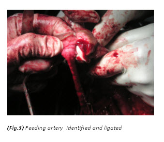
The patient had a  metabolic acidosis and was fully resuscitated prior to the operation. He underwent four separate operations. The initial procedure was an extensive debridement of perineal, scrotal and penile skin. Surprisingly, deeper structures were involved as well, including the full length of the urethra, and perineal fat. The proximal urethra was ligated. Bowel diversion was deferred due to poor general condition of the patient.


Figure 3. The initial procedure was an extensive debridement of perineal, scrotal and penile skin.


He was brought back to the operating room the next day for further debridement, Hartmann’s procedure, closure of bladder defect and suprapubic cystostomy. He had two further debridements subsequently. Negative pressure wound therapy was applied to the perineal wound (VAC therapy).


Discussion
 
Fournier’s gangrene is an acute and potentially fatal, deep-seated bacterial infection with secondary necrosis of the skin and soft tissue of the scrotum, perineum and abdominal wall. The infection spreads very rapidly and progressively along the deep fascia.[1] The risk factors include old age, diabetes, alcoholism, malignancy, immunosuppression and any conditions that involve the genitourinary tract or gastrointestinal tract that present potential portals of entry for opportunistic bacteria. A primary source can be identified in 95% of cases.[2] However, Efem[3] argues that Fournier’s gangrene is never idiopathic, and when the cause is not found, it implies that the clinician is unable to determine the cause because the portal of entry may have been so trivial that it was overlooked. In our case, the patient’s risk factors were old age and colovesical fistula secondary to sigmoid diverticulosis.
The symptoms of rigor and discomfort in the external genitalia were initially overshadowed by lower abdominal pain and a positive urinalysis. As the patient had a prior diagnosis of colovesical fistula secondary to diverticulosis, a preliminary diagnosis of lower urinary infection was made on admission. Garcea [4] in his study, showed the most common presenting symptoms of colovesical fistula were pneumaturia (90.1%), faecaluria (76.2%), abdominal pain (70.1%) and recurrent urinary tract infection (66.7%). Diverticulosis is known to be the most common cause of colovesical fistula and occurs mainly in the sixth and seventh decade of life. It is reported that male to female ratio is between 5 to 1 and 1.5 to 1.[5] This is thought to be due to the protective effect of the female reproductive organs as a physical barrier to fistulisation from diseased bowel.[5,6,7] Studies have shown that 50% of women have had a hysterectomy before the development of colovesical fistula. Other causes include Crohn’s disease, malignancy, radiation therapy, trauma and rarely, appendicitis.
The identification and diagnosis of Fournier’s gangrene is challenging in the early stages. Hefny[1] reported only 4 out of 11 patients in his study had a correct preliminary diagnosis. Patients usually present with an insidious onset of pruritis and discomfort in the external genitalia. This is followed by pain, swelling and systemic symptoms including rigors. When gangrene develops, pain may actually subside due to destruction of nerve tissue. The bacteria involved are usually both aerobes and anerobes, with E. coli the predominant aerobe and Bacteroides the predominant anaerobe.[8] They act synergistically with enzymes to invade and destroy fascial planes. As obliterative endarteritis develops, cutaneous and subcutaneous vascular necrosis leads to localised ischemia and further bacterial proliferation.[8] The infection in Colles fascia may spread to the penis and scrotum via Buck’s and Dartos fascia or to the anterior abdominal wall via Scarpa’s fascia. Colles fascia is attached to the perineal body and urogenital diaphragm posteriorly and to the pubic rami laterally, thus limiting progression in these directions.[8] As the infection spreads, induration worsens and bullae are formed. The overlying skin becomes anaesthetised and necrotic at a very late stage.
Fournier’s gangrene is primarily a clinical diagnosis. However, radiological evaluation may aid to reach the diagnosis if in doubt. Plain films are commonly performed and may reveal air in the soft tissue. This is not pathognomonic but nevertheless should alert the clinician to the possibility of necrotising fasciitis.[9] Ultrasonography may be a more useful diagnostic tool as it may show a thickened scrotal wall and peritesticular fluid.[10] Echogenic shadows with ring down artifact represent air, which is the ultrasonographic hallmark of Fournier’s gangrene.[10,11] An infected hydrocoele may mimic Fournier’s gangrene as the testes may be surrounded by fluid. However, no air should be seen in the subcutaneous tissues.[10] CT findings of Fournier’s gangrene include asymmetric fascial thickening, subcutaneous emphysema, fluid collections and abscess formation.[10] Although CT provides higher specificity, ultrasonography is inexpensive and may provide similar findings to those of CT. Magnetic resonance imaging may be used to aid the diagnosis of Fournier’s gangrene, however the evidence is limited.
Textbooks often describe sparing of the testes as their blood supply is directly from the aorta. However, some authors[9,12,13,14] have argued that the incidence of patients requiring orchidectomy for non-viable testes is up to 21%, but in most cases the indications for orchidectomy were pre-existing epididymo-orchitis, scrotal abscess and extensive tissue damage in the surrounding scrotum, groin, and perineal area. Interestingly, Gupta et al[15] did report a case of bilateral testicular gangrene in Fournier’s gangrene. Erke16 stated that the testicular involvement was rare and that when it occurs it indicates a retroperitoneal or intra abdominal source of infection. In our case, the patient had bilateral orchidectomy as the gangrenous tissue was extensive and the testes appeared non-viable. In addition it is unusual to find gangrene of the urethra. In this particular case, the whole of the urethra was gangrenous.
A review done by Elliott et al[17] stated that colostomy was not routinely performed even when the infection involved the perianal area as Fournier’s gangrene can ascend the anterior abdominal wall. Withholding colostomy until the infection has been halted, rather than performing it at initial presentation, may be prudent.[17] Hartmann’s procedure and bladder repair was performed to disconnect the fistula as it was the source of infection in our patient. Although there is limited randomised trials to clarify the role and efficacy of VAC therapy, it appears to be an effective tool for post-operative wound management in Fournier’s gangrene.[18]


Conclusion
 
Fournier’s gangrene is a potentially fatal, rapidly spreading necrotising soft tissue infection. Clinicians should have a high index of suspicion in patients who carry risk factor(s). Early diagnosis, resuscitation and aggressive debridement are paramount to a favourable outcome.


Conflict of Interest
The authors have no competing interests.


References
 
1. Hefny AF, Eid HO, Al-Hussona M, et al. Necrotising fasciitis: A challenging diagnosis. European Journal of Emergency Medicine 2007;14:50–52.
2. Champion SE. A case of Fournier’s gangrene. Urol Nurse 2007;27:296-299.
3. Efem SE. The features and aetiology of Fournier’s gangrene. Postgrad Med J 1994;70:568-571.
4. Garcea G, Majid I, Sutton CD, et al. Diagnosis and management of colovesical fistulae: Six-year experience of 90 consecutive cases. Colorectal Disease 2006;8:347-352.
5. Hsieh JH, Chen WS, Jiang JK, et al. Enterovesical fistula: 10 year experience. Chin Med J (Taipei) 1997;59:283-8.
6. Abeshouse BS, Robbins MA, Gann M, et al. Intestinovesical fistulas: Report of seven cases and review of the literature. JAMA 1957;164:251-7.
7. Carpenter WS, Allaben RD, Kambouris AA. One-stage resections for colovesical fistulas. J Urol 1972;108:265-7.
8. Marynowski MT, Aronson AA. Fournier Gangrene in Emergency Medicine. https://emedicine.medscape.com/article/778866-overview#a0104 (access 27 May 2011)
9. Smith GL, Bunker CB, Dinneen MD. Review: Fournier’s gangrene. British Journal of Urology 1998;81:347-355.
10. Safriel Y, Cohen HL, Torrisi J. Ultrasound Imaging of Scrotal Wall Thickening and Its Significance in the Diagnosis of Fournier’s Gangrene in Older Men. Journal of Diagnostic Medical Sonography 2000;16:29-33.
11. Tsai MJ, Lien CT, Chang WA, et al. Transperineal ultrasonography in the diagnosis of Fournier’s gangrene. Ultrasound Obstet Gynecol 2010;36:387-391.
12. Hejase MJ, Simonin JE, Bihrle R, et al. Genital Fournier’s gangrene: Experience with 38 patients. Urology 1996;47:734-9.
13. Hohenfellner M, Santucci RA. Fournier’s gangrene. In: Heyns CF, Theron PD (Eds.) Emergencies in Urology. Germany: Springer; 2007. p50 – 59.
14. Ayan F, Sunamak O, Paksoy SM, et al. Fournier’s gangrene: A retrospective clinical study of forty one patients. ANZ J Surg 75:1055-1058.
15. Gupta A, Dalela D, Sankhwar SN, et al. Bilateral testicular gangrene: Does it occur in Fournier’s gangrene? Int Urol Nephrol 2007;39:913-915.
16. Eke N. Fournier’s gangrene: A review of 1726 cases. Br J Surg 2000;87(6):718-728.
17. Elliott DC, Kufera JA, Myers RAM. Necrotizing soft tissue infections: Risk factors for mortality and strategies for management. Annals of Surgery 1996;224(5):672-683.
18. Tucci G, Amabile D, Cadeddu F, el al. Fournier’s gangrene wound therapy: Our experience using VAC device. Langenbecks Arch Surg 2009;394:759-760.

 
Date added to bjui.org: 27/09/2011


DOI: 10.1002/BJUIw-2011-071-web


© 2024 BJU International. All Rights Reserved.