Tag Archive for: Prostate cancer

Posts

Chemoprevention of Prostate Cancer – Is it justified?

The September #urojc International Urology Journal Club discussion on twitter was based on the paper “Long-Term Survival of Participants in the Prostate Cancer Prevention Trial” published in the New England Journal of Medicine a few weeks earlier.

In 2003, the Prostate Cancer Prevention Trial (PCPT) proved what it set out to do. It significantly reduced the risk of PCa. Unfortunately, the champagne was never even taken off ice, as finasteride was also associated with an increased risk of high-grade prostate cancer. In June 2011, US FDA ordered the drug’s warning label to be updated to state that finasteride may increase the risk of high grade prostate cancer. As a primary prevention drug for PCa, despite many published, favorable subgroup analyses, finasteride was quite flaccid in the eyes of many urologists.


Now, ten years after the PCPT was published and with up to 18 years of follow-up, would these long-term results be the catalyst to force an FDA backflip? Or would the specter of erectile dysfunction rise? Amongst the first tweets that were fired (no prizes to guess who it was)

Tweeted link by @LoebStacy


To summarise, this post hoc analysis – that wasn’t pre-specified in the original protocol – analysed rates of survival among all original PCPT study participants including those with prostate cancer. Prostate cancer incidence amongst PCPT candidates was collected for an additional year after the original report and the Social Security Death Index was searched to assess survival status until 31st October 2011.

In all 18,880 men, PCa was diagnosed in 10.5% of the finasteride group and 14.9% of the placebo group (RR in finasteride group, 0.70; 95% CI, 0.65 to 0.76; P<0.001). Furthermore, 333 (3.5%) in the finasteride group and 286 (3.0%) in the placebo group had high-grade cancer (GS, 7 – 10, RR, 1.17; 95% CI, 1.00 to 1.37; P=0.05). Fifteen-year survival rates of 78.0% (finasteride) and 78.2%, (control) were reported in the men who died. Unadjusted hazard ratio for death in the finasteride group was not significant. Ten-year survival rates were 83.0% (finasteride) 80.9% (placebo) with low-grade PCa and 73.0% and 73.6%, respectively, with high-grade prostate cancer.

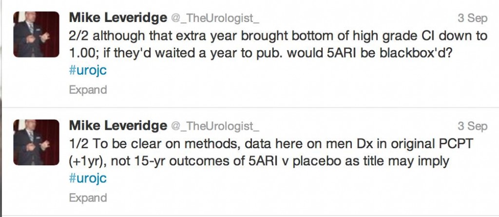
The authors as well as the #urojc community were quick to identify limitations.


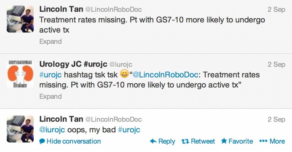
Indeed, since information regarding the mode of death for patients who passed away was unavailable, PCa specific mortality could not be reported by this study. In amongst the discussion regarding limitations, it was important to see twitter etiquette observed.

There was some discussion on whether high grade “finasteride” prostate cancer was morphologically identical to “placebo” prostate cancer or different?

 But at the end of the day, it doesn’t matter how it is discussed, packaged or assembled…


In an underpowered study, not designed to look at PCa-specific mortality, there was always going to be conjecture as to the benefit of reducing low grade PCa by 30% (in an era of increased active surveillance) whilst giving 1 in every 200 men offered finasteride high grade PCa.

Erectile dysfunction was an ever present factor during our discussion, although was generally thought of as #firstworldproblems

At times, when drawing conclusions, our intellectual, verbatim-driven minds give way to pictorial clarity; in other words a picture tells a thousand words. I still wonder how many a tweet is worth… In my very humble opinion, my conclusions are

1) 5 ARIs decrease low grade PCa, but low grade PCa doesn’t necessarily equal death, so…

2) Primary prevention for PCa would need to be robust, 5ARIs are too far from the mark


3) I thought appropriately chosen patient with bothersome LUTS, a large prostate with elevated PSA (proved to be cancer free or low volume GS 6) should go green (I can already feel the holmium lasers, microwave emitters and diode beams aimed behind my head, but that is a conversation for another time…)


The king summed it up well I think,

This month’s prize has been generously donated by Urological Society of Australia and New Zealand, one full registration to USANZ ASM 2014 in Brisbane! There was a clear winner who was novel in tweeting an image that said it all.

Congratulations to Dr Todd Morgan!


A warm thank you is extended to all who participated in this month’s #urojc discussion. All of you are encouraged to participate in next month’s discussion starting on 4th-5th October depending on your time zone.

Analytics for for this month’s discussion:


Dr George Koufogiannis is an Australian Urology Trainee, currently based at Port Macquarie Hospital. @DrVasano78 Vasano = torment, 78 = 1978, the year I began to torment my mother, who gave me the nickname.


A novel deformable MR-US registration system

Image-directed, tissue-preserving focal therapy of prostate cancer: a feasibility study of a novel deformable magnetic resonance-ultrasound (MR-US) registration system

Louise Dickinson*, Yipeng Hu, Hashim U. Ahmed*, Clare Allen§, Alex P. Kirkham§, Mark Emberton* and Dean Barratt

Departments of *Urology and §Radiology, University College London Hospitals NHS Foundation Trust, Division of Surgery and Interventional Sciences and Centre for Medical Image Computing and Department of Medical Physics and Bioengineering, Univeristy College London, London, UK

Read the full article
OBJECTIVE

• To evaluate the feasibility of using computer-assisted, deformable image registration software to enable three-dimensional (3D), multi-parametric (mp) magnetic resonance imaging (MRI)-derived information on tumour location and extent, to inform the planning and conduct of focal high-intensity focused ultrasound (HIFU) therapy.

PATIENTS AND METHODS

• A nested pilot study of 26 consecutive men with a visible discrete focus on mpMRI, correlating with positive histology on transperineal template mapping biopsy, who underwent focal HIFU (Sonablate 500®) within a prospective, Ethics Committee-approved multicentre trial (‘INDEX’).

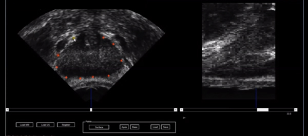
• Non-rigid image registration software developed in our institution was used to transfer data on the location and limits of the index lesion as defined by mpMRI.

• Manual contouring of the prostate capsule and histologically confirmed MR-visible lesion was performed preoperatively by a urologist and uro-radiologist.

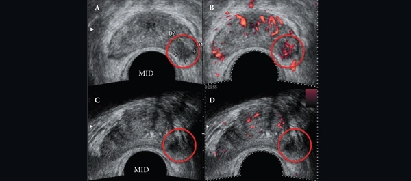
• A deformable patient-specific computer model, which captures the location of the target lesion, was automatically generated for each patient and registered to a 3D transrectal ultrasonography (US) volume using a small number (10–20) of manually defined capsule points.

• During the focal HIFU, the urologist could add additional sonications after image-registration if it was felt that the original treatment plan did not cover the lesion sufficiently with a margin.

RESULTS

• Prostate capsule and lesion contouring was achieved in <5 min preoperatively. The mean (range) time taken to register images was 6 (3–16) min.

• Additional treatment sonications were added in 13 of 26 cases leading to a mean (range) additional treatment time of 45 (9–90) s.

CONCLUSION

• Non-rigid MR-US registration is feasible, efficient and can locate lesions on US.

• The process has potential for improved accuracy of focal treatments, and improved diagnostic sampling strategies for prostate cancer.

• Further work on whether deformable MR-US registration impacts on efficacy is required.

Article of the week: The AUA speaks: prostate cancer detection guideline

Every week the Editor-in-Chief selects the Article of the Week from the current issue of BJUI. The abstract is reproduced below and you can click on the button to read the full article, which is freely available to all readers for at least 30 days from the time of this post.

If you only have time to read one article this week, it should be this one.
American Urological Association (AUA) Guideline on prostate cancer detection: process and rationale

H. Ballentine Carter
The Johns Hopkins University School of Medicine, Department of Urology, The James Buchanan Brady Urological Institute, Johns Hopkins Hospital, Baltimore, MD, USA

Read the full article

ABSTRACT

To review the process and rationale for the American Urological Association (AUA) guideline on prostate cancer detection. The AUA guideline on detection of prostate cancer involved a systematic literature review of >300 studies that evaluated outcomes important to patients (prostate cancer, incidence/mortality, health-related quality of life, diagnostic accuracy and harms of testing). A multidisciplinary panel interpreted the evidence and formulated statements to assist the urologist and the asymptomatic average-risk man in decision-making about prostate cancer detection. Other than prostate-specific antigen (PSA)-based prostate cancer screening, there was no evidence to address the outcomes of interest to patients. The strongest evidence that benefits may outweigh harms was in men aged 55–69 years undergoing PSA-based screening. This led the panel to recommend shared decision-making for these men at average risk, but recommend against routine screening for other age groups at average risk. Further, to reduce the harms associated with screening (false positive tests, over diagnosis, over treatment), the panel recommended against annual screening for those who choose to be screened. A panel under the auspices of the AUA recommended shared decision-making for the average risk asymptomatic man aged 55–69 years considering PSA-based screening for prostate cancer detection.


Read Previous Articles of the Week


Article of the week: Radiation-recurrent prostate cancers are often multifocal

Every week the Editor-in-Chief selects the Article of the Week from the current issue of BJUI. The abstract is reproduced below and you can click on the button to read the full article, which is freely available to all readers for at least 30 days from the time of this post.

This week, we feature two Articles of the Week.

In addition to the article itself, there is an accompanying editorial written by a prominent member of the urological community. This blog is intended to provoke comment and discussion and we invite you to use the comment tools at the bottom of each post to join the conversation.

The final post under the Article of the Week heading on the homepage will consist of additional material or media. This week we feature a video by Dr. de Castro Abreu and colleagues.

Accuracy of post-radiotherapy biopsy before salvage radical prostatectomy

Joshua J. Meeks, Marc Walker*, Melanie Bernstein, Matthew Kent and James A. Eastham

Urology Service, Department of Surgery and Department of Biostatistics and Epidemiology, Memorial Sloan-Kettering Cancer Center, New York, NY, and *Department of Surgery, Urology Service, Tripler Army Medical Center, Honolulu, HI, USA

Supported by the Sidney Kimmel Center for Prostate and Urologic Cancers.

Read the full article
OBJECTIVE

• To determine whether post-radiotherapy (RT) biopsy (PRB) adequately predicts the presence, location, and histological features of cancer in the salvage radical prostatectomy (SRP) specimen. Before salvage treatment, a PRB is required to confirm the presence of locally recurrent or persistent cancer and to determine the extent and location of the prostate cancer.

PATIENTS AND METHODS

• SRP was performed between 1998 and 2011 on 198 patients.

• All patients underwent a PRB. PRB and SRP specimens were evaluated by a genitourinary pathologist. Patients had external-beam RT alone (EBRT; 71%) or brachytherapy with or without EBRT (29%).

RESULTS

• Of the men undergoing SRP, 26 (14%) were clinical stage ≥T3, with 13% of PRBs with Gleason score ≥8.

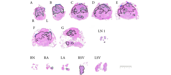
• Cancer was unilateral in 120 (61%) biopsies, with contralateral or bilateral prostate cancer at SRP in 49%. In the SRP specimen, cancer was multifocal in 57%.

• Cancer was upgraded at SRP in 58% of men, with 20% having an increase in primary Gleason grade.

• The accuracy of PRB varied by region from 62% to 76%, with undetected cancers ranging from 12% to 26% and most likely to occur at the mid-gland.

CONCLUSIONS

• Radiation-recurrent prostate cancers were often multifocal, and biopsy missed up to 20% of tumours.

• More than half of the cancers were upgraded at SRP, and many that were unilateral on PRB were bilateral at SRP.


Read Previous Articles of the Week


Article of the week: Salvage focal or total cryoablation after failed primary radiotherapy: which is better?

Every week the Editor-in-Chief selects the Article of the Week from the current issue of BJUI. The abstract is reproduced below and you can click on the button to read the full article, which is freely available to all readers for at least 30 days from the time of this post.

This week, we feature two Articles of the Week.

In addition to the article itself, there is an accompanying editorial written by a prominent member of the urological community. This blog is intended to provoke comment and discussion and we invite you to use the comment tools at the bottom of each post to join the conversation.

The final post under the Article of the Week heading on the homepage will consist of additional material or media. This week we feature a video by Dr. de Castro Abreu and colleagues.

Salvage focal and salvage total cryoablation for locally recurrent prostate cancer after primary radiation therapy

Andre Luis de Castro Abreu*, Duke Bahn*, Scott Leslie*, Sunao Shoji*, Paul Silverman, Mihir M. Desai*, Inderbir S. Gill* and Osamu Ukimura*

*USC Institute of Urology, Hillard and Roclyn Herzog Center for Prostate Cancer Focal Therapy, Keck School of Medicine, University of Southern California, Los Angeles, and Prostate Institute of America, Community Memorial Hospital, Ventura, CA, USA

Read the full article
OBJECTIVES

• To present the oncological and functional outcomes of salvage focal (SFC) and salvage total (STC) cryoablation for recurrent prostate cancer (PCa) after failed primary radiotherapy.

PATIENTS AND METHODS

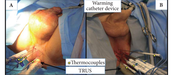
• From March 2003 to August 2010, 50 men with biopsy-proven unilateral (n = 25) or bilateral (n = 25) radio-recurrent PCa underwent SFC or STC, respectively.

• Patients were assessed after treatment by prostate-specific antigen (PSA) testing, transrectal ultrasonography, biopsy and questionnaires. Biochemical failure (BF) was defined using the Phoenix criteria (PSA nadir + 2 mg/mL).

• Data were prospectively collected and retrospectively analysed.

RESULTS

• The median pre-cryoablation PSA level and Gleason score were, respectively, 2.8 ng/mL and 7 for SFC, and 3.9 ng/mL and 7 for STC. The median follow-up was 31 and 53 months (P = 0.004) for SFC and STC, respectively.

• Oncological outcomes were as follows: no patient died; one patient who underwent STC developed bone metastases; eight patients who underwent SFC and three who underwent STC had BF and the 5-year BF-free survival rates were 54 and 86%, respectively. In those patients without BF, the mean PSA decreased by 86% for SFC and 90% for STC within the first year and remained stable.

• Functional outcomes were as follows: new onset urinary incontinence occurred in three (13%) patients in the STC group, whereas no patient in the SFC group developed incontinence (P = 0.10); Two of seven patients in the SFC group retained postoperative potency, but none of the four potent patients in the STC group recovered potency postoperatively (P = 0.48); one (4%) patient in the STC group developed a recto-urethral fistula, but none occurred in the SFC group (P = 0.48).

CONCLUSIONS

• SFC and STC are feasible and safe with acceptable mid-term oncological outcomes. For carefully selected patients, SFC is an option that could be associated with lower treatment-related morbidity compared with STC.

• Although longer follow-up and more patient numbers are needed, our initial oncological and functional outcomes of SFC and STC are encouraging.


Read Previous Articles of the Week


Editorial: Salvaging failed radiation therapy: does the tumour location permit a less toxic approach?

In the introduction to their manuscript in this issue of the BJUI, Meeks et al. outline a significant challenge for physicians managing prostate cancer: from the estimated 240 000 diagnosed annually (USA) to the 120 000 choosing radiation, to the 40 000 estimated biochemical failures in the first 5 years who may benefit from additional local therapy to avoid local and/or systemic progression. The basis of these calculations was from conventional beam radiation, and although we expect dose-escalation strategies to perform better, the ideal management strategy remains to be identified. Indeed, Zelefsky et al. showed that there was a higher risk of metastatic disease with external beam radiation therapy than with surgery for high-risk prostate cancer, although there was some confounding of the results due to the differences in salvage treatment. This confounding may be the key point: more acceptable salvage options may promote optimal local control and fewer progressions.

Certainly, the concern with salvage therapy after failed radiation is the toxicity, and the concept of achieving less urinary incontinence with cryotherapy or even focal cyrotherapy is attractive, as outlined by de Castro Abreu et al. in this issue. In their parallel cohorts of total and focal salvage cryotherapy, urinary incontinence occurred in three (13%) of the 25 salvage total and zero of the 25 salvage focal therapies, and there was only one fistula in either series. However, the cancer control outcomes are different among these non-randomised and non-comparable cohorts: 87% disease-free survival for patients with bilateral disease treated with total cryotherapy and 54% disease-free survival for patients with unilateral disease treated with focal cryotherapy. These comparisons are limited, but one could hypothesise that salvage total therapy has improved disease control over salvage focal therapy.

Returning to the Meeks et al. study, a cohort of 198 patients with biopsy confirmed radiation recurrence underwent a salvage prostatectomy at a single institution. Pre-treatment biopsies showed 48% and 13% Gleason sums 7 and 8–10, respectively, and multifocal location in 61% (92/151 patients). Salvage prostatectomies showed 56% advanced pathological stage and 35% Gleason 8–10, and multifocal location in 57%. In comparing specific biopsy locations to radical prostatectomy mapping, undetected cancers from biopsy ranged from 12% to 26%, and 58% upgrading. In patients with unilaterally localised biopsies, final pathology was unilateral in only half – a statistic that matches the PSA failure rate from focal therapy in the de Castro Abreu et al.’s study. The authors point to a non-radiated biopsy-to-prostatectomy study and by comparison conclude that the accuracy of biopsy in radiated prostates is actually greater, perhaps due to the smaller radiated gland. But let’s be clear – both groups had significant rates of multifocal disease and inaccuracies between biopsy and radical prostatectomy.

These two BJUI studies provide a developing agenda of what we know and do not know about salvage therapy for failed radiation:

  • Local failure after radiation selects patients who probably have significant disease in terms of volume, stage, and grade, and should not be confused with the over-detection of low-volume, low-grade disease seen in primary treatments for PSA-screened disease.
  • Salvage focal therapy for unilateral disease by biopsy may be less morbid but may be only 50% effective.
  • The link between metastatic progression and PSA failure after failed salvage focal therapy is unknown, and completion treatment of the other side could be studied.
  • The additive accuracy of post-radiation biopsy plus imaging is not established.
  • We are basing most of our treatment recommendations on tumour morphology (histopathology, location, size) and surrogates (PSA failure definitions) rather than biology and survival.
  • The current management of post-radiation local failure should consider total gland treatments as the standard and focal therapies as experimental.

John W. Davis and Seungtaek Choi*
Departments of Urology and *Radiation Oncology, UT MD Anderson Cancer Center, Houston, TX, USA

Article by Meeks et al.
Article by de Castro Abreu et al.

Video: Cryoablation after failed primary radiotherapy: study finds encouraging results

Salvage focal and salvage total cryoablation for locally recurrent prostate cancer after primary radiation therapy

Andre Luis de Castro Abreu*, Duke Bahn*, Scott Leslie*, Sunao Shoji*, Paul Silverman, Mihir M. Desai*, Inderbir S. Gill* and Osamu Ukimura*

*USC Institute of Urology, Hillard and Roclyn Herzog Center for Prostate Cancer Focal Therapy, Keck School of Medicine, University of Southern California, Los Angeles, and Prostate Institute of America, Community Memorial Hospital, Ventura, CA, USA

Read the full article
OBJECTIVES

• To present the oncological and functional outcomes of salvage focal (SFC) and salvage total (STC) cryoablation for recurrent prostate cancer (PCa) after failed primary radiotherapy.

PATIENTS AND METHODS

• From March 2003 to August 2010, 50 men with biopsy-proven unilateral (n = 25) or bilateral (n = 25) radio-recurrent PCa underwent SFC or STC, respectively.

• Patients were assessed after treatment by prostate-specific antigen (PSA) testing, transrectal ultrasonography, biopsy and questionnaires. Biochemical failure (BF) was defined using the Phoenix criteria (PSA nadir + 2 mg/mL).

• Data were prospectively collected and retrospectively analysed.

RESULTS

• The median pre-cryoablation PSA level and Gleason score were, respectively, 2.8 ng/mL and 7 for SFC, and 3.9 ng/mL and 7 for STC. The median follow-up was 31 and 53 months (P = 0.004) for SFC and STC, respectively.

• Oncological outcomes were as follows: no patient died; one patient who underwent STC developed bone metastases; eight patients who underwent SFC and three who underwent STC had BF and the 5-year BF-free survival rates were 54 and 86%, respectively. In those patients without BF, the mean PSA decreased by 86% for SFC and 90% for STC within the first year and remained stable.

• Functional outcomes were as follows: new onset urinary incontinence occurred in three (13%) patients in the STC group, whereas no patient in the SFC group developed incontinence (P = 0.10); Two of seven patients in the SFC group retained postoperative potency, but none of the four potent patients in the STC group recovered potency postoperatively (P = 0.48); one (4%) patient in the STC group developed a recto-urethral fistula, but none occurred in the SFC group (P = 0.48).

CONCLUSIONS

• SFC and STC are feasible and safe with acceptable mid-term oncological outcomes. For carefully selected patients, SFC is an option that could be associated with lower treatment-related morbidity compared with STC.

• Although longer follow-up and more patient numbers are needed, our initial oncological and functional outcomes of SFC and STC are encouraging.

Is Gleason 6 really cancer?

The recently published Viewpoint of the National Cancer Institute working group on “Overdiagnosis and Overtreatment in Cancer” by Esserman and colleagues [1] raises continued discussion as to whether some lesions currently classified as carcinomas should have the designation of “cancer” removed, based on low rates of progression, death, and other adverse outcomes. Pertinent to those interested in urology, a central example in the article is prostatic adenocarcinoma.

One simple answer to this question is that to a small extent, a subgroup of prostatic lesions has already been reclassified as not cancer: In current practice, needle biopsy or radical prostatectomy specimens with an overall Gleason score (GS) of 5 or less are now quite rare in current practice. This shift is due in part to modern updates to the Gleason grading system [2], under which many tumors now reach thresholds for GS6 or above. However, at least some lesions previously considered adenocarcinoma with a low overall GS would now be categorized as atypical adenomatous hyperplasia or adenosis in the era of immunohistochemistry for markers of prostatic basal cells. Nonetheless, the current and more controversial debate surrounds whether some (or all?) tumors currently classified as GS6 could be recategorized as not “cancer”.

Arguments against removing the cancer designation from some prostatic adenocarcinomas:

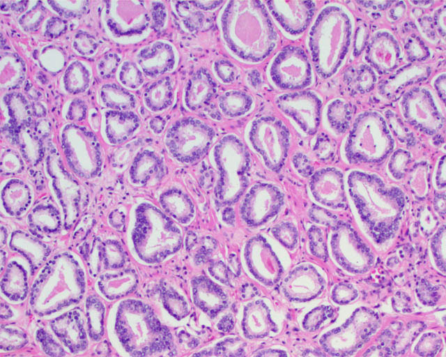
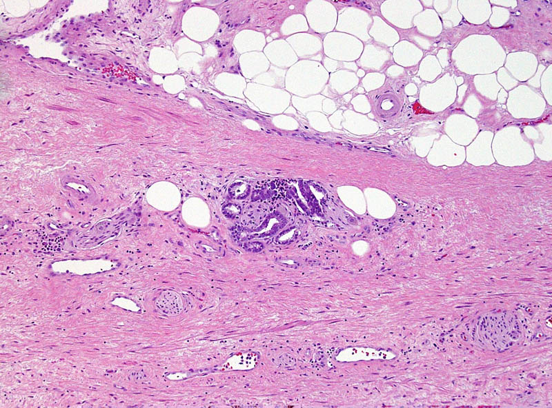
A major difficulty from the pathologic standpoint in adopting a non-cancer nomenclature for some tumors (such as GS6 adenocarcinomas) is that the Gleason pattern 3 component of a GS 3+3=6 tumor (small, round prostatic glands that lack a basal cell layer and infiltrate between benign glands) is for all intents and purposes identical to the Gleason pattern 3 component of a GS 3+4=7 or higher prostate cancer. These similarities are not limited exclusively to the microscopic appearance but also include a number of immunohistochemical and molecular features, as summarized in a recent article addressing this question [3]. Therefore, no pathologic features are as yet defined that ideally predict whether Gleason pattern 3 glands in a biopsy specimen represent a pure GS6 tumor or a component of higher-grade tumor in which the high-grade component is not represented. Not surprisingly, it is not unusual for tumors with GS6 on needle biopsy to be upgraded to GS7 at radical prostatectomy [3], particularly when a high tumor volume is present in the needle biopsy.

Gleason pattern 3 glands from a GS7 tumor, identical to those of a GS6 tumor.

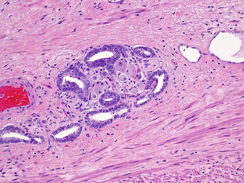
To compare to other cancers with low risk of aggressive behavior, basal cell carcinoma and squamous cell carcinoma of the skin similarly show locally infiltrative properties, supporting their classification as carcinomas by a classical pathologic definition. Despite that the word “carcinoma” continues to be used for these tumors, most patients are not concerned that they have a life-threatening disease and these lesions are even excluded from the American Cancer Society statistics regarding cancers [4]. In the same way, Gleason pattern 3 glands exhibit infiltrative growth by extending between benign glands, invading nerves, and sometimes extending outside of the prostate. This difference in mindset regarding some types of “cancers” could be considered supportive evidence for the assertion in the recent Melbourne Consensus Statement that uncoupling prostate cancer diagnosis from intervention may be more appropriate than removing its “cancer” nomenclature.


This small GS6 adenocarcinoma was an incidental finding in a radical cystoprostatectomy specimen for bladder cancer but surprisingly extended into periprostatic fat via this focus of perineural invasion.

Supporting removal of the cancer designation from some prostatic adenocarcinomas:

A valid argument of the NCI Viewpoint is that a neoplasm should have a substantive rate of progression and patient death if it is to be considered a cancer. Likewise, others have questioned whether low-volume GS6 tumors fulfill other molecular and pathogenetic hallmarks of cancer, such as unlimited replicative potential and other features [5].

In general, benign and malignant neoplasms can be regarded as having some prototypical gross and microscopic pathologic characteristics, such as a circumscribed vs infiltrative growth and homogeneous vs pleomorphic cell population. However, differentiating benign from malignant lesions also relies heavily on parameters specific to the organ involved. Clear cell renal cell carcinoma, another genitourinary tract tumor, often does not possess these prototypical features of malignancy. Tumors often form a well-circumscribed mass without an “invasive” growth pattern and they often are composed of a uniform population of cells. However, based on known behavior of these tumors, their status as a malignancy is not in doubt. Conversely, renal oncocytoma is a benign neoplasm that shares some of these general features (a round mass composed of a homogeneous population of renal tubular cells). Occasionally oncocytomas appear infiltrative by extending into the perinephric fat or renal vein, yet their status as benign is also not the subject of debate. If some prostate cancers do not have a substantial likelihood of resulting in progression and death, they may not meet an important criterion for a diagnosis of cancer, despite that other features, such as infiltration of tissues, invasion of nerves, and loss of the basal cell layer are characteristic of a malignant neoplasm.

Since a diagnosis of GS6 by needle biopsy is not always predictive of a radical prostatectomy overall GS6, a major challenge to such an approach would be to determine where such a cutoff could be drawn between “cancer” and “not cancer” [5]. If based on tumor volume, it would be difficult to conceptualize that a small amount of GS6 glands would be regarded as a benign lesion, whereas a large amount of identical glands would represent a malignant lesion. Alternatively, the presence of Gleason pattern 4 could used as the point of differentiation (GS7 or above). In the endometrium, a disorganized proliferation of crowded glands with some cytologic features of cancer is regarded as complex atypical hyperplasia. Diagnosis of adenocarcinoma is then reserved for proliferations with a confluent growth of these glands, similar to the threshold for recognizing a component of cribriform glands as Gleason pattern 4. A limitation to such an approach, however, is that a substantial fraction of patients with a needle biopsy GS6 are upgraded to GS7 at radical prostatectomy, as discussed above. Likewise, the ability to treat and monitor GS6 adenocarcinoma nonsurgically is not quite analogous to that of endometrial hyperplasia.

Higher magnification of image 2 shows Gleason pattern 3 glands invading a nerve with ganglion cells.

Other points of discussion

The NCI Viewpoint also suggests that high-grade prostatic intraepithelial neoplasia (HGPIN) no longer be considered cancer or even neoplasia.  A comparison to ductal carcinoma in situ (DCIS) of the breast for this argument is somewhat flawed, as HGPIN neither contains the word “carcinoma” nor is justification for treatment in and of itself. Its status as a risk factor for a future cancer even remains debated. The proposal to remove “neoplasia” from HGPIN is also a confusing one, particularly as cervical cancer is noted as an example of the successful application of screening, in which “cervical intraepithelial neoplasia” is the preferred term for precancerous lesions. The authors suggest the designation “indolent lesions of epithelial origin” (IDLE) for cancers in this category to convey their low likelihood of aggressive behavior. However, would recognizing the status of these lesions as at least premalignant neoplasms be more appropriate?

Likely a typographical error in the Viewpoint is that the authors also cite reclassification of urothelial papilloma as papillary urothelial neoplasm of low malignant potential [1]. Since urothelial papilloma has never been considered a malignant neoplasm, the authors likely meant reclassifying “grade 1 urothelial carcinoma” to papillary urothelial neoplasm of low malignant potential.

References
[1]        Esserman LJ, Thompson IM, Reid B. Overdiagnosis and Overtreatment in Cancer: An Opportunity for Improvement. JAMA. 2013 Jul 29:

[2]        Epstein JI, Allsbrook WC, Jr., Amin MB, Egevad LL. The 2005 International Society of Urological Pathology (ISUP) Consensus Conference on Gleason Grading of Prostatic Carcinoma. Am J Surg Pathol. 2005 Sep: 29:1228-42

[3]        Carter HB, Partin AW, Walsh PC, et al. Gleason score 6 adenocarcinoma: should it be labeled as cancer? J Clin Oncol. 2012 Dec 10: 30:4294-6

[4]        Siegel R, Naishadham D, Jemal A. Cancer statistics, 2013. CA Cancer J Clin. 2013 Jan: 63:11-30

[5]        Ahmed HU, Arya M, Freeman A, Emberton M. Do low-grade and low-volume prostate cancers bear the hallmarks of malignancy? Lancet Oncol. 2012 Nov: 13:e509-17


Sean Williamson is Senior Staff Pathologist in the Department of Pathology and Laboratory Medicine, Henry Ford Health System, Detroit MI, USA. @Williamson_SR

Step-by-Step: Percutaneous suprapubic tube bladder drainage in RARP

Percutaneous suprapubic tube bladder drainage after robot-assisted radical prostatectomy: a step-by-step guide

Khurshid R. Ghani, Quoc-Dien Trinh, Jesse D. Sammon, Wooju Jeong, Andrea Simone, Ali Dabaja, Stacey Dusik, James O. Peabody and Mani Menon

Vattikuti Urology Institute, Henry Ford Health System, Detroit, MI, USA

Read the full article
OBJECTIVE

• To describe our technique of maintaining bladder drainage after robot-assisted radical prostatectomy (RARP) using a percutaneous suprapubic tube (PST) in place of a urethral catheter.

METHODS

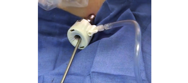
• A watertight anastomosis permits placement of the PST. Contraindications include morbid obesity, concomitant inguinal hernia mesh repair, anticoagulation therapy, limited hand dexterity in the patient, bladder neck reconstruction and extensive adhesiolysis at RARP.

• The necessary equipment includes a 14-F PST balloon catheter set, a three-way connector, a connecting tube, a suture passer, 1/0 polypropylene sutures on a CT1 needle, a sterile plastic button, adhesive and steri-strips.

RESULTS

• The important steps for PST placement are: Step 1: robot-assisted placement of a bladder wall anchor suture; Step 2: transferring the bladder wall suture to anterior abdominal skin; Step 3: guided placement of the PST under robotic vision; Step 4: securing the PST within the bladder and abdominal wall; Step 5. postoperative care: clamping the PST on postoperative day 5, recording each void and post-void residual urine volumes in a patient diary, removal of the PST on postoperative day 7 after 48 h of voiding with residual urine <100 mL per void.

CONCLUSION

• We provide a concise step-by-step guide for placement of a PST during RARP as well as important management aspects for the successful adoption of this technique.

Article of the week: Long-term study finds excellent outcomes after RARP

Every week the Editor-in-Chief selects the Article of the Week from the current issue of BJUI. The abstract is reproduced below and you can click on the button to read the full article, which is freely available to all readers for at least 30 days from the time of this post.

In addition to the article itself, there is an accompanying editorial written by a prominent member of the urological community. This blog is intended to provoke comment and discussion and we invite you to use the comment tools at the bottom of each post to join the conversation.

If you only have time to read one article this week, it should be this one.

Long-term evaluation of survival, continence and potency (SCP) outcomes after robot-assisted radical prostatectomy (RARP)

Vincenzo Ficarra*, Marco Borghesi*, Nazareno Suardi§, Geert De Naeyer*, Giacomo Novara, Peter Schatteman*, Ruben De Groote*, Paul Carpentier* and Alexander Mottrie*

*OLV Robotic Surgery Institute, Aalst, Belgium, University of Padova, Padova, University of Bologna, Bologna, and §Vita-Salute University San Raffaele, Milan, Italy

Read the full article
OBJECTIVE

• To report combined oncological and functional outcome in a series of patients who underwent robot-assisted radical prostatectomy (RARP) for clinically localised prostate cancer in a single European centre after 5-year minimum follow-up according to survival, continence and potency (SCP) outcomes.

PATIENTS AND METHODS

• We extracted from our prostate cancer database all consecutive patients with a minimum follow-up of 5 years after RARP. Biochemical failure was defined as a confirmed PSA concentration of >0.2 ng/mL.

• All patients alive at the last follow-up were evaluated for functional outcomes using the Expanded Prostate Cancer Index Composite (EPIC) and Sexual Health Inventory for Men (SHIM) questionnaires.

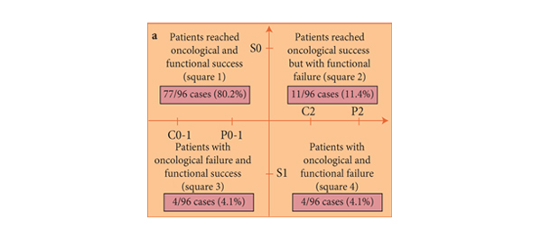
• Oncological and functional outcomes were reported according to the SCP system. Specifically, patients were classified as using no pad (C0), using one pad for security (C1), and using ≥1 pad (C2) (not including the prior definition).

• Patients potent (SHIM score of >17) without any aids were classified as P0 category; patients potent (SHIM score of >17) with use of phosphodiesterase type 5 inhibitorsas P1; and patients with erectile dysfunction (SHIM score of <17) as P2 category. Patients who did not undergo a nerve-sparing technique, who were not potent preoperatively, who were not interested in erections, or who did not have sexual partners were classified as Px category.

RESULTS

• The 3-, 5- and 7-year biochemical recurrence-free survival rates were 96.3%; 89.6% and 88.3%, respectively.

• At follow-up, 146 (79.8%) were fully continent (C0), 20 (10.9%) still used a safety pad (C1) and 17 (9.3%) were incontinent using ≥1 pad (C2).

• Excluding Px patients, 52 patients (47.3%) were classified as P0; 41 patients (37.3%) were classified as P1 and 17 patients (15.5%) were P2.

• In patients preoperatively continent and potent, who received a nerve-sparing technique and did not require any adjuvant therapy, oncological and functional success was attained by 77 (80.2%) patients.

• In the subgroup of 67 patients not evaluable for potency recovery (Px), oncological and continence outcomes were attained in 46 patients (68.7%).

CONCLUSIONS

• Oncological and functional success was attained in a high percentage of patients who underwent RARP at ≥5 years follow-up.

• Interestingly, this study confirmed that excellent oncological and functional outcomes can be obtained in the ‘best’ category of patients, i.e. those preoperatively continent and potent and with tumour characteristics suitable for a nerve-sparing technique.


Read Previous Articles of the Week


© 2024 BJU International. All Rights Reserved.