Tag Archive for: Prostate cancer

Posts

Article of the Month: Does RARP benefit patients with oligometastatic PCa?

Every Month, the Editor-in-Chief selects an Article of the Month from the current issue of BJUI. The abstract is reproduced below and you can click on the button to read the full article, which is freely available to all readers for at least 30 days from the time of this post.

In addition to the article itself, there is an accompanying editorial written by a prominent member of the urological community. This blog is intended to provoke comment and discussion and we invite you to use the comment tools at the bottom of each post to join the conversation.

If you only have time to read one article this week, it should be this one.

Does robot-assisted radical prostatectomy benefit patients with prostate cancer and bone oligometastases?

Won Sik Jang, Myung Soo Kim, Won Sik Jeong, Ki Don Chang, Kang Su Cho, Won Sik Ham, Koon Ho Rha, Sung Joon Hong and Young Deuk Choi

Department of Urology, Urological Science Institute, Yonsei University College of Medicine, Seoul, Korea

Abstract

Objective

To investigate the peri-operative and oncological outcomes of robot-assisted radical prostatectomy (RARP) in patients with oligometastatic prostate cancer (PCa).

Patients and Methods

We retrospectively reviewed the records of 79 patients with oligometastatic PCa treated with RARP or androgen deprivation therapy (ADT) between 2005 and 2015 at our institution. Of these 79 patients, 38 were treated with RARP and 41 were treated with ADT without local therapy. Oligometastatic disease was defined as the presence of five or fewer hot spots detected by preoperative bone scan. We evaluated peri-operative outcomes, progression-free survival (PFS), and cancer-specific survival (CSS). We analysed data using Kaplan–Meier methods, with log-rank tests and multivariate Cox regression models.

Results

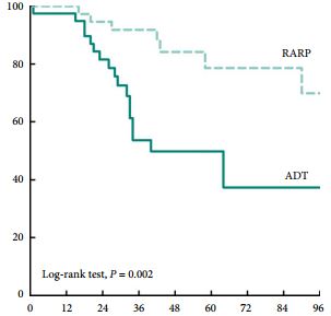
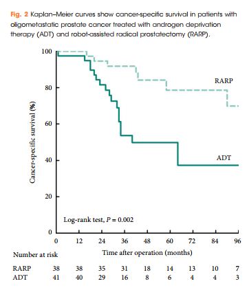
Patients treated with RARP experienced similar postoperative complications to those previously reported in RP-treated patients, and fewer urinary complications than ADT-treated patients. PFS and CSS were longer in RARP-treated compared with ADT-treated patients (median PFS: 75 vs 28 months, P = 0.008; median CSS: not reached vs 40 months, P = 0.002). Multivariate analysis further identified RARP as a significant predictor of PFS and CSS (PFS: hazard ratio [HR] 0.388, P = 0.003; CSS: HR 0.264, P = 0.004).

Conclusions

We showed that RARP in the setting of oligometastatic PCa is a safe and feasible procedure which improves oncological outcomes in terms of PFS and CSS. In addition, our data suggest that RARP effectively prevents urinary tract complications from PCa. The study highlights results from expert surgeons and highly selected patients that cannot be extrapolated to all patients with oligometastatic PCa; to confirm our findings, large, prospective, multicentre studies are required.

Article of the Week: Comparison of transperineal mpMRI/fusPbx and sysPbx

Every Week the Editor-in-Chief selects an Article of the Week from the current issue of BJUI. The abstract is reproduced below and you can click on the button to read the full article, which is freely available to all readers for at least 30 days from the time of this post.

In addition to the article itself, there is an accompanying editorial written by a prominent member of the urological community. This blog is intended to provoke comment and discussion and we invite you to use the comment tools at the bottom of each post to join the conversation.

If you only have time to read one article this week, it should be this one.

Prospective comparison of transperineal magnetic resonance imaging/ultrasonography fusion biopsy and transrectal systematic biopsy in biopsy-naïve patients


Angelika Borkowetz*, Boris Hadaschik†‡, Ivan Platzek§, Marieta Toma, Georgi TosevTheresa Renner*, Roman Herout*, Martin Baunacke*, Michael Laniado §, Gustavo Baretton, Jan Philipp Radtke, Claudia Kesch, Markus Hohenfellner† , Michael Froehner*, Heinz-Peter Schlemmer**, Manfred Wirth* and Stefan Zastrow*


*Department of Urology, Technische Universitat Dresden, Dresden, Germany, Department of Urology, University Hospital Heidelberg, Heidelberg, Germany, Department of Urology, University Hospital Essen, Essen, Germany, §Department of Radiology and Interventional Radiology, Technische Universitat Dresden, Dresden, Germany, Department of Pathology, Technische Universitat Dresden, Dresden, Germany, and **Department of Radiology, German Cancer Research Centre (DKFZ), Heidelberg, Germany


Read the full article

Abstract

Objectives

To evaluate the value of multiparametric magnetic resonance imaging (mpMRI) in the detection of significant prostate cancer (PCa) and to compare transperineal MRI/ultrasonography fusion biopsy (fusPbx) with conventional transrectal systematic biopsy (sysPbx) in biopsy-naïve patients.

Patients and Methods

This multicentre, prospective trial investigated biopsy-naïve patients with suspicion of PCa undergoing transperineal fusPbx in combination with transrectal sysPbx (comPbx). The primary outcome was the detection of significant PCa, defined as Gleason pattern 4 or 5. We analysed the results after a study period of 2 years.

Results

The study included 214 patients. The median (range) number of targeted and systematic cores was 6 (2–15) and 12 (6–18), respectively. The overall PCa detection rate of comPbx was 52%. FusPbx detected more PCa than sysPbx (47% vs 43%; P = 0.15). The detection rate of significant PCa was 38% for fusPbx and 35% for sysPbx (P = 0.296). The rate of missed significant PCa was 14% in fusPbx and 21% in sysPbx. ComPbx detected significantly more significant PCa than fusPbx and sysPbx alone (44% vs 38% vs 35%; P < 0.005). In patients presenting with Prostate Imaging Reporting and Data System (PI-RADS) 4 and 5 lesions there was a higher detection rate of significant PCa than in patients presenting with PI-RADS ≤3 lesions in comPbx (61% vs 14%; P < 0.005).

Conclusions

For biopsy-naïve men with tumour-suspicious lesions in mpMRI, the combined approach outperformed both fusPbx and sysPbx in the detection of overall PCa and significant PCa. Thus, biopsy-naïve patients may benefit from sysPbx in combination with mpMRI targeted fusPbx.

Editorial: The new frontier of prostate biopsy: determining the role of image-guidance in moving the needle

One of the most pressing topics in urological oncology concerns the role of MRI/ultrasonography (US)-fusion guided biopsies in detecting prostate cancer. The literature on this emerging technology is permeated by questions regarding when it should be used, how it should be performed, and which patients stand to benefit. The stakes are high to figure this out, as real patients will continue to suffer from missteps made in diagnosis and treatment of prostate cancer whilst we seek to improve our detection methods.

In this issue of BJUI, Borkowetz et al. [1] compare prostate cancer detection rates between MRI/US-fusion targeted and conventional systematic biopsies. They prospectively enrolled a cohort of biopsy-naïve men who had an elevated PSA level and/or an abnormal DRE. All men received both a transperineal MRI/US-fusion biopsy and a systematic TRUS-guided biopsy. They found that combining both approaches led to improved detection rates for clinically significant and overall prostate cancer compared to either method alone.

A strength of this study [1] is its focus on biopsy-naïve men, for which data on the comparison of biopsy techniques is relatively limited. Siddiqui et al. [2] were instrumental in demonstrating that targeted MRI/US-fusion biopsy, compared to the systematic TRUS biopsy, was associated with improved diagnostic accuracy for higher-risk tumours and decreased detection of low-risk disease. However, the large majority of men in the study had received a prior biopsy, making it difficult to generalise the results to biopsy-naïve men. Focusing on biopsy-naïve men is of great importance – men referred for a biopsy after an elevated PSA level or abnormal DRE increasingly face conflicting opinions about the next best step in diagnostic evaluation, especially given the recent influx of imaging and biomarker tests that aim to guide this decision point.

One group previously compared MRI-guided and systematic TRUS biopsy in a large group of biopsy-naïve of men and found the MRI-guided in-bore technique to be superior in detecting significant cancers and avoiding insignificant cancers [3]. However, that study was limited by both its definition of ‘significant cancer’ (it included Gleason 3 + 3 disease) and the variance in the definition of ‘low-risk’ cancer between MRI-guided and TRUS biopsies. By contrast, the present authors uniformly defined significant cancer as Gleason ≥3 + 4. This is just one example highlighting the widespread disagreement over the parameters used to measure the efficacy of targeted-biopsy techniques in cancer detection. Continued incorporation of standardised scales such as the Prostate Imaging Reporting and Data System (PI-RADS), which was also used in the accompanying article, will help to mitigate some of this disagreement in future studies. Of course, these standardised scales are still subject to inter- and intra-observer variability, which may decrease with further clarification of the grading systems, as well as appropriate reader training [4].

Whilst Borkowetz et al. [1] helped to fill important gaps in the literature, their study was not without limitations. They included PI-RADS 2 scores for targeted biopsies although many urologists would consider these lesions insignificant and not worth targeting. Furthermore, comparing a transperineal MRI/US-fusion with a transrectal systematic approach makes it difficult to separate the true effect of the targeted approach from that of the anatomical approach. Using both methods in each patient also precludes any comparison of complication rates between the biopsy approaches, an important clinical endpoint for both the urologists administering the biopsies and the patients enduring the complications.

Methodology aside, the new frontier of prostate biopsy technique still relies on a basic triad of efficacy: (i) improved accuracy of cancer detection, (ii) reduced complication rates, and (iii) manageable cost and practicality of widespread implementation. For the first tenet, the recently published PROstate MRI Imaging Study (PROMIS) trial cemented the ability of multi-parametric MRI to detect clinically significant prostate cancer with greatly improved sensitivity and negative predictive value over TRUS [5]. The ongoing randomised PRostate Evaluation for Clinically Important Disease: Sampling Using Image-guidance Or Not? (PRECISION) trial will hopefully add to the step taken by Borkowetz et al. [1], by comparing MRI-targeted and systematic TRUS biopsies on the outcomes of accuracy in cancer detection, adverse events, patient health-related quality of life, and cost [6]. Continued investigation in all of these areas will be crucial to guiding how we move the needle in prostate cancer diagnostics.

Sean A. Fletcher, Sebastian Berg and Quoc-Dien Trinh


Division of Urological Surgery, Center for Surgery and Public Health, Brigham and Womens Hospital, Harvard Medical School, Boston, MA, USA


References


1 Borkowetz AHadaschik BPlatzek I et al. Prospective comparison of transperineal magnetic resonance imaging/ultrasonography fusion biopsy and transrectal systematic biopsy in biopsy-naive patients. BJU Int 2018;121: 5360


2 Siddiqui MM, Rais-Bahrami STurkbey B et al. Comparison of MR/ultrasound fusion-guided biopsy with ultrasound-guided biopsy for the diagnosis of prostate cancer. JAMA2015; 313: 3907


3 Pokorny MRde Rooij MDuncan E et al. Prospective study of diagnostic accuracy comparing prostate cancer detection by transrectal ultrasound-guided biopsy versus magnetic resonance (MR) imaging with subsequent MR-guided biopsy in men without previous prostate biopsies. Eur Urol 2014; 66: 229


5 Ahmed HUEl-Shater Bosaily A,Brown LC et al. Diagnostic accuracy of multi-parametric MRI and TRUS biopsy in prostate cancer (PROMIS): a paired validating confirmatory study. Lancet 2017; 389:81522


6 ClinicalTrials.gov. PRostate Evaluation for Clinically Important Disease: Sampling Using Image-guidance or Not? (PRECISION), Identification No. NCT02380027 (2017). Available at: https://clinicaltrials.gov/ct2/show/NCT02380027. Accessed October 2017.


Article of the Week: Prostate Health Index density improves detection of clinically significant prostate cancer

Every Week the Editor-in-Chief selects an Article of the Week from the current issue of BJUI. The abstract is reproduced below and you can click on the button to read the full article, which is freely available to all readers for at least 30 days from the time of this post.

In addition to the article itself, there is an accompanying editorial written by a prominent member of the urological community. This blog is intended to provoke comment and discussion and we invite you to use the comment tools at the bottom of each post to join the conversation.

Finally, the third post under the Article of the Week heading on the homepage will consist of additional material or media. This week we feature a video discussing the paper.

If you only have time to read one article this week, it should be this one.

Prostate Health Index density improves detection of clinically significant prostate cancer

Jeffrey J. Tosoian*, Sasha C. Druskin*, Darian Andreas*, Patrick Mullane*, Meera Chappidi*, Sarah Joo*, Kamyar Ghabili*, Mufaddal Mamawala*, Joseph Agostino*, Herbert B. Carter*, Alan W. Partin*, Lori J. Sokoll*§ and Ashley E. Ross*§


*Department of Urology, James Buchanan Brady Urological Institute, Johns Hopkins University School of Medicine, Baltimore, MD, Virginia Commonwealth University School of Medicine, Richmond, VA, Department of Pathology, and §Sidney Kimmel Comprehensive Cancer Center, Johns Hopkins University School of Medicine, Baltimore, MD, USA
Read the full article

Abstract

Objectives

To explore the utility of Prostate Health Index (PHI) density for the detection of clinically significant prostate cancer (PCa) in a contemporary cohort of men presenting for diagnostic evaluation of PCa.

Patients and Methods

The study cohort included patients with elevated prostate-specific antigen (PSA; >2 ng/mL) and negative digital rectal examination who underwent PHI testing and prostate biopsy at our institution in 2015. Serum markers were prospectively measured per standard clinical pathway. PHI was calculated as ([{−2}proPSA/free PSA] × [PSA]½), and density calculations were performed using prostate volume as determined by transrectal ultrasonography. Logistic regression was used to assess the ability of serum markers to predict clinically significant PCa, defined as any Gleason score ≥7 cancer or Gleason score 6 cancer in >2 cores or >50% of any positive core.

Results

Of 118 men with PHI testing who underwent biopsy, 47 (39.8%) were found to have clinically significant PCa on biopsy. The median (interquartile range [IQR]) PHI density was 0.70 (0.43–1.21), and was 0.53 (0.36–0.75) in men with negative biopsy or clinically insignificant PCa and 1.21 (0.74–1.88) in men with clinically significant PCa (P < 0.001). Clinically significant PCa was detected in 3.6% of men in the first quartile of PHI density (<0.43), 36.7% of men in the IQR of PHI density (0.43–1.21), and 80.0% of men with PHI density >1.21 (P < 0.001). Using a threshold of 0.43, PHI density was 97.9% sensitive and 38.0% specific for clinically significant PCa, and 100% sensitive for Gleason score ≥7 disease. Compared with PSA (area under the curve [AUC] 0.52), PSA density (AUC 0.70), %free PSA (AUC 0.75), the product of %free PSA and prostate volume (AUC 0.79), and PHI (AUC 0.76), PHI density had the highest discriminative ability for clinically significant PCa (AUC 0.84).

Conclusions

Based on the present prospective single-centre experience, PHI density could be used to avoid 38% of unnecessary biopsies, while failing to detect only 2% of clinically significant cancers.

Article of the Week: Association of HDI with global bladder, kidney, prostate and testis cancer

Every Week the Editor-in-Chief selects an Article of the Week from the current issue of BJUI. The abstract is reproduced below and you can click on the button to read the full article, which is freely available to all readers for at least 30 days from the time of this post.

In addition to the article itself, there is an accompanying editorial written by a prominent member of the urological community. This blog is intended to provoke comment and discussion and we invite you to use the comment tools at the bottom of each post to join the conversation.

If you only have time to read one article this week, it should be this one.

Association of Human Development Index with global bladder, kidney, prostate and testis cancer incidence and mortality

Alyssa K. Greiman*, James S. Rosoff† and Sandip M. Prasad*


*Department of Urology, Medical University of South Carolina, Charleston, SC, Department of Urology, Yale School of Medicine, New Haven, CT, and Department of Surgery, Ralph M. Johnson VA Medical Center, Charleston, SC, USA


Read the full article

Abstract

Objectives

To describe contemporary worldwide age-standardized incidence and mortality rates for bladder, kidney, prostate and testis cancer and their association with development.

Materials and Methods

We obtained gender-specific, age-standardized incidence and mortality rates for 184 countries and 16 major world regions from the GLOBOCAN 2012 database. We compared the mortality-to-incidence ratios (MIRs) at national and regional levels in males and females, and assessed the association with socio-economic development using the 2014 United Nations Human Development Index (HDI).

Results

Age-standardized incidence rates were 2.9 (bladder) to 7.4 (testis) times higher for genitourinary malignancies in more developed countries compared with less developed countries. Age-standardized mortality rates were 1.5–2.2 times higher in more vs less developed countries for prostate, bladder and kidney cancer, with no variation in mortality rates observed in testis cancer. There was a strong inverse relationship between HDI and MIR in testis (regression coefficient 1.65, R2 = 0.78), prostate (regression coefficient −1.56, R2 = 0.85), kidney (regression coefficient −1.34, R2 = 0.74), and bladder cancer (regression coefficient −1.01, R2 = 0.80).

Conclusion

While incidence and mortality rates for genitourinary cancers vary widely throughout the world, the MIR is highest in less developed countries for all four major genitourinary malignancies. Further research is needed to understand whether differences in comorbidities, exposures, time to diagnosis, access to healthcare, diagnostic techniques or treatment options explain the observed inequalities in genitourinary cancer outcomes.

Editorial: Human development and its impact on genitourinary cancers

Using the extensive data from the WHO International Agency for Research on Cancer and the United Nations Human Development Report, Greiman et al. [1] aimed to investigate how human development is associated with incidence and mortality of genitourinary cancers. Even though they generate some interesting descriptive findings, we have to remain critical of these descriptive statistics and carefully assess what needs to be investigated next.

Firstly, despite having highlighted the need for attention to indicators of longevity, education, and income per head when assessing human development, the human development index (HDI) is a rather crude measurement. As a geometric mean of normalised indices for each of these three domains, the HDI simplifies but only captures part of what human development entails. Important indicators of health care such as inequalities, poverty, human security, and empowerment are not reflected in the HDI (www.hdr.undp.org). In the context of cancer incidence and mortality this is an important limitation, as it has for instance been shown that socioeconomic status affects early phase cancer trial referrals, which can be considered as a proxy for access to health care [2]. This inequality has been hypothesised to be linked to more comorbidities and lower education in those who are most deprived – a complex interaction which may not be completely captured by the HDI.

Secondly, registration of incidence and mortality of cancers may vary substantially between countries based on both medical practice and governance. These differences are important when trying to generate hypotheses following the ecological study of Greiman et al. [1]. In the case of bladder cancer, for instance, mortality has been estimated to be 17% in the Netherlands, compared to 22% in the USA, and 50% in the UK. As cancer treatments are expected to be similar in these developed countries, it has been thought that a lower registration of non-muscle-invasive bladder cancer in the UK could explain this higher proportion [3]. Thus, discrepancies in cancer registration, even between developed countries, may limit our awareness of cancer burden.

Thirdly, the study design suffers from ‘ecological fallacy’. The latter refers to the inability to draw causal inference about the effect of the HDI on genitourinary cancer at the individual level, in conjunction with the underlying problem of heterogeneity of exposure levels [4]. This limitation was not mentioned by Greiman et al. [1], but affects their conclusions. The lack of information on, for instance, smoking data, comorbidities, and ethnicity make it difficult to understand how development is affecting cancer incidence or mortality. It would have been interesting to also investigate cancers other than genitourinary cancers because a comparison of different tumour types might have shed light on differences in medical practice or risk factors across countries and help tease out the ecological effect of human development.

Despite the aforementioned limitations, the descriptive analysis by Greiman et al. [1] can be helpful for generating hypotheses – as also outlined by the authors. This ecological effect of human development on incidence and mortality rates of genitourinary cancers is particularly relevant when evaluating the impacts of prevention and intervention programmes for these cancers. Their findings suggest that further investigation is required to examine the hypothesis regarding human development and incidence/mortality of genitourinary cancers. To further elucidate this association, methodological challenges will need to be overcome, as HDI assessment has been criticised for being too crude. Nevertheless, it should be possible to collect more detailed information to allow for an understanding of which components of a country’s collective resources affect cancer incidence and mortality the most, e.g. differences in resources used for cancer detection and treatment.

Mieke Van Hemelrijck
Division of Cancer Studies, Translational Oncology and Urology Research (TOUR), Kings College London, London, UK


References


1 Greiman AKRosoff JSPrasad SM. Association of Human Development Index with global bladder, kidney, prostate and testis cancer incidence and mortality. BJU Int2017; 120: 799-807


2 Mohd Noor A Sarker DVizor S et al. Effect of patient socioeconomic status on access to early-phase cancer trials. J Clin Oncol 2013; 31: 224– 30.


3 Boormans JLZwarthoff EC. Limited funds for bladder cancer research and what can we do about it. Bladder Cancer 2016; 2: 4951


4 Morgenstern H . Ecologic studies in epidemiology: concepts, principles, and methods. Annu Rev Public Health 1995; 16: 618


Article of the Week: Evaluation of targeted and systematic biopsies using MRI and US image-fusion guided transperineal prostate biopsy

Every Week the Editor-in-Chief selects an Article of the Week from the current issue of BJUI. The abstract is reproduced below and you can click on the button to read the full article, which is freely available to all readers for at least 30 days from the time of this post.

In addition to the article itself, there is an accompanying editorial written by a prominent member of the urological community. This blog is intended to provoke comment and discussion and we invite you to use the comment tools at the bottom of each post to join the conversation.

If you only have time to read one article this week, it should be this one.

Multicentre evaluation of targeted and systematic biopsies using magnetic resonance and ultrasound image-fusion guided transperineal prostate biopsy in patients with a previous negative biopsy


Nienke L. Hansen*†‡, Claudia Kesch§, Tristan Barrett, Brendan Koo, Jan P. Radtke§**, David Bonekamp** , Heinz-Peter Schlemmer**, Anne Y. Warren‡††, Kathrin Wieczorek‡‡Markus Hohenfellner§, Christof Kastner§§ and Boris Hadaschik§


*Department of Diagnostic and Interventional Radiology, University Hospital RWTH Aachen, Aachen, Germany, CamPARI Clinic, Addenbrookes Hospital and University of Cambridge, Cambridge, UK, Department of Diagnostic and Interventional Radiology, University Hospital Cologne, Cologne§Department of Urology, University Hospital Heidelberg, Heidelberg, Germany, Department of Radiology, Addenbrookes Hospital and University of Cambridge, Cambridge, UK, **Department of Radiology, DKFZ, Heidelberg, Germany, ††Department of Pathology, AddenbrookeHospital and University of Cambridge, Cambridge, UK, ‡‡Institute of Pathology, University of Heidelberg, Heidelberg, Germany, and §§Department of Urology, Addenbrookes Hospital and University of Cambridge, Cambridge, UK


Read the full article

Abstract

Objectives

To evaluate the detection rates of targeted and systematic biopsies in magnetic resonance imaging (MRI) and ultrasound (US) image-fusion transperineal prostate biopsy for patients with previous benign transrectal biopsies in two high-volume centres.

Patients and Methods

A two centre prospective outcome study of 487 patients with previous benign biopsies that underwent transperineal MRI/US fusion-guided targeted and systematic saturation biopsy from 2012 to 2015. Multiparametric MRI (mpMRI) was reported according to Prostate Imaging Reporting and Data System (PI-RADS) Version 1. Detection of Gleason score 7–10 prostate cancer on biopsy was the primary outcome. Positive (PPV) and negative (NPV) predictive values including 95% confidence intervals (95% CIs) were calculated. Detection rates of targeted and systematic biopsies were compared using McNemar’s test.

Results

The median (interquartile range) PSA level was 9.0 (6.7–13.4) ng/mL. PI-RADS 3–5 mpMRI lesions were reported in 343 (70%) patients and Gleason score 7–10 prostate cancer was detected in 149 (31%). The PPV (95% CI) for detecting Gleason score 7–10 prostate cancer was 0.20 (±0.07) for PI-RADS 3, 0.32 (±0.09) for PI-RADS 4, and 0.70 (±0.08) for PI-RADS 5. The NPV (95% CI) of PI-RADS 1–2 was 0.92 (±0.04) for Gleason score 7–10 and 0.99 (±0.02) for Gleason score ≥4 + 3 cancer. Systematic biopsies alone found 125/138 (91%) Gleason score 7–10 cancers. In patients with suspicious lesions (PI-RADS 4–5) on mpMRI, systematic biopsies would not have detected 12/113 significant prostate cancers (11%), while targeted biopsies alone would have failed to diagnose 10/113 (9%). In equivocal lesions (PI-RADS 3), targeted biopsy alone would not have diagnosed 14/25 (56%) of Gleason score 7–10 cancers, whereas systematic biopsies alone would have missed 1/25 (4%). Combination with PSA density improved the area under the curve of PI-RADS from 0.822 to 0.846.

Conclusion

In patients with high probability mpMRI lesions, the highest detection rates of Gleason score 7–10 cancer still required combined targeted and systematic MRI/US image-fusion; however, systematic biopsy alone may be sufficient in patients with equivocal lesions. Repeated prostate biopsies may not be needed at all for patients with a low PSA density and a negative mpMRI read by experienced radiologists.

Editorial: Getting to the right biopsy in the right patient at the right time

Guidelines now recommend performing multiparametric MRI (mpMRI) and targeted prostate biopsies in men with a history of prior negative biopsy and continued concern for significant cancer. This new approach to prostate re-biopsy is aimed at improving prostate cancer detection. However, several important clinical factors may help clinicians’ fine-tune the process of repeated prostate biopsy. In this month’s issue of the BJUI, Hansen et al. [1] present a multicentre study of patients with prior negative TRUS biopsy undergoing MRI/TRUS-fusion transperineal biopsy.

In the study, 487 men undergo mpMRI and transperineal biopsy with detection of clinically significant (Gleason score 7–10) cancer as the primary outcome. Several factors are evaluated to compare cancer detection rates, including systematic biopsies, targeted biopsies, PSA density (PSAD), and Prostate Imaging Reporting and Data System (PI-RADS) version 1 score. From their cohort, a suspicious lesion (PIRADS 3–5) was identified in 343 (70%) patients. Prostate cancer was detected in 249 (51%), with 149 (31%) having Gleason score 7–10 cancer. Potentially missed significant cancers from the anterior prostate were found in 27% (40/149). Cancer was detected in 28% (40/144) of patients with PI-RADS 1–2 lesions, with 8% (11/144) being Gleason score 7–10. For patients with PI-RADS 3–5 lesions, cancer was identified in 61% (209/343) with 40% (138/343) being Gleason score 7–10. For patients with PI-RADS 3–5 lesions, systematic biopsies alone failed to detect 13/138 significant cancers, while targeted biopsies missed 24/138 cancers. The combination of systematic and targeted biopsies was significantly better for Gleason score 7–10 prostate cancer detection than either alone. The addition of a PSAD threshold of 0.15 ng/mL/mL for the detection of Gleason score 7–10 resulted in a significant improvement in the area under the curve (0.846) of the receiver operating characteristic curve for PSAD groups and PI-RADS score.

Getting the right biopsy: In this study [1], patients with a prior negative TRUS biopsy underwent TRUS-fusion transperineal biopsy. Having two approaches to prostate biopsy can be advantageous when evaluating men with prior negative biopsies. Historical studies have found comparable prostate cancer detection between transrectal and transperineal biopsies for men undergoing both initial biopsy [2] and saturation re-biopsy [3]. However, the detection of anterior lesions has remained a persistent challenge from the transrectal approach. As in the current study [1], use of transperineal biopsy can detect cancer in up to 30% of tumours that would otherwise be missed on extended template TRUS biopsy [4]. Although attempts to reach anterior lesions from the transrectal approach may be feasible [5], the transperineal approach is felt to provide better sampling in comparison [6].

Getting the right patient: Patient-specific factors such as PI-RADS lesions 3–5 and PSAD have become increasing utilised for stratifying patients who may benefit from additional biopsies using image guidance. As the authors suggest, patients with negative imaging may consider deferring repeat biopsy, particularly those with reassuring PSADs (<0.15 ng/mL/mL). In their study [1], only 4% (6/144) of men with negative mpMRI and a PSAD of <0.15 ng/mL/mL harboured clinically significant cancer (five Gleason score 3 + 4 and one Gleason score 8). Patients with concerning PSAD, but negative mpMRI and those with lesions identified in the peripheral zone could have the option to undergo repeated, fusion-directed TRUS or transperineal biopsy. For patients with lesions identified in the anterior prostate, a transperineal prostate biopsy may provide the highest detection rate.

At the right time: Now that high quality prostate MRI is becoming more widely available; men with a prior negative biopsy should strongly consider the benefit of repeated biopsy after prostate imaging. In addition to identifying suspicious lesions, calculating PSAD has been found to improve the likelihood of detecting clinically significant prostate cancer. Without additional testing, a personalised biopsy plan can be created.

A thorough discussion of the prescribed biopsy approach and the likelihood of detecting a significant cancer is the final step to the right biopsy in the right patient at the right time.

Kelly Stratton


Department of Urology, University of Oklahoma College of Medicine, Oklahoma City, OK, USA


Read the full article


1 Hansen NL, Kesch C, Barrett T et al. Multicentre evaluation of target and systematic biopsies using magnetic resonance and ultrasound image-fusion guided transperineal prostate biopsy in patients with a previous negative biopsy. BJU Int 2016; 120: 6318


2 Hara R, Jo Y, Fujii T et al. Optimal approach for prostate cancer detection as initial biopsy: prospective randomized study comparing transperineal versus transrectal systematic 12-core biopsy. Urology 2008; 71: 1915


3 Abdollah F, Novara G, Briganti A et al. Trans-rectal versus trans- perineal saturation rebiopsy of the prostate: is there a difference in cancer detection rate? Urology 2011; 77: 9215


4 KomaiY, Numao N, Yoshida S et al. High diagnostic ability of multiparametric magnetic re onance imaging to detect anterior prostate cancer miss ed by transrectal 1 2-core biopsy. JUrol2013; 190: 867 7


5 Volkin D, Turkbey B, Hoang AN et al. Multiparametric magnetic resonance imaging (MRI) and subsequent MRI/ultrasonography fusion-guided biopsy increase the detection of anteriorly located prostate cancers. BJU Int 2014; 114: E439


6 Borkowetz A, Platzek I, Toma M et al. Direct comparison of multiparametric magnetic resonance imaging (MRI) results with nal histopathology in patients with proven prostate cancer in MRI/ ultrasonography-fusion biopsy. BJU Int 2016; 118: 21320


Article of the Week: Effect of MetS on serum PSA levels is concealed by enlarged prostate

Every Week the Editor-in-Chief selects an Article of the Week from the current issue of BJUI. The abstract is reproduced below and you can click on the button to read the full article, which is freely available to all readers for at least 30 days from the time of this post.

In addition to the article itself, there is an accompanying editorial written by a prominent member of the urological community. This blog is intended to provoke comment and discussion and we invite you to use the comment tools at the bottom of each post to join the conversation.

Finally, the third post under the Article of the Week heading on the homepage will consist of additional material or media. This week we feature a video discussing the paper.

If you only have time to read one article this week, it should be this one.

Actual lowering effect of metabolic syndrome on serum prostate-specific antigen levels is partly concealed by enlarged prostate: results from a large-scale population-based study

Sicong Zhao*, Ming Xia*, Jianchun Tang† and Yong Yan*


*Department of Urology, and Department of Cardiology, Beijing Shijitan Hospital, Capital Medical University, Beijing, China


Read the full article

Abstract

Objectives

To clarify the lowering effect of metabolic syndrome (MetS) on serum prostate-specific antigen (PSA) levels in a Chinese screened population.

Subjects and Methods

A total of 45 540 ostensibly healthy men aged 55–69 years who underwent routine health check-ups at Beijing Shijitan Hospital between 2008 and 2015 were included in the study. All the men underwent detailed clinical evaluations. PSA mass density was calculated (serum PSA level × plasma volume ÷ prostate volume) for simultaneously adjusting plasma volume and prostate volume. According to the modified National Cholesterol Education Programme–Adult Treatment Panel (NCEP-ATP) III criteria, patients were dichotomized by the presence of MetS, and differences in PSA density and PSA mass density were compared between groups. Linear regression analysis was used to evaluate the effect of MetS on serum PSA levels.

Results

When larger prostate volume in men with MetS was adjusted for, both PSA density and PSA mass density in men with MetS were significantly lower than in men without MetS, and the estimated difference in mean serum PSA level between men with and without MetS was greater than that before adjusting for prostate volume. In the multivariate regression model, the presence of MetS was independently associated with an 11.3% decline in serum PSA levels compared with the absence of MetS. In addition, increasing number of positive MetS components was significantly and linearly associated with decline in serum PSA levels.

Conclusion

The actual lowering effect of MetS on serum PSA levels was partly concealed by the enlarged prostate in men with MetS, and the presence of MetS was independently associated with lower serum PSA levels. Urologists need to be aware of the effect of MetS on serum PSA levels and should discuss this subject with their patients.

Editorial: Anomalous observation with regard to PCa in cancer research

In science, reports showing data deviating from what is expected are called anomalous observations. Metabolic syndrome (MetS) is a promoter of cancer at almost all sites [1]; however, when it comes to prostate cancer (PCa), a series of reports have been published showing an inverse relationship between MetS and its aspects and incident PCa. This lack of coherence in cancer research seriously hampers efforts to fight cancer disorders. It is therefore crucial to find an explanation for this incoherence.

In the search for a reasonable explanation for this anomalous observation, a hypothesis has been formulated, based on the study by Häggström et al. [2], and stating that the PSA-driven diagnostic procedure in PCa, which creates low-stage incident PCa material, is the culprit. The PSA-driven diagnostic procedure introduces several bias mechanisms, which tend to protect men with MetS from being diagnosed with PCa. Thus, men with MetS and its aspects are under-represented in PCa populations generated by PSA-driven diagnostics, thereby creating a distorted incident PCa population. This hypothesis also predicts that high-stage PCa, as well as non-localized and lethal PCa, are not subject to these bias mechanisms, as a minor reduction in the PSA level is of no importance for the PCa diagnosis at these high PSA levels. Finally, the hypothesis predicts that the link between MetS and incident PCa is stage-dependent. A study testing this hypothesis is now in progress.

Several studies have reported that men with MetS had lower PSA levels compared with men without MetS. Zhao et al. [3] address this specific question in this issue of BJUI and confirm that the presence of MetS was independently associated with a lower PSA level and that the enlarged prostate gland, which is an aspect of MetS, partly concealed an even greater PSA level reduction [3]. The findings indicate that a bias mechanism inverses the link between MetS and incident PCa and support the above-mentioned hypothesis.

In short, the following bias mechanisms have been described. MetS is associated with greater body fat with increased aromatase activity, resulting in a reduced testosterone level, which, in turn, is related to a reduced PSA level, as the production of PSA is under androgen control. Another possible bias mechanism, leading to men with MetS being diagnosed less often with PCa, is that these men are more likely to be obese. It is well established that men with a higher BMI also have larger plasma volumes and therefore have greater haemodilution of the PSA production, resulting in a lower PSA level. This means that incident PCa is diagnosed less often in men with MetS, as their PSA level is lower. MetS is also associated with an enlarged prostate gland volume, which means that fewer incident PCas are diagnosed, given the same tumour volume and the same number of biopsies. Another bias mechanism is that a high proportion of men with high socio-economic status undergo PSA testing in the PSA era. It is well established that men with a high socio-economic status have a lower prevalence of MetS and therefore have higher PSA levels, as indicated by the present report in the BJUI [3], and an elevated risk of PCa. Thus, multiple bias mechanisms seem to conceal low-stage PCa in the PSA era.

If it could be confirmed that the negative relationship between MetS and incident PCa is a spurious observation as a result of bias mechanisms, this would open the door for the MetS hypothesis regarding the promotion of multiple cancer disorders. This door has previously been closed by findings in a series of reports of an inverse relationship between MetS and its aspects and incident prostate cancer. Furthermore, this could lead to increased efforts to fight the metabolic aberrations of MetS. It is now well established that MetS and its aspects could be reduced by changes in lifestyle, including physical activity and diet. The most convincing evidence of the effect of diet on MetS comes from studies involving decreased intake of carbohydrates and increased intake of unsaturated fats. Recently, leading authorities in nutrition, endocrinology and metabolism presented a critical review and concluded that carbohydrate restriction is the single most effective intervention to reduce all features of MetS [4]. Another review concluded that carbohydrate restriction is one of the few common interventions that target all features of MetS [5]. This conclusion has recently been confirmed in a meta-analysis by Mansoor et al. [6].

In conclusion, new knowledge challenges the anomalous observation of PCa showing a negative relationship between MetS and PCa. The credibility of the hypothesis that MetS is an important promoting factor for cancer at almost all sites is strengthened. MetS could be treated effectively with a low carbohydrate and high fat diet.

Jan Hammarsten, MD, PhD
Department of Urology, Institute of Clinical SciencesUniversity of Gothenburg, Gothenburg, Sweden


Read the full article


References


1 Esposito K, Chiodini P, Colao AM et al. Metabolic syndrome and risk of cancer. Diabetes Care 2012; 35: 240211 


2Haggstrom C, Stocks T, Ulmert D et al. Prospective study on metabolic factors and risk of prostate cancer. Cancer 2012; 118: 6199206


3 Zhao S, Xia M, Tang J et al. The actual lowering effect of metabolic syndrome on serum prostate-specic antigen levels is partly concealed by enlarged prostate: results from large-scale population-based study. BJU Int 2017; 120: 4829


4 Feinman RD, Pogozelski WK, Astrup A et al. Dietary carbohydrate restriction as the rst approach in diabetes management: critical review and evidence base. Nutrition 2015;31: 113


5 Accurso A, Bernstein RK, Dahlqvist A et al. Dietary carbohydrate restriction in type 2 diabetes mellitus and metabolic syndrome: time for critical appraisal. Nutrition & Metabolism 2008; 5: 9


6 Mansoor N, Vinknes UJ , Veierod MB et al. Effects of low-carbohydrate diets v. low fat diets on body weight and cardiovascular risk factors: meta-analysis of randomized controlled trials. Br J Nutrition 2016; 115: 4667


© 2024 BJU International. All Rights Reserved.