Tag Archive for: renal cell carcinoma

Posts

Editorial: External validation of Karakiewicz models: do they hold up?

Cancer-specific survival (CSS) in patients with RCC depends on important prognostic factors including specific clinical signs or symptoms, tumour-related factors and various laboratory findings. To better predict prognosis and aid patient counselling, several investigators have developed tools which have greatly enhanced our ability to predict outcomes in patients with RCC. For instance, Kattan et al. [1] have combined manner of presentation, tumour histology, tumour size and pathological stage to develop a nomogram that predicts cancer-free survival after nephrectomy. The stage, size, grade, and necrosis (SSIGN) score is another predominant model that provides individualized information for patients with clear-cell RCC. It incorporates the 1997 TNM stage, tumour size, nuclear grade and presence of tumour necrosis to predict recurrence and survival after radical nephrectomy [2]. The Karakiewicz nomogram [3] was developed to predict CSS based on multi-institutional data. The preoperative nomogram includes patient age, gender, clinical stage, presence of metastases, tumour size and symptom classification. The postoperative one includes TNM stage, tumour size, Fuhrman grade, histological subtype and local symptoms. Tan et al. [4] compared several prognostic systems (the Karakiewicz, Kattan and Sorbellini nomograms, and the Leibovich model) and concluded that in terms of individual counselling, the postoperative Karakiewicz nomogram is likely to be more useful than other models and provides excellently calibrated CSS estimates; however, before a prediction tool becomes popular in clinical use, it is crucial to perform internal and external validation to prove its generalizability. For example, the UCLA integrated staging system (UISS) helps to identify patients with localized or metastatic disease at low, intermediate, and high risk of disease progression and has been validated internally and externally [5].

The present study by Cindolo et al. [6] aims to assess the accuracy and generalizability of the pre- and postoperative Karakiewicz nomograms in predicting CSS. It is a retrospective study involving >3000 patients from multiple European and US centres between 1992 and 2010. They include high-, mid- and low-volume institutes, as well as different populations. This helps to provide a heterogenous study cohort to better reflect the real clinical situation and hence to improve the reproducibility of the nomogram. The preoperative and postoperative models have a good predictive ability with a stratified C-index of 0.784 and 0.842, respectively, and the latter discriminates substantially better. The authors conclude that the Karakiewicz nomograms proved to have excellent accuracy and generalizability.

With more RCC therapeutic options including surveillance, ablation, surgery and systemic therapies, better prediction tools are needed to help clinical decision-making. A wealth of literature now supports the hypothesis that nomograms and artificial neural networks are superior to classic TNM staging systems in risk assessment; therefore, these predictive tools are important to guide the counselling, treatment and follow-up of patients with RCC.

Peggy Chu1 and Ringo Wing-Hong Chu2
1Department of Surgery, Tuen Mun Hospital, and 2Department of Surgery, Kwong Wah Hospital, Hong Kong, China

References
  1. Kattan MW, Reuter V, Motzer RJ et al. A postoperative prognostic nomogram for renal cell carcinoma. J Urol 2001; 166: 63–7
  2. Frank I, Blute ML, Cheville JC et al. An outcome prediction model for patients with clear cell renal cell carcinoma treated with radical nephrectomy based on tumor stage, size, grade and necrosis: the sign score. J Urol 2002; 168: 2395–400
  3. Karakiewicz PI, Briganti A, Chun FK et al. Multi-institutional validation of a new renal cancer-specific survival nomogram. J Clin Oncol 2007; 25: 1316–22
  4. Tan MH, Li H, Choong CV et al. The Karakiewicz nomogram is the most useful clinical predictor for survival outcomes in patients with localized renal cell carcinoma. Cancer 2011; 117: 5314–24
  5. Cindolo L, Chiodini P, Gall C et al. Validation by calibration of the UCLA integrated staging system prognostic model for nonmetastatic renal cell carcinoma after nephrectomy. Cancer 2008; 113: 65–71
  6. Cindolo L, Chiodini P, Brookman-May S et al. Assessing the accuracy and generalizability of the preoperative and postoperative Karakiewicz nomograms for renal cell carcinoma: results from a multicentre European and US study. BJU Int 2013; 112: 578–584.

Article of the week: Fit to a T-cell: measuring host immunity in renal cell carcinoma

Every week the Editor-in-Chief selects the Article of the Week from the current issue of BJUI. The abstract is reproduced below and you can click on the button to read the full article, which is freely available to all readers for at least 30 days from the time of this post.

In addition to the article itself, there is an accompanying editorial written by a prominent member of the urological community. This blog is intended to provoke comment and discussion and we invite you to use the comment tools at the bottom of each post to join the conversation.

Finally, the third post under the Article of the Week heading on the homepage will consist of additional material or media. This week we feature a video of Dr. Scala discussing her article.

If you only have time to read one article this week, it should be this one.

Regulatory T cells, interleukin (IL)-6, IL-8, Vascular endothelial growth factor (VEGF), CXCL10, CXCL11, epidermal growth factor (EGF) and hepatocyte growth factor (HGF) as surrogate markers of host immunity in patients with renal cell carcinoma

Marianeve Polimeno, Maria Napolitano, Susan Costantini*, Luigi Portella, Arianna Esposito, Francesca Capone*, Eliana Guerriero*, AnnaMaria Trotta, Serena Zanotta, Luigi Pucci, Nicola Longo, Sisto Perdonà, Sandro Pignata, Giuseppe Castello* and Stefania Scala

Oncological Immunology, National Cancer Institute ‘G. Pascale’, *National Cancer Institute ‘G. Pascale’ Cancer Research Center, Mercogliano, Avellino, Genitourinary Oncology and Rare Cancer Center, Federico II University, Department of Urology, National Cancer Institute ‘G. Pascale’, Naples, Italy

M.P. and M.N. contributed equally to this work.

Read the full article
OBJECTIVE

• To identify a phenotype that could be informative and prognostic in patients with renal cell carcinoma (RCC) peripheral blood was evaluated for TH1, TH2, regulatory T cells (Tregs), natural killer (NK) and NKT cells and for cytokines/chemokines.

PATIENTS AND METHODS

• Peripheral blood from 77 patients with RCC and 40 healthy controls was evaluated by flow cytometry using monoclonal antibodies against CD4, CD25, FoxP3, CD45RA, CD45RO, CD152, CD184, CD279, CD3, CD16, CD56, CD161, CD158a, CD4, CD26, CD30, CD183 and CD184.

• A concomitant evaluation of 38 molecules was conducted in patients’ serum using a multiplex biometric ELISA-based immunoassay.

RESULTS

• The number of NK cells CD3/CD16+, CD3/CD16+/CD161+ (NK) and CD3/CD16+/CD161+/CD158a+ (NK- Kir 2+) was greater in the patients with RCC (P < 0.05); and the number of Treg cells CD4+/CD25high+/FOXP3+ and the subset CD4+/CD25high+/FOXP3+/CD45RA+ (naïve) and CD45R0+(memory) cells, were greater in the patients with RCC (P < 0.001).

• An increase in the following was observed in the serum of patients with RCC compared with healthy controls: interleukin (IL)-4, IL-6, IL-8, IL-10, G-CSF, CXCL10, CXCL11, hepatocyte growth factor (HGF) and vascular endothelial growth factor (VEGF). According to Ingenuity Pathway Analysis (IPA), CXCL10, IL-6, IL-8, epidermal growth factor (EGF), HGF and VEGF were associated with a network that controls cellular movement, tissue development and cellular growth.

• Kaplan–Meier analysis for disease-free survival showed that high numbers of CD4+/CD25high+/FOXP3+/CD45RA+ (Treg naïve) and low numbers of CD3/CD16+/CD161+/CD158a+ (NK-Kir+) cells predict short disease-free survival in patients with RCC.

CONCLUSION

• Concomitant evaluation of Treg (CD4+/CD25high+/FOXP3+ and CD4+/CD25high+/FOXP3+/CD45RA+) and of six soluble factors (IL-6, IL-8 ,VEGF, CXCL10, CXCL11, EGF, HGF) might be a surrogate marker of host immunity in patients with RCC.


Read Previous Articles of the Week


Editorial: Regulatory T cells in renal cell carcinoma: additional fuel to the bonfire of debate

In the developing immune system, all T cells are positively selected in the neonatal thymus for the ability to recognize self-antigens, the major histocompatibility complex (MHC) proteins. Thus, the mature T-cell repertoire is trained to ‘see’ foreign pathogens ‘complexed’ with those self-antigens (‘MHC-presentation’). Fundamentally, this requirement predisposes mammalian systems to the development of autoimmune diseases, as all T cells are self-reactive. That such diseases are the exception rather than the rule is attributable to a small population (∼2–5%) of circulating T cells, termed ‘regulatory T cells’ (Tregs), that suppress the activation and function of many other immune cells. The fine balance between Tregs and other pro-inflammatory cells is essential for maintaining self-tolerance while allowing immunological reactivity against danger signals such as foreign antigens (mostly pathogens) and malignant cells. Many pathogens co-evolving alongside the mammalian immune system have learned to ‘hijack’ this balance to propagate disease or to inhibit their own clearance, notably Leishmaniasis, malaria, tuberculosis, HIV, hepatitis C virus and Helicobacter pylori. In these scenarios, an excess of Tregs induced by the pathogens prevents their clearance and establishes infective chronicity.

Likewise, in malignant diseases, such as pancreatic and ovarian cancer, an excess of Tregs is thought to contribute to failure of the immune system to clear neoplastic cells. Whether the tumour environment appropriates the regulatory function of Tregs to propagate its own survival in a manner akin to infectious agents, or whether Tregs infiltrate larger tumours in which there is more chronic inflammation is unclear. Nevertheless, a correlation between higher Treg numbers and poorer outcomes is a common feature of malignancies. In this issue of the BJUI Polimeno et al. add evidence to the debate over whether Treg numbers in RCC are associated with worse outcomes. While previous publications both support (Cancer Immunol Immunother 2007, BJU Int 2009) and refute (Clin Cancer Res 2007) this assertion, the data presented by Polimeno et al. identify not only increased circulating Treg numbers in patients with RCC but also find an association betweenTreg numbers, especially those that express the naïve T-cell marker CD45RA, and both larger tumour load and worse prognosis. In the same dataset, as expected, the authors also find that a shorter disease-free survival was evident in patients with lower numbers of tumoricidal natural killer cells. In the serum, patients with RCC had higher concentrations of soluble factors involved in cell growth and movement, such as epidermal growth factor, hepatocyte growth factor, vascular endothelial growth factor and interferon γ-induced protein 10 (also known as CXCL10), and markers of active inflammation, such as interleukins 6 and 8.

These observations suggest several broad possibilities: (i) that the ‘Tregs’ identified in the tumour environment and circulation are not Tregs but are in fact other activated T cells that temporarily express the same surface markers as bona fide Tregs; (ii) that Tregs in the context of RCC are unable to control tumour-associated inflammation; (iii) that Tregs contribute to tumour survival by inhibiting clearance of neoplasms by other immune cells, resulting in chronic inflammation; and/or (iv) that Tregs are actively contributing to the inflammation by converting to pro-inflammatory phenotypes, as has been demonstrated by several groups. These possibilities can be differentiated by isolating Tregs from the tumour environment or local draining lymph nodes and testing their functional characteristics in vitro; however, the fact that CD45RA+ Tregs were independently associated with worse outcomes makes (i) and (ii) less likely, as such cells are less likely to be recently activated and are inherently less plastic than other populations of Tregs.

In our opinion, the clinical value of the data presented in this paper and those of others, even if the underlying biology is poorly understood, should next be determined in a prospective study to see whether the immunological ‘fingerprint’ in peripheral blood can correctly identify those patients who are more likely to do poorly, targeting them for closer monitoring and/or more aggressive therapy.

Behdad Afzali and Giovanna Lombardi
Medical Research Council Centre for Transplantation, King’s College London, King’s Health Partners, Guy’s Hospital, and National Institute for Health Research Biomedical Research Centre at Guy’s and St Thomas’ NHS Foundation Trust and King’s College London, Guy’s Hospital, London, UK

Read the full article

Video: Host immunity in renal cell carcinoma: call on the Tregs

Regulatory T cells, interleukin (IL)-6, IL-8, Vascular endothelial growth factor (VEGF), CXCL10, CXCL11, epidermal growth factor (EGF) and hepatocyte growth factor (HGF) as surrogate markers of host immunity in patients with renal cell carcinoma

Marianeve Polimeno, Maria Napolitano, Susan Costantini*, Luigi Portella, Arianna Esposito, Francesca Capone*, Eliana Guerriero*, AnnaMaria Trotta, Serena Zanotta, Luigi Pucci, Nicola Longo, Sisto Perdonà, Sandro Pignata, Giuseppe Castello* and Stefania Scala

Oncological Immunology, National Cancer Institute ‘G. Pascale’, *National Cancer Institute ‘G. Pascale’ Cancer Research Center, Mercogliano, Avellino, Genitourinary Oncology and Rare Cancer Center, Federico II University, Department of Urology, National Cancer Institute ‘G. Pascale’, Naples, Italy

M.P. and M.N. contributed equally to this work.

Read the full article
OBJECTIVE

• To identify a phenotype that could be informative and prognostic in patients with renal cell carcinoma (RCC) peripheral blood was evaluated for TH1, TH2, regulatory T cells (Tregs), natural killer (NK) and NKT cells and for cytokines/chemokines.

PATIENTS AND METHODS

• Peripheral blood from 77 patients with RCC and 40 healthy controls was evaluated by flow cytometry using monoclonal antibodies against CD4, CD25, FoxP3, CD45RA, CD45RO, CD152, CD184, CD279, CD3, CD16, CD56, CD161, CD158a, CD4, CD26, CD30, CD183 and CD184.

• A concomitant evaluation of 38 molecules was conducted in patients’ serum using a multiplex biometric ELISA-based immunoassay.

RESULTS

• The number of NK cells CD3/CD16+, CD3/CD16+/CD161+ (NK) and CD3/CD16+/CD161+/CD158a+ (NK- Kir 2+) was greater in the patients with RCC (P < 0.05); and the number of Treg cells CD4+/CD25high+/FOXP3+ and the subset CD4+/CD25high+/FOXP3+/CD45RA+ (naïve) and CD45R0+(memory) cells, were greater in the patients with RCC (P < 0.001).

• An increase in the following was observed in the serum of patients with RCC compared with healthy controls: interleukin (IL)-4, IL-6, IL-8, IL-10, G-CSF, CXCL10, CXCL11, hepatocyte growth factor (HGF) and vascular endothelial growth factor (VEGF). According to Ingenuity Pathway Analysis (IPA), CXCL10, IL-6, IL-8, epidermal growth factor (EGF), HGF and VEGF were associated with a network that controls cellular movement, tissue development and cellular growth.

• Kaplan–Meier analysis for disease-free survival showed that high numbers of CD4+/CD25high+/FOXP3+/CD45RA+ (Treg naïve) and low numbers of CD3/CD16+/CD161+/CD158a+ (NK-Kir+) cells predict short disease-free survival in patients with RCC.

CONCLUSION

• Concomitant evaluation of Treg (CD4+/CD25high+/FOXP3+ and CD4+/CD25high+/FOXP3+/CD45RA+) and of six soluble factors (IL-6, IL-8 ,VEGF, CXCL10, CXCL11, EGF, HGF) might be a surrogate marker of host immunity in patients with RCC.

Procedure: Robot-assisted laparoscopic PN

A prospective comparison of surgical and pathological outcomes obtained after robot-assisted or pure laparoscopic partial nephrectomy in moderate to complex renal tumours: results from a French multicentre collaborative study

Alexandra Masson-Lecomte1,2,3, Karim Bensalah5,6, Elise Seringe2,3, Christophe Vaessen1,2, Alexandre de la Taille4,7, Nicolas Doumerc8,9, Pascal Rischmann8,9, Franck Bruyère10,11, Laurent Soustelle12,13, Stéphane Droupy12,13 and Morgan Rouprêt1,2

1Department of Urology, Pitié Salpétrière, Assistance Publique – Hôpitaux de Paris, Paris, 2Université Paris 6, Paris, 3Department of Statistics, Pitié Salpétrière, Assistance Publique – Hôpitaux de Paris, Paris, 4Department of Urology, Henri Mondor, Assistance Publique – Hôpitaux de Paris, Paris, 5Department of Urology, CHU de Reims, Reims, 6Université de Reims Champagnes-Ardenne, Marne, 7Université Paris-Est Creteil, Marne, 8Department of Urology, CHU Rangueil, Toulouse, 9Université Toulouse 3, Toulouse, 10Department of Urology, CHU Bretonneau, Tours, 11Université François-Rabelais, Tours, 12Department of Urology, CHU Caremeau, Nimes, 13Université Montpellier 1, Montpellier, France

Read the full article
OBJECTIVE

• To prospectively compare the surgical and pathological outcomes obtained with robot-assisted laparoscopic partial nephrectomy (RAPN) or laparoscopic PN (LPN) for renal cell carcinoma in a multicentre cohort.

PATIENTS AND METHODS

• Between 2007 and 2011, 265 nephron-sparing surgeries were performed at six French urology departments. The patients underwent either RAPN (n = 220) or LPN (n = 45) procedures.

• The operative data included operative duration, warm ischaemia time (WIT) and estimated blood loss (EBL). The postoperative outcomes included length of stay (LOS), creatinine variation (Modification of Diet in Renal Disease group), Clavien complications and pathological results.

• The complexity of the renal tumour was classified using the R.E.N.A.L. nephrometry scoring system. Student’s t-test and chi-squared tests were used to compare variables.

RESULTS

• The median follow-ups for the RAPN and LPN groups were 7 and 18 months, respectively (P < 0.001).

• Age and American Society of Anesthesiology score were significantly higher in the LPN group (P = 0.02 and P = 0.004, respectively).

• These variables were lower in the RAPN group: WIT [mean (SD) 20.4 (9.7) vs 24.3 (15.2) min; P = 0.03], operative duration [mean (SD) 168.1 (55.5) vs 199.7 (51.2) min; P < 0.001], operating room occupation time [mean (SD) 248.3 (66.7) vs 278.2 (71.3) min; P = 0.008], EBL [mean (SD) 244.8 (365.4) vs 268.3 (244.9) mL; P = 0.01], use of haemostatic agents [used in 78% of RAPNs and 100% of LPNs; P < 0.001] and LOS [mean (SD) 5.5 (4.3) vs 6.8 (3.2) days; P = 0.05).

• There were no significant differences between pre- and postoperative creatinine levels, pathology report or complication rates between the groups. The main limitation was due to the study’s non-randomised design.

CONCLUSION

• RAPN is not inferior to pure LPN for perioperative outcomes (i.e. EBL, operative duration, WIT, LOS). Only a randomised study with a longer follow-up can now provide further insight into oncological outcomes.


Renal cell carcinoma in a setting of chronic lithium toxicity

A case of renal cell carcinoma on a background of acquired cystic disease due to chronic lithium toxicity is described. The possible pathogenetic mechanisms that may have lead to the development of the renal cell carcinoma in this setting are explored.

Authors: Zardawi, Ibrahim; Nagonkar, Santoshi; Patel, Purvish
Corresponding Author: Zardawi, Ibrahim


Lithium salts are widely used in the treatment of affective disorders of the bipolar type. Lithium is a nephrotoxic substance and patients undergoing long-term lithium treatment can present with acute lithium intoxication, chronic renal disease or nephrogenic diabetes insipidus.1,2 Lithium causes structural renal damage with tubular loss, fibrosis, inflammation, glomerular sclerosis and cyst formation.3 Cysts appear to predispose the kidney to renal cell carcinoma and an association between the two conditions has been well documented.4 A case of renal cell carcinoma on a background of acquired cystic disease due to chronic lithium toxicity is described. The possible pathogenetic mechanisms that may have lead to the development of the renal cell carcinoma in this setting are explored.

A 72 year old obese female with an 18 year history of a schizoaffective disorder, presented in March 2011 with painless haematuria. She had been on Lithium for approximately 12 years, from 1993 to 2005. She was diagnosed in 2005 with Type 2 Diabetes Mellitus and in 2008 with chronic kidney disease thought to be due to chronic interstitial nephritis from long term Lithium use. She has been a heavy user of non-steroidal anti-inflammatory drugs but is a non-smoker. Her past medical history included hysterectomy and appendectomy. On examination, she was found to be mildly hypertensive with a blood pressure of 155/90 mm Hg. She was haemodynamically stable with no pallor, bruising, petechiae or purpura and no signs of mucosal bleeding. Her abdomen was soft and non-tender and there were no masses or organomegaly. Her medications at presentation included Aripiprazole 10mg daily, Sodium Valproate 500mg bd, Atorvastatin 20mg daily, Thyroxin 50mcg daily and Telmisartan Hydrochlorothiazide 80/12.5 mg daily.

Investigations revealed a raised blood urea at 16.5 mmol/L (reference range (3.0-10.0 mmol/L) and serum Creatinine of 205 umol/L (reference range 30-100 umol/L). Renal ultrasound showed a mass (4.5x 4.7x 4.5 cm) with cystic and solid components and increased vascularity in the upper pole of the left kidney. No other renal masses were detected but both kidneys contained multiple cysts. A mobile echogenic mass (3.4cm X 2.8 cm in size), thought to be a blood clot, was detected in the bladder. No significant abnormalities were detected on cystoscopy and retrograde ureteroscopy, except for a blood clot which was removed during the procedure. No bladder or ureteric masses or stones were identified. Bladder biopsy showed active (acute and chronic) inflammation but no urothelial dysplasia or malignancy. A left renal tumour, indenting the renal sinus and extending into the renal vein with the presence of thrombosis was detected on Magnetic Resonance Imaging (MRI). Numerous cysts, mostly less that 5mm in diameter were identified in both kidneys. There was no evidence of metastatic disease in the abdomen or pelvis. A left laparoscopic nephrectomy was performed. The patient has been well post nephrectomy with no clinical or radiological evidence of tumour recurrence.

The macroscopic specimen consisted of a kidney with attached peri-nephric fat. The kidney (140x85x40mm) contained numerous cysts up to 30mm in maximum diameter (Fig. 1). A relatively circumscribed, partly necrotic tumour with haemorrhage necrosis, 60 x 55mm was noted in the upper part of the kidney with extension into the renal vein and peri-hilar fat (Fig. 1). Microscopically, the renal tumour was a Fuhrman grade 2 clear cell carcinoma. The tumour had extended into the peri-hilar adipose tissue and the renal vein. The remainder of the kidney showed numerous simple cysts, each lined with a layer of flattened cuboidal cells (Fig. 2). Cysts were present throughout the renal parenchyma with involvement of cortical and medullary tissues.

Lithium salts are widely used in the treatment of affective disorders of the bipolar type. A frequent side effect of long-standing lithium treatment is kidney damage. Compared with the general population, patients on long-term Lithium salts are 6 times more likely to develop chronic renal failure. In a recent Swedish study, plasma creatinine levels of >150 µmol/L (normal range 30-100 µmol/L) were detected in 12% of patients on long-term lithium treatment and renal replacement therapy was required in 5.3% of the lithium treated population.5 Patients undergoing long-term lithium treatment can present with acute lithium intoxication, chronic renal disease or nephrogenic diabetes insipidus.1,2 Lithium causes structural renal damage with tubular loss, fibrosis, inflammation, glomerular sclerosis and cyst formation.3,4

Cysts appear to predispose the kidney to renal cell carcinoma and an association between the two conditions has been well documented.5 The patient presented here, is obese, had been on lithium for 12 years and has misused non-steroidal anti-inflammatories for a long period of time. The above combination is important when considering renal cell carcinoma in this setting because a relationship, albeit indirect, can be established between these medications and renal cell carcinoma. Renal biopsy from patients on long-term lithium treatment often shows chronic interstitial nephritis with associated tubular atrophy, cortical and medullary fibrosis, glomerular sclerosis, tubular dilatation and cyst formation.6,7 This patient had numerous renal cysts and macroscopically these cysts were indistinguishable from the cysts of adult polycystic kidney disease. These cysts, which were deemed to be acquired (acquired cystic disease), were attributed to lithium toxicity.

Renal cell carcinoma is a group of malignancies that arise from the renal tubular epithelium. Although smoking, obesity and hypertension are the main risk factors for renal cell cancer in both sexes, exposure to environmental chemicals and prolonged intake of phenacetin-containing analgesics also play a role in the aetiology of renal cell carcinoma.8 An association between renal cell carcinoma and acquired and hereditary cysts has been well documented.9 The majority of renal neoplasms in the setting of acquired cystic kidney disease have been in patients on chronic haemodialysis for several years. Tumours also occur in patients with chronic renal failure who have not been dialysed. The number of cysts is important for establishing a diagnosis of renal cell carcinoma associated with acquired cystic disease. At least 5 cysts are necessary for a diagnosis of acquired cystic disease.8 This patient, who had not been dialysed, fulfils the criteria for acquired renal disease. Although renal tumours are found in 25% of patients with acquired cystic disease, not all lesions are histologically malignant and only 5% metastasise. This observation is different to the findings in this case, which is a high stage tumour with a large tongue of tumour extending into the main renal vein. In some cases of acquired cystic disease, atypical proliferative epithelial changes, harbouring cytogenetic abnormalities, representing early neoplastic transformation have been found. Some clear cell renal cell carcinomata arising in the setting of acquired cystic disease has been shown to have von Hipple-Lindau (VHL) gene mutations.8 The cysts in this patient are simple and there is no epithelial hyperplasia or atypia, therefore no precancerous proliferative changes are demonstrable morphologically. It is worthwhile remembering that renal cell carcinomas are rare in adult polycystic kidneys and probably occur with no more greater frequency than might be expected based on the prevalence of the two conditions.8

A small but consistent excess of renal cell carcinoma has been established with exposure to phenacetin-containing analgesics which also cause cancer of the renal pelvis.8 This patient has abused many types of analgesics for a long period of time and had often consumed large quantities of these analgesics.

The risk of renal cell carcinoma increases steadily with increasing body mass index (BMI).10 The incidence of renal cell carcinoma in obese people with a BMI of >29 kg/m2 is double that of normal individuals. In Europe one quarter of kidney cancers in both sexes are attributable to excess weight, particularly in women8. A real association would be supported by oestrogen-mediated carcinogenesis that is documented in animal models. Conversely, it could be a confounding effect of excess body weight that is often increased in women who have had many children. Our patient’s BMI is close to 30 kg/m2. The mechanism by which obesity predisposes to renal cell cancer in humans is unclear, although hormonal factors are thought to play a role. An alternative explanation is that hypertension or metabolic complications of obesity may result in kidney damage that increases susceptibility to carcinogens or promoting agents. The association with obesity persists after controlling for other risk factors, such as a history of hypertension or kidney disease and intake of meat, fat or protein.10

The incidence of renal cell carcinoma is significantly increased in people with a history of hypertension that is independent of obesity and tobacco smoking.9 The association with the use of diuretics instead is likely to be due to the associated hypertension.

Lithium is commonly used in the treatment of bipolar mental illness. It is nephrotoxic and long-term use of lithium can lead to chronic renal failure through tubular damage with cyst formation. Kidneys with multiple cysts are at risk of renal cell carcinoma. In this case, it is difficult to determine if long term Lithium use is responsible for the renal cell carcinoma as other contributing factures to renal cell carcinoma, such hypertension, obesity and analgesic abuse are also present. Despite this, patients on long-term lithium therapy should undergo regular renal function and imaging tests.

References
1. Hansen HE. Renal toxicity of lithium. Drugs 1981; 22: 461–476.
2. Timmer RT, Sands JM: Lithium intoxication. J Am Soc Nephrol. 1999;10: 666–674.
3. Hansen HE, Hestbech J, Olsen S, Amdisen A. Renal function and renal pathology in patients with lithium-induced impairment of renal concentrating ability. Proc Eur Dial Transplant Assoc. 1977;14:518-527.
4. Bendz H, Schön S, Attman PO, Aurell M. Renal failure occurs in chronic lithium treatment but is uncommon. Kidney Int. 2010;77:219-224.
4. Hurst FP, Jindal RM, Fletcher JJ, Dharnidharka V, Gorman G, Lechner B, Nee R, Agodoa LY, Abbott KC. Incidence, predictors and associated outcomes of renal cell carcinoma in long-term dialysis patients. Urology. 2011;77:1271-1276.
6. Markovitz GS, Radhakrishnan J, Kammbham N, Valeri AM, Hines WH, D’Agati VD. Lithium nephrotoxicity: a progressive combined glomerular and tubulointerstitial nephropathy. J Am Soc Nephrol 2000; 11:1439–1448.
7. Farres MT, Ronco P, Saadoun D, Remy P, Vincent F, Khalil A, Le Blanche AF. Chronic Lithium Nephropathy: MR Imaging for Diagnosis. Radiology 2003; 229:570–574.
8. WHO classification of tumours: Pathology and genetics of tumours of the urinary system and male genital organs. Edited by Eble JN, Sauter G, Epstein JI, Sesterhenn IA. IARC press, Lyon 2004.
9. McCredie M, Stewart JH, Day NE. Different roles for phenacetin and paracetamol in cancer of the kidney and renal pelvis. Int J Cancer. 1993;53:245-249
10. Chow WH, McLaughlin JK, Mandel JS, Wacholder S, Niwa S, Fraumeni JF (Jr). Obesity and risk of renal cell cancer. Cancer Epidemiol Biomarkers Prev 1996;5:17-21.
11. Setiawan VW, Daniel O. Stram DO, Nomura AMY, Kolonel LN and Brian E. Henderson BE. Risk Factors for Renal Cell Cancer: The Multiethnic Cohort. Am. J. Epidemiol. 2007; 166: 932-940.

062fig1


Figure 1. Numerous cysts in the kidney (left panel) with part of a tumour in the right upper corner of the kidney. A large haemorrhagic tumour in the upper pole of the kidney (right panel) with renal vein invasion by tumour (arrow).

062fig2


Figure 2. Kidney with simple cysts, tubular atrophy and interstitial fibrosis with mild inflammation (left panel) and clear cell renal cell carcinoma within the renal vein, as arrowed in the right panel.


Date added to bjui.org: 02/12/2012

DOI: 10.1002/BJUIw-2012-062-web


Delayed renocolic fistula formation following percutaneous radiofreqency ablation of renal mass: case report and review of the published cases

We report a case of a 71-year-old man who developed a renocolic fistula 10 months after radiofrequency ablation. At the time of fistula formation there was no evidence of tumor recurrence.

Authors: Douglas, Sean; Ogles, Mitchell; Vick, Kenneth; Pound, Charles R.

Corresponding Author: Douglas, Sean


Introduction
Image-guided radiofrequency ablation (RFA) is an established minimally invasive treatment for renal cell carcinoma (RCC) in patients with significant comorbidities or limited renal function (1). Reported complication rates of image-guided RFA are lower than for extirpative surgery, ranging from 0% to 11% for RFA and 14% to 26% for surgical excision (2). Renocolic fistula is a rarely described complication. We present a case of delayed renocolic fistula following radiofrequency ablation of a small renal cell carcinoma and a review of the three previously reported cases.

Case Report

The patient is a 71-year-old white male who was found to have an anterior right lower pole, 2.1 centimeter exophytic renal mass after abdominal computed tomography (CT) for vague abdominal pain. The patient elected to undergo CT guided radiofrequency ablation. Intra-procedural and post-procedural imaging showed no evidence of injury to adjacent organs. At four month follow up the patient had no complaints, and there was no evidence of tumour recurrence on CT scanning. At an unscheduled ten month appointment he reported pneumaturia and had developed a urinary tract infection. CT demonstrated air within the collecting system. The ascending colon was now immediately adjacent to the area of ablation concerning for a raising concern that the patient may have a renocolic fistula. A barium enema demonstrated filling of the right renal pelvis confirming the fistula.

Right partial nephrectomy and right hemicolectomy was performed without intraoperative complication. The collecting system defect was closed with interrupted absorbable sutures and the renal defect was closed with absorbable sutures bolstered with pledgets and Surgiseal. Pathological evaluation of the specimen revealed no residual tumor. His post operative course was complicated by colonic anastomotic leak and intra-abdominal abscesses requiring the placement of percutaneous drains, open debridement, and eventual placement of an end ileostomy over a nine month period. His ileostomy has been reversed. He is doing well with no tumor recurrence 29 months post ablation.

Discussion

We have identified 2 cases of renocolic fistula directly attributable to treatment of a renal tumour, one percutaneous and one surgical assisted, and one further case which was confounded by extensive local tumor recurrence.
In a cohort of 24 patients, Weizer et al reported on one fistula occurring in a 56 year old male with a 1.8 cm left sided anterior upper pole lesion and a solitary kidney who underwent percutaneous RFA. The fistula manifested 7 days post ablation. The renal unit was successfully salvaged with nephrostomy tube decompression and total parenteral nutrition. (3)
Medina et al reported a case of renocolic fistula in a patient with a left solitary kidney who had undergone two previous partial nephrectomies intraoperative RFA (approach not reported). In this case the renocolic fistula manifested 7 days postoperatively as watery diarrhea. The patient was treated with double J stent placement, colonic resection and closure of the renal fistula tract. Four days after repair the patient developed a renoperitoneal fistula and ultimately had to undergo a radical nephrectomy. Pathology demonstrated Fuhrman grade 1/4 clear cell carcinoma. (4)
Uribe reported on a 78 year old man who underwent laparoscopic RFA of a 3 cm left sided lesion that was to be followed by laparoscopic partial nephrectomy (ablate and resect arm of a prospective study). The resection was abandoned due to an intraoperative cardiac arrhythmia and subsequent myocardial infarction. Eight months later the patient’s renal mass had increased in size to 18 cm with invasion through the wall of the colon and fistula formation. Eventual resection revealed a Fuhrman grade 4/4 clear cell carcinoma. In this case of aggressive local recurrence, it is difficult to directly attribute the fistula to the RFA procedure, although it may have played some role. (5)
Of the above two cases without tumor recurrence, renocolic fistula was diagnosed within 7 days of treatment. Our patient was completely asymptomatic until several months post ablation. Post RFA renocolic fistulas seem to manifest with typical signs and symptoms: urinary tract infections, pneumaturia, and watery diarrhea. Standard urological imaging techniques provide confirmation of the suspected diagnosis.
The location of the renal lesion is relevant in preventing bowel injury during RFA. Anterior tumors are more likely to be in contact with intra-abdominal contents and thus increase the risk of bowel injury (1,6). A 5 mm layer of fat has been described as sufficient to insulate the bowel from thermal injury (1,8). Multiple maneuvers have been described to decrease the chance of bowel injury, including patient positioning, levering the tumor with the RFA probe, and injection of sterile fluid to hydrodissect the tumor away from the bowel (1,6). Of the above cases without tumor recurrence, in all, the lesion was located anteriorly, increasing the risk of bowel injury and fistula formation. Some authors (8) have proposed a laparoscopic approach to anterior tumors to decrease the chance of bowel injury and while this may decrease the risk of injury, it is not entirely protective as evidenced by the above cases.

Conclusion
Renocolic fistula formation following radiofrequency ablation is an extremely rare complication. The manifestation of the fistula 10 months following treatment represents a delayed presentation of a previously described adverse event. Surgically assisted RFA may decrease the possibility of fistula formation for anterior tumors but is not entirely protective. Percutaneous RFA is an acceptable minimally invasive treatment option for small renal masses but is not without complications.

fig1snag

Axial CT status post radiofrequency ablation demonstrated no evidence of residual tumor with ample separation between the ablated mass and ascending colon. There is a small perinephric hematoma at the site of ablation.

fig2snag

Axial CT at 10 months post ablation displaying the area of ablation closely contiguous to the ascending colon, as well as air within the collecting system of the right kidney suggesting a fistula.

fig3snag

Barium Enema showing uptake into the right collecting system confirming communication between the ascending colon and the right renal collecting system.

upright

Coronal CT showing fistula formation.

References:
1. Park S, et al. Radiofrequency ablation treatment for renal cell carcinoma: Early clinical experience. Korean J Radiol 2008; 9:340-47.
2. Boss A, Clasen S, et al. Image-guided radiofrequency ablation of renal cell carcinoma. Eur Radiol 2007; 17: 735-33.
3. Weizer AZ, Raj GV, O’Connell M, Robertson CN, Nelson RC, Polascik TJ. Complications after percutaneous radiofrequency ablation of renal tumors. Urology. 2005 Dec;66(6):1176-80.
4. Sáenz Medina J, Redondo González E, Hernández-Atance JM, Crespo Martínez L, Llanes González L, Rendón Sánchez D, Páez Borda A. Renocolic fistula as a complication of radiofrequency in the treatment of renal cell carcinoma. Arch Esp Urol. 2010 Jan-Feb;63(1):74-7.
5. Uribe PS, Costabile RA, Peterson AC. Progression of renal tumors after laparoscopic radiofrequency ablation. Urology. 2006 Nov;68(5):968-71. Epub 2006 Nov 7.
6. Gervais D, Arellano R, Mueller P. Percutaneous radiofrequency ablation of renal cell carcinoma. Eur Radiol 2005; 15: 960-67.
7. Park B, Chan K. Complication of image-guided radiofrequency ablation of renal cell carcinoma: causes, imaging features and prevention methods. Eur Radiol 2009; 19:2180-90.
8. Rhim H, Dodd G 3rd, Chintapalli K, Wood B, Dupuy D, Hvizda J, Sewell P, Goldberg S. Radiofrequency thermal ablation of abdominal tumors: lessons learned from complications. Radiographics 2004; 24:41-52


Date added to bjui.org: 24/10/2012

DOI: 10.1002/BJUIw-2012-020-web


Papillary Cell Carcinoma in Post transplant Dialysis dependent patient – presenting as retroperitoneal hematoma

We present the case of a middle age male with ESRD with post transplant graft rejection, who presented with fever and left flank pain. Histopathology revealed a papillary cell carcinoma. Papillary cell carcinoma presenting as haemorrhage has not been previously reported in literature.


Authors: Nadeem, Mehwash; Ather, M Hammad; Sulaiman, M Nasir

Corresponding Author: Nadeem, Mehwash

For Correspondence and reprint requests

Dr Mehwash Nadeem
Dept of Surgery
Aga Khan University
P O Box 3500, Stadium Road
Karachi 74800, Pakistan
E mail [email protected]


Abstract
Renal cell carcinoma (RCC) is one of the known complications of end stage renal disease (ESRD) but these patients have favourable outcome in comparison to the general population. Spontaneous bleeding in renal tumours is a rare presentation and reported only anecdotally. We present the case of a middle age male with ESRD with post transplant graft rejection, who presented with fever and left flank pain. On investigation he was found to have a left perinephric collection. Initially he was managed conservatively, but subsequently underwent surgical exploration due to persistent flank pain. At operation, he was found to have a retroperitoneal hematoma. Histopathology revealed a papillary cell carcinoma. Papillary cell carcinoma presenting as haemorrhage has not been previously reported in literature.

Key Messages
Patients with ESRD on haemodialysis (HD) have a 100 times greater risk of developing RCC so these patients should be screened annually. There should be a high index of suspicion to diagnose these tumours and more specifically identifying these tumours as a cause of haemorrhage.

Introduction

Renal cell carcinoma (RCC) in patients with dialysis dependant end stage renal disease (ESRD) is about 100 times more common than in the general population. The histopathological type and features are distinct with a clear relation to the duration that a patient has remained haemodialysis (HD) dependent. Clear cell carcinoma is the most common renal tumour in the in general population as well as in patients with ESRD, papillary cell carcinoma represents a small fraction of cases and has not previously been reported in literature to present as haemorrhage.

Case History
The patient is a 48 year old man with co morbidities of hypertension, Hepatitis C, Crohn’s disease and ESRD (on thrice weekly haemodialysis). He underwent renal transplant in 2003 after remaining on haemodialysis (HD) for one year. Four years later he had graft rejection and again become dialysis dependent. He presented complaining of a high grade intermittent fever responding to antipyretics and vague left flank pain which was non-radiating and aggravated on movement. On examination he had a blood pressure of 100/60 mmHg, pulse of 68 beats/minute and he was afebrile. On abdominal examination, there was a scar in the right iliac fossa, the graft was palpable but not tender and there was mild tenderness in the left flank. His haemoglobin was 12gm/dl with a total leukocyte count of 12.3. A non contrast enhanced CT scan (CT KUB) showed extensive fat stranding around the left kidney with an area of high density representing a collection, the corticomedullary differentiation was lost in all three kidneys and there were small calculi in the renal graft (Figure 1 and 2). The patient was admitted with a probable diagnosis of acute pyelonephritis. The possibility of drain placement was discussed, but due to multi-focality and the small size of the collection, the decision was made for conservative management. He was started on Imipenim to which his fever responded but his haemoglobin dropped to 4gm/dl without any obvious source of bleeding. His CT scan was discussed with a radiologist and they raised suspicion of small areas of haemorrhage in the left kidney rather than a collection, along with a mixed density mass in the upper pole of the right kidney which could represent a complex cyst. A contrast enhanced CT scan of his abdomen was performed which demonstrated similar findings without any evidence of ongoing bleeding. His haemoglobin stabilized at 8gm/dl after the transfusion of 2 units of packed cells but he continued to have left flank pain despite epidural analgesia. Due to persistent pain, the drop in haemoglobin and his contralateral kidney showing a solid mass highly suspicious of a cancer an elective surgical exploration was planned. Per-operatively, the patient was found to have a huge retroperitoneal hematoma on left side crossing the mid line, from which approximately 2 litres of blood clots were evacuated. The graft kidney was small and shrunken with a stone in the renal pelvis. The right kidney contained an upper pole mass. Bilateral native and graft nephrectomy was done. The patient’s post operative course was uneventful. He was discharged home on the 6th post operative day with advice on alternate day HD. Histopathology showed a papillary renal cell carcinoma in right kidney, with foci of similar tumour seen in the left kidney as well. Extensive infarct in the left kidney was seen, the margins of the graft kidney as well as the ureteric margins of both kidneys were tumour free. Since the disease was organ confined, no further treatment was given to the patient. The patient was last seen three months ago and was well.

Discussion
End stage renal disease (defined in the Framingham Study as a GFR that is < 60% of the normal level) developed in about 9% of the subjects in the Framingham Study cohort during an 18-year follow-up period(1). The data of National Health and Nutrition Examination Surveys of 1988 to 1994 and 1999 to 2004 suggest that the prevalence of chronic kidney disease increased from 10 to 13% over the 10-year period from 1994 to 2004(2). The rate of renal replacement therapy over the 5-year observation period in a study was 1.1%, 1.3%, and 19.9%, respectively (3).
Patients with end-stage renal disease (ESRD) on dialysis have more than a 100 times greater risk of RCC than age-matched healthy controls, (4-5) and the risk is considered to be progressively higher in patients with a longer duration of dialysis (6-7). A multicenter retrospective study of more than 1200 patients by Neuzillet et al has reported that RCC occurring in patients with ESRD has many favourable clinical, pathologic, and outcome features compared with those diagnosed in patients from the general population. In the ESRD group, RCC occurred in younger patients (55+/-12 yrs vs. 62+/-12 yrs; p < 0.0001) and more often in male patients (3.2 men vs.1.6 women; p < 0.0001). Tumours were more frequently discovered incidentally in the ESRD group (87% vs.44%; p < 0.0001) (8). In this series, the incidental RCC diagnosis rate was in 87% compared with 44% in the general population. Such a high rate (75-100%) of incidental diagnosis had already been reported (9-10). This may be due to either to the constitutional low aggressiveness of ESRD tumours, related in part to their favourable histological phenotype, or to early diagnosis of renal tumours in the ESRD setting (due to screening) (8).
The spectrum of histological types of RCC arising in ESRD is distinct from that of sporadic RCC. Tumours arising in the setting of ESRD show microscopic features that are either similar to sporadic cases (clear cell, papillary and chromophobe RCC) or unique to ESRD, including acquired cystic disease (ACD)-associated RCC, and clear cell-papillary RCC (11). Clear cell carcinomas are less frequent (59% vs. 89%) and papillary carcinomas are more frequent (37%) than in the general population (7%) but with clear cell still being the most common (8).
A spontaneous retroperitoneal haemorrhage (SRH) is defined as a retroperitoneal haemorrhage that occurs without proceeding trauma or any underlying pathology. The majority of the patients respond to conservative treatment, with stopping of the offending agent (anticoagulants), judicious resuscitation with fluids and blood products and other supportive care. Surgery or radiological intervention is performed if there is evidence of continued bleeding, but these two modalities have some disadvantages. Spontaneous rupture of a renal neoplasm is a rare entity. Most commonly reported are angiomyolipomas. Other renal tumours reported in literature presenting as retroperitoneal hematoma are leiomyosarcoma (12-13) and metastatic gestational trophoblastic tumour (14). Papillary cell carcinoma of renal origin has not yet been reported in the literature presenting as retroperitoneal hematoma.
In our patient triple nephrectomy was dictated by three different indications. The left kidney was removed because it was the source of pain, bleeding and relative hemodynamic instability, the right kidney was removed as it was harbouring a solid mass suspicious of malignancy and the graft was removed as it had been rejected and also contained a calculus.

Conclusion
Annual screening of patients with ESRD should be done for RCC and ACKD. Although spontaneous rupture of renal neoplasm is a rare entity, there should be a high index of suspicion for bleeding when patients with ESRD present with flank pain in order to avoid a fatal outcome. Presentation of papillary cell carcinoma as a ruptured renal neoplasm has not been reported in literature as yet.

Fig 1 050


Figure 1: white arrow showing mixed density mass and red arrow showing extensive perinephric stranding

Fig 2 050


Figure 2: Image of right upper pole mixed density mass. Arrow showing area of collection around left kidney References:

References
1. Fox CS, Larson MG, Leip EP, Culleton B, Wilson PW, Levy D. Predictors of new-onset kidney disease in a community-based population. JAMA. 2004 Feb 18;291(7):844-50.
2. Coresh J, Selvin E, Stevens LA, Manzi J, Kusek JW, Eggers P, et al. Prevalence of chronic kidney disease in the United States. JAMA. 2007 Nov 7;298(17):2038-47.
3. Keith DS, Nichols GA, Gullion CM, Brown JB, Smith DH. Longitudinal follow-up and outcomes among a population with chronic kidney disease in a large managed care organization. Arch Intern Med. 2004 Mar 22;164(6):659-63.
4. Choyke PL. Acquired cystic kidney disease. Eur Radiol. 2000;10(11):1716-21.
5. Hora M, Hes O, Reischig T, Urge T, Klecka J, Ferda J, et al. Tumours in end-stage kidney. Transplant Proc. 2008 Dec;40(10):3354-8.
6. Vamvakas S, Bahner U, Heidland A. Cancer in end-stage renal disease: potential factors involved -editorial. Am J Nephrol. 1998;18(2):89-95.
7. Peces R. Malignancy and chronic renal failure. Saudi J Kidney Dis Transpl. 2003 Jan-Mar;14(1):5-14.
8. Neuzillet Y, Tillou X, Mathieu R, Long JA, Gigante M, Paparel P, et al. Renal cell carcinoma (RCC) in patients with end-stage renal disease exhibits many favourable clinical, pathologic, and outcome features compared with RCC in the general population. Eur Urol. 2011 Aug;60(2):366-73.
9. Kojima Y, Takahara S, Miyake O, Nonomura N, Morimoto A, Mori H. Renal cell carcinoma in dialysis patients: a single center experience. Int J Urol. 2006 Aug;13(8):1045-8.
10. Neuzillet Y, Lay F, Luccioni A, Daniel L, Berland Y, Coulange C, et al. De novo renal cell carcinoma of native kidney in renal transplant recipients. Cancer. 2005 Jan 15;103(2):251-7.
11. Tickoo SK, dePeralta-Venturina MN, Harik LR, Worcester HD, Salama ME, Young AN, et al. Spectrum of epithelial neoplasms in end-stage renal disease: an experience from 66 tumor-bearing kidneys with emphasis on histologic patterns distinct from those in sporadic adult renal neoplasia. Am J Surg Pathol. 2006 Feb;30(2):141-53.
12. Grasso M, Blanco S, Fortuna F, Crippa S, Di Bella C. Spontaneous rupture of renal leiomyosarcoma in a 45-year-old woman. Arch Esp Urol. 2004 Oct;57(8):870-2.
13. Moazzam M, Ather MH, Hussainy AS. Leiomyosarcoma presenting as a spontaneously ruptured renal tumor-case report. BMC Urol. 2002 Nov 19;2:13.
14. Vijay RK, Kaduthodil MJ, Bottomley JR, Abdi S. Metastatic gestational trophoblastic tumour presenting as spontaneous subcapsular renal haematoma. Br J Radiol. 2008 Sep;81(969):e234-7


Date added to bjui.org: 09/10/2012

DOI: 10.1002/BJUIw-2012-050-web


Fat-containing renal mass with small areas of calcification: surgical excision reveals renal cell carcinoma with osseous metaplasia

We report a case of a  fat containing renal mass, which was suggestive of angiomyolipoma and percutaneous ablation was initially considered. 


Authors: A.Pai, S. Kumar, N. Onwu, A. Jones, Royal Berkshire Hospital
Corresponding Author: A. Pai, Department of Urology, Royal Berkshire Hospital, Reading. E-mail: [email protected]

Abstract 


We report a case of a  fat containing renal mass, which was suggestive of angiomyolipoma and percutaneous ablation was initially considered.  The presence of tiny calcifications made us reconsider our differential diagnosis; a robotic partial nephrectomy revealed a clear cell renal cell carcinoma.


Introduction


It is widely accepted that evidence of macroscopic fat within a renal mass on CT is suggestive of angiomyolipoma (1).  We describe a case where a 5cm fat-containing renal tumour was consistent with an angiomyolipoma and percutaneous ablation was initially recommended. Tiny areas of intratumoral calcification made us reconsider our management.  A robotic partial nephrectomy was carried out, which revealed a clear cell renal cell carcinoma with osseous metaplasia. Despite the current consensus that angiomyolipomas over 4cm should be considered for percutaneous ablation (2) , this case shows that when there is diagnostic uncertainty, surgical excision should be considered as first line management.


Case Report


53 year old mechanical engineer presented as a general surgical patient with left sided abdominal pain.  He underwent an ultrasound scan, with the incidental finding of a well defined heterogeneous solid mass in the upper pole of the right kidney.


A contrast CT scan confirmed a 5cm solid mass arising from the upper pole of the right kidney, separate from the adrenal gland.  The lesion contained multiple hypoattenuating foci suggestive of macroscopic fat and a small amount of intratumoral calcification was shown (Figure 1).


Figure 1 Axial (Figure 1a), coronal (Figure 1b) CT images with IV contrast enhancement, showing a right renal mass with multiple foci of fat.  Intratumoral microcalcifications are present.


Figure 1b. 


The images were highly suggestive of an angiomyolipoma (AML) and since the lesion was bigger than 4cm, percutaneous ablation was initially recommended.  However, the presence of a tiny amount of calcification visible within the tumour raised the possibility of a renal cell carcinoma (RCC).
A robotic right partial nephrectomy was carried out.  Pathological examination revealed solid architecture with areas of metaplastic bone and adipose tissue associated with haemorrhage and degenerative changes with the tumour (Figure 2).


Figure 2. Photomicrograph of surgical specimen (haematoxylin eosin stain; original magnification: x100). Clear cell renal cell carcinoma with foci of metaplastic bone and adipose tissue. 


The features were consistent with a clear cell renal cell carcinoma, Fuhrman Grade 3, with clear resection margins.


Discussion


Although it is clear that radiologically detectable fat is strongly associated with angiomyolipoma, there have been exceptions.  In particular, there have been reports of fat-containing RCC’s (3-5).   The engulfing of perinephric fat and cholesterol necrosis misinterpreted as macroscopic fat are the two most common mechanisms of fat deposition within RCC (4).  The third, and rarest mechanism, is osseous metaplasia of the nonepithelial stromal portion of the tumour, with  growth of fatty marrow elements and trabeculae (1,4).  However this usually happens in cases of osseous metaplasia without calcification. Since this case contained calcification, this is unlikely to be the cause of the fat seen in this tumour.
In the reported cases of RCC with osseous metaplasia, the majority have shown some evidence of macroscopic calcification (3,5).  In the present case, there was a tiny amount of calcification.  Since AML only rarely shows evidence of calcification (4), it is clear that RCC should be considered in any fat-containing mass with calcification.
The advancement of percutaneous ablation for AML means it is imperative that the correct provisional diagnosis is made based on imaging.  We suggest that in fat-containing lesions larger than 4 cm where there is uncertainty over the diagnosis of AML,  diagnostic renal mass biopsy or surgical excision, should be considered as first line, invasive management.


References 


1. Hélénon O, Merran S, Paraf F, Melki P, Correas JM, Chrétien Y, et al. Unusual fat-containing tumors of the kidney: a diagnostic dilemma. Radiographics. 1997 Feb;17(1):129-144.
2. Kothary N, Soulen MC, Clark TWI, Wein AJ, Shlansky-Goldberg RD, Crino PB, et al. Renal angiomyolipoma: long-term results after arterial embolization. J Vasc Interv Radiol. 2005 Jan;16(1):45-50.
3. Hélénon O, Chrétien Y, Paraf F, Melki P, Denys A, Moreau JF. Renal cell carcinoma containing fat: demonstration with CT. Radiology. 1993 Aug;188(2):429-430.
4. Richmond L, Atri M, Sherman C, Sharir S. Renal cell carcinoma containing macroscopic fat on CT mimics an angiomyolipoma due to bone metaplasia without macroscopic calcification. Br J Radiol. 2010 Aug;83(992):e179-181.
5. Roy C, Tuchmann C, Lindner V, Guth S, Vasilescu C, Saussine C, et al. Renal cell carcinoma with a fatty component mimicking angiomyolipoma on CT. Br J Radiol. 1998 Sep;71(849):977-979.


Date added to bjui.org: 02/11/2011 


DOI: 10.1002/BJUIw-2011-066-web


Potentially Fatal Lingual Angioedema in Everolimus Treated Metastatic Renal Cell Carcinoma

We describe a case of an acute onset of severe everolimus-induced lingual angioedema in a 70 year-old man, who received everolimus 10 mg in the treatment of mRCC.

Authors: Martijn van Dorp1, Sevilay Altintas1, Johan Feyen2, Luc Vanuytrecht3

1Department of Medical Oncology, University Hospital Antwerp, Antwerp, Belgium.
2Department of Orthopaedic Surgery, AZ St Dimpna, Geel, Belgium.
3Department of Head and Neck Surgery, AZ St Dimpna, Geel, Belgium.

 
Corresponding Author: Martijn van Dorp, University Hospital Antwerp, Wilrijkstraat 10, 2650 Edegem, Antwerp, Belgium. Tel: +3238214973. Fax: +3238214102. E-mail: [email protected]


Abstract
Everolimus is commonly used as a second line therapeutic option in the treatment of metastatic renal cell carcinoma (mRCC). Angioedema is a well described adverse event of everolimus treatment in the transplant area, where it is used as an immunosuppressant. However this is an extremely rare adverse event when everolimus is used in the treatment of mRCC.
We describe a case of an acute onset of severe everolimus-induced lingual angioedema in a 70 year-old man, who received everolimus 10 mg in the treatment of mRCC. Complete resolution occurred when everolimus was withdrawn. After re-administration of a reduced dose no recurrence was observed during a follow-up period of 11 months. During and after the onset of lingual angioedema, we opted not to terminate the administration of an angiotensin-converting enzyme (ACE) inhibitor, showing that everolimus was synergistic for the occurrence of this event.
We concluded that the occurrence of lingual angioedema in this particular case was an adverse event associated with everolimus treatment. In the literature, as in our case, a strong association between the co-administration of an ACE inhibitor and an inhibitor of the mammalian target of rapamycin (mTOR) is described.


Introduction 
Targeted therapy is now the standard treatment for metastatic renal cell carcinoma (mRCC). In recent years, there have been a series of phase III randomized trials showing benefit of targeted therapy, i.e. the small molecule multitargeted tyrosine kinase inhibitors sunitinib [1], sorafenib [2] and pazopanib [3], the vascular endothelial growth factor inhibitor bevacizumab [4] and the mammalian target of rapamycin (mTOR) inhibitors everolimus [5] and temsirolimus [6].
Everolimus demonstrated a significant improvement in progression free survival of 4.9 months versus 1.9 months for placebo in the phase III RECORD-1 study [5]. Upcoming are a phase II RECORD-2 study [7], everolimus and bevacizumab versus interferon alfa and bevacizumab in first-line, and a phase II RECORD-3 study [8], everolimus as first-line therapy followed by second-line sunitinib versus sunitinib as first-line followed by second-line everolimus. Usage of everolimus is an important contributor in the treatment of mRCC. Other tumor types in which mTOR inhibitors are explored include breast cancer, GIST, gastric carcinoma, hepatocellular carcinoma and B-cell lymphoma. [9]
The most common grade 3 or 4 adverse events for mTOR inhibitors are hyperlipidaemia, hyperglycaemia, stomatitis, non-infectious pneumonitis and myelosuppression. [5, 6] In this case we report a potentially fatal [10] but rare toxicity: lingual angioedema.


Case
 
We present a 70-year old white male who underwent a nephrectomy for a pT1bN0M0 renal cell carcinoma of the clear cell type, who developed diffuse skin, bone, lung and lymph node metastases 14 years later. After radiotherapy to a single bone metastasis in the tibia, he initially started on sorafenib 400 mg bid. One and a half years later the patient progressed and at that time he opted to participate into the RAD001 expanded access trail. He received oral everolimus 10 mg qd.
114 days after initiation of this therapy, he experienced a progressive swelling and a burning sensation of the tongue at 3:00 am. Within an hour he presented to the emergency department with a vast, swollen and oedematous tongue, as a result of which he wasn’t able to close his mouth (Figure 1).


Figure 1. Lingual angioedema five hours after presentation


He was unable to speak and had  stridorous breathing. Physical examination revealed diffuse bilateral swelling and redness of the base of the mouth, the soft palate and the tongue. He did not have hives, pruritus, nor were there signs of peripheral edema. The patient did not have a history of allergic reactions and these symptoms occurred for the first time in his life. His concurrent medications included quinapril 5 mg qd, indapamide 2.5 mg qd and atorvastatine 20 mg qd.
At the time of presentation, total and differential leukocyte counts, thrombocyte counts, liver enzymes and coagulation factors were within the normal range. However, there was a marked rise in the fibrin degradation products and iron deficiency anaemia.
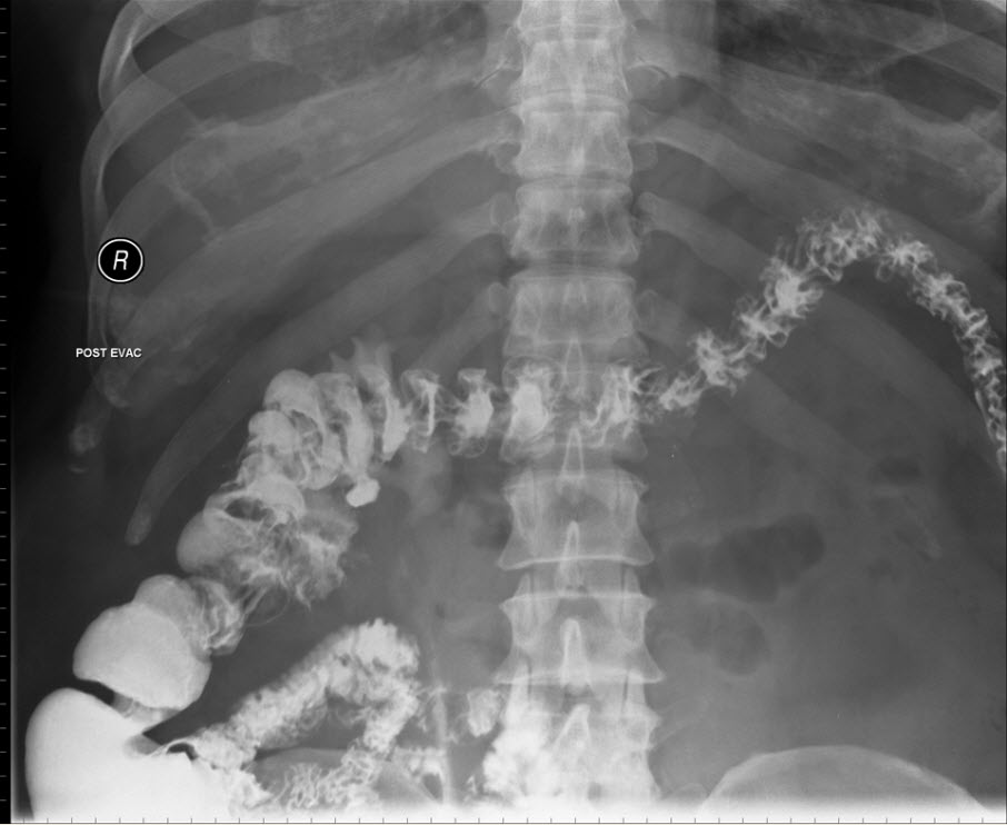
Indirect laryngoscopy revealed diffuse nasopharyngeal, oropharyngeal and laryngeal swelling without critical airway obstruction. Chest X-ray showed no significant abnormalities. He was diagnosed with lingual angioedema.
 
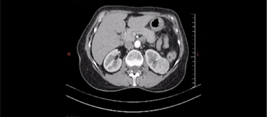
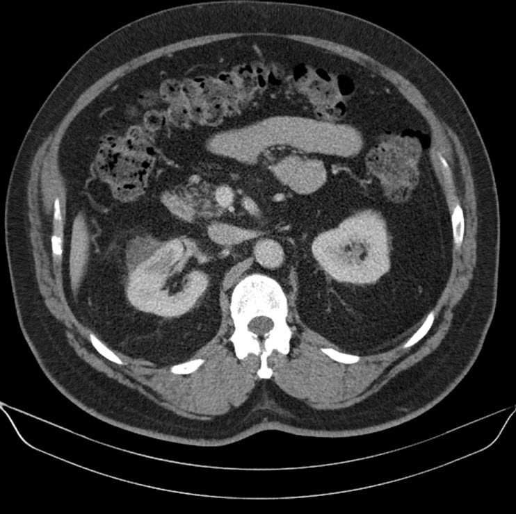
Everolimus was discontinued and intravenous treatment with methylprednisolone 125 mg, ranitidine 50 mg, promethazine 25 mg and cetirizine 10 mg was started and improved the patients’ symptoms. The dosages of his other medications, including quinapril, were not altered. Although the progression of the swelling was clearly brought to a halt five hours after administration of medication, there was no decrease noted in the volume of the tongue. Therefore intravenous hydrocortisone 250 mg in combination with ranitidine 50 mg was administrated and a CT scan was performed. The CT scan (Figure 2) revealed narrowing of the naso- and oropharynx, a dilated soft palate and an unmistakable voluminous tongue.


Figure 2. Computed tomography showing nearly complete obstruction of the oropharynx and obvious swelling of the tongue
 
 
He remained hospitalised for four more days in which time he received a declining dose of methylprednisolone, starting at 40 mg qd. His tongue returned to normal 18 hours after commencement. He was discharged with methylprednisolone 16mg qd for three more days.
Everolimus was re-administered nine days after the initial event at a reduced dose of 5 mg per day. More than 11 months after reinitiation, there has been no recurrence.


Discussion
 
To our knowledge this is the second reported case of lingual angioedema as a result of using a mTOR inhibitor in the treatment of mRCC. Mackenzie and Wood [11] described a 61-year old man treated with everolimus 10 mg qd in second line and who developed lingual angioedema after 21 days of treatment. Interestingly this patients’ concurrent medication also contained an angiotensin-converting enzyme (ACE) inhibitor, ramipril 10mg qd.  Everolimus was initially stopped, but resumed one week later with no recurrence of the noticed side-effect.
In transplant medicine, there is longstanding clinical experience with mTOR inhibitors, where sirolimus and everolimus have been used as immunosuppressants. Everolimus is administrated at a dosage of 3 mg qd, as part of triple therapy, together with a calcineurin inhibitor and prednisone. Calcineurin inhibitors (like cyclosporine and tacrolimus) and everolimus are both metabolized by the CYP3A4 isoenzyme system, and their concomitant administration increases everolimus exposure by 2- to 3-fold. [12]
The largest series reports on 309 german kidney transplant recipients who had received mTOR inhibitors as immunosuppressants. Nine patients developed angioedema after a mean period of 123 days under combined therapy of an mTOR- and an ACE inhibitor. Six of these nine patients ceased the administration of the ACE inhibitor, no recurrence took place. In the three patients who continued to take the ACE inhibitor, angioedema recurred. All patients continued their mTOR inhibitor after the onset of the angioedema. In seven patients the everolimus or sirolimus levels were measured at the time of the event and six of them had increased plasma concentration levels. After replacement of the ACE inhibitor by an angiotensin receptor blocker (ARB) no recurrence was noticed. [13]
Stallone et al [14] were in 2004 the first to report the development of lingual angioedema in five kidney transplant recipients, one month after initiating the combination everolimus and an ACE inhibitor. Interestingly, in these five patients both drugs were administered at high dosages. The sirolimus dosage was decreased and the ACE inhibitor was stopped, but later reintroduced without recurrence of the assumed side effect. The authors hypothesized a dosage dependent synergistic effect when combining both drugs.
No reports of temsirolimus associated with lingual angioedema were found. Of note, temsirolimus is not used in the transplant population and is a newer agent.
Angioedema is a well-known side effect of ACE inhibitors. The OCTAVE trial reported the occurrence of ACE inhibitor induced angioedema in 0,68% out of >12,000 patients. [15] 99% of all cases present within the first year of therapy, of which a clear majority present within the first 8 weeks. [15, 16] Our patient had taken quinapril, in a low dose, for 2 years and 9 months without any adverse clinical events.
The concomitant administration of everolimus and inhibitors or inducers of the CYP3A4 enzyme system may affect everolimus blood levels. Concomitant administration of typical CYP3A4 inhibitors (ketoconazole [17] and erythromycin [18]) has been shown to significantly increase everolimus maximum concentration and everolimus area under the curve.
ACE inhibitors are also metabolized by the CYP3A4 enzyme system. CYP3A4 interaction could be an important part in linking the occurrence of angioedema to the concomitant administration of everolimus and an ACE inhibitor.
Furthermore, the incidence of angioedema is greater in the transplant setting. This might be because of the additional CYP3A4 interactions induced by the administration of calcineurin inhibitors.


Conclusion
 
In recent years different types of cancer are being treated by targeted therapy, given the relatively favorable side effects in comparison to chemotherapy. An impressive advance, especially in renal cell cancer, is made in increasing overall survival and disease free survival due to the specialized form of inhibiting tumor proliferation and differentiation.
In an area where targeted therapy is being examined and experimented on all types of cancer, one must never forget to think about possible drug interactions and side effects.
We describe the occurrence of lingual angioedema due to everolimus treatment. As in our patient, literature revealed a strong association with concomitant administration of ACE inhibitors. We demonstrated that interrupting everolimus therapy, while continuing the ACE inhibitor, led to a resolution of lingual angioedema. Furthermore we showed that a rechallenge with a reduced dose of everolimus within two weeks after the initial event was safe and the patient could be treated for a further 11 months.
Dosage of everolimus and drug interaction most probably play an important role in the pathogenesis of lingual angioedema.


References
 
1. Motzer RJ, Hutson TE, Tomczak P et al. Sunitinib versus interferon alfa in metastatic renal cell carcinoma. N Engl J Med 2007; 356:115-124.
2. Escudier B, Eisen T, Stadler WM et al. Sorafenib in advanced clear-cell renal-cell carcinoma. N Engl J Med 2007; 356:125-134.
3. Sternberg Co, Davis ID, Mardiak J et al. Pazopanib in locally advanced or metastatic renal cell carcinoma: results of a randomized phase III trail. J Clin Oncol 2010; 8:1061-1068.
4. Escudier B, Pluzanska A, Koralewski P et al. Bevacizumab plus interferon alfa-2a for treatment of metastatic renal cell carcinoma: a randomised, double-blind phase III trial. Lancet 2007; 370:2103-2111.
5. Motzer RJ, Escudier B, Oudard S et al. Efficacy of everolimus in advanced renal cell carcinoma: a double-blind, randomised, placebo-controlled phase III trial. Lancet 2010; 372:449-456.
6. Hudes G, Carducci M, Tomczak P et al. Temsirolimus, interferon alfa, or both for advanced renal-cell carcinoma. N Engl J Med 2007; 356:2271-2281.
7. NCT00719264: A randomized, open-label, multi-center phase II study to compare bevacizumab plus RAD001 versus interferon alfa-2a plus bevacizumab for the first-line treatment of patients with metastatic clear cell carcinoma of the kidney. Available at: https://www.clinicaltrials.gov. Last accessed Dec 22, 2009.
8. NCT00903175: Efficacy and safety comparison of RAD001 versus sunitinib in the first-line and second-line treatment of patients with metastatic renal cell carcinoma. Available at: https://www.clinicaltrials.gov. Last accessed Jun 23, 2009.
9. Chan H, Grossman AB, Bukowski RM. Everolimus in the treatment of renal cell carcinoma and neuroendocirne tumors. Adv Ther 2010; 278:495-511.
10. Ulmer JL, Garvey MJ. Fatal angioedema associated with lisinopril. Ann Pharmacother 1992; 26:1245-6.
11. M. Mackenzie, L. A. Wood. Lingual angioedema associated with everolimus. Acta Oncologica 2010; 49:107-109.
12. Schaffer SA, Ross HJ. Everolimus: efficacy and safety in cardiac transplantation. Expert Opin Drug Saf 2010; 9:843-854.
13. Duerr M, Glander P, Iekmann F et al. Increased incidence of angioedema with ACE inhibitors in combination with mTOR inhibitors in kidney transplant recipients. Clin J Am Soc Nephrol 2010; 5:703-708.
14. Stallone G, Infante B, Di Paolo S et al. Sirolimus and angiotensin-converting enzyme inhibitors together induce tongue oedema in renal transplant recipients. Nephrol Dial Transplant 2004; 19:2906-2908.
15. Kostis JB, Kim HJ, Rusnak J et al. Incidence and characteristics of angioedema associated with enalapril. Arch Intern Med 2005; 165:1637-1642.
16. Schuster C, Reinhart WH, Hartmann K et al. Angioedema induced by ACE inhibitors and angiotensin II-receptor antagonists: analysis of 98 cases. Schweiz Med Wochenschr 1999; 129:362.
17. Kovarik JM, Beyer D, Bizot MN et al. Blood concentrations of everolimus are markedly increased by ketoconazole. J Clin Pharmacol 2005; 45:514-158.
18. Kovarik JM, Beyer D, Bizot MN et al. Effect of multiple-dose erythromycin on everolimus pharmacokinetics. Eur J Clin Pharmacol 2005; 61:35-38.

The authors have declared no conflicts of interest.


Date added to bjui.org: 20/10/2011


DOI: 10.1002/BJUIw-2011-069-web


© 2024 BJU International. All Rights Reserved.