Tag Archive for: simulation

Posts

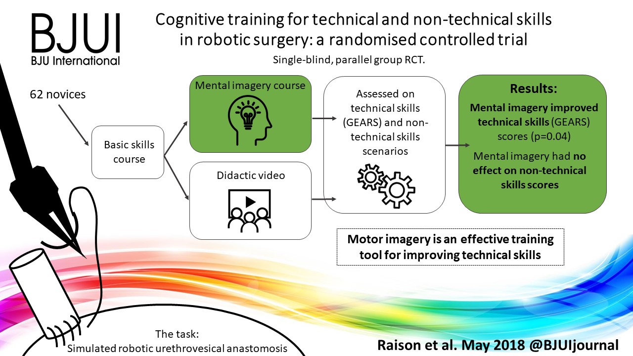
Article of the month: SIMULATE: Protocol and curriculum development of the first multicentre international randomized controlled trial assessing the transferability of simulation‐based surgical training

Every month, the Editor-in-Chief selects an Article of the Month from the current issue of BJUI. The abstract is reproduced below and you can click on the button to read the full article, which is freely available to all readers for at least 30 days from the time of this post.

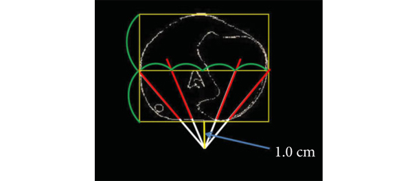
There is also a visual abstract created by a member of the team.

If you only have time to read one article this month, we recommend this one. 

Simulation in Urological Training and Education (SIMULATE): Protocol and curriculum development of the first multicentre international randomized controlled trial assessing the transferability of simulation‐based surgical training

Abdullatif Aydin*, Kamran Ahmed*, Mieke Van Hemelrijck, Hashim U. Ahmed§–, Muhammad Shamim Khan* ** and Prokar Dasgupta* ** on behalf of the SIMULATE Trial Group**

*MRC Centre for Transplantation, King’s College London, London, Department of Urology, King’s College Hospital NHS Foundation Trust, London,School of Cancer and Pharmaceutical Studies, King’s College London, London, §Department of Surgery and Cancer, Imperial College London, Department of Urology, Imperial College Healthcare NHS Trust, London, and **Department of Urology, Guy’s and St Thomas’ NHS Foundation Trust, London, UK

Abstract

Objectives

To report the study protocol for the first international multicentre randomized controlled trial investigating the effectiveness of simulation‐based surgical training and the development process for an evidence‐based training curriculum, to be delivered as an educational intervention.

Participants and Methods

This prospective, international, multicentre randomized controlled clinical and educational trial will recruit urology surgical trainees who must not have performed ≥10 of the selected index procedure, ureterorenoscopy (URS). Participants will be randomized to simulation‐based training (SBT) or non‐simulation‐based training (NSBT), the latter of which is the current sole standard of training globally. The primary outcome is the number of procedures required to achieve proficiency, where proficiency is defined as achieving a learning curve plateau of 28 or more on an Objective Structured Assessment of Technical Skills (OSATS) assessment scale, for three consecutive operations, without any complications. All participants will be followed up either until they complete 25 procedures or for 18 months. Development of the URS SBT curriculum took place through a two‐round Delphi process.

The SIMULATE ureteroscopy training curriculum, designed to be delivered as the educational intervention. The curriculum begins with introductory didactic lectures followed by virtual reality and dry‐lab simulation for semi‐rigid and flexible URS, respectively. If feasible, this is to be followed by full immersion simulation and cadaveric training, respectively. fURS, flexible ureterorenoscopy; NTS, non‐technical skills; OR, operating room; UO, ureteric access; URS, ureteroscopy; VR, virtual reality.

Results

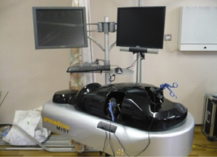
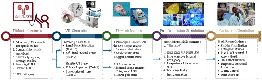
A total of 47 respondents, consisting of trainees (= 24) with URS experience and urolithiasis specialists (= 23), participated in round 1 of the Delphi process. Specialists (= 10) finalized the content of the curriculum in round 2. The developed interventional curriculum consists of initial theoretic knowledge through didactic lectures followed by select tasks and cases on the URO‐Mentor (Simbionix, Lod, Israel) VR Simulator, Uro‐Scopic Trainer (Limbs & Things, Bristol, UK) and Scope Trainer (Mediskills, Manchester, UK) models for both semi‐rigid and flexible URS. Respondents also selected relevant non‐technical skills scenarios and cadaveric simulation tasks as additional components, with delivery subject to local availability.

Conclusions

SIMULATE is the first multicentre trial investigating the effect and transferability of supplementary SBT on operating performance and patient outcomes. An evidence‐based training curriculum is presented, developed with expert and trainee input. Participants will be followed and the primary outcome, number of procedures required to proficiency, will be reported alongside key clinical secondary outcomes, (ISCRTN 12260261).

Video: Cognitive training for technical and non‐technical skills in robotic surgery

Cognitive training for technical and non‐technical skills in robotic surgery: a randomised controlled trial

Read the full article

Abstract

Objective

To investigate the effectiveness of motor imagery (MI) for technical skill and non‐technical skill (NTS) training in minimally invasive surgery (MIS).

Subjects and Methods

A single‐blind, parallel‐group randomised controlled trial was conducted at the Vattikuti Institute of Robotic Surgery, King’s College London. Novice surgeons were recruited by open invitation in 2015. After basic robotic skills training, participants underwent simple randomisation to either MI training or standard training. All participants completed a robotic urethrovesical anastomosis task within a simulated operating room. In addition to the technical task, participants were required to manage three scripted NTS scenarios. Assessment was performed by five blinded expert surgeons and a NTS expert using validated tools for evaluating technical skills [Global Evaluative Assessment of Robotic Skills (GEARS)] and NTS [Non‐Technical Skills for Surgeons (NOTSS)]. Quality of MI was assessed using a revised Movement Imagery Questionnaire (MIQ).

Results

In all, 33 participants underwent MI training and 29 underwent standard training. Interrater reliability was high, Krippendorff’s α = 0.85. After MI training, the mean (sd) GEARS score was significantly higher than after standard training, at 13.1 (3.25) vs 11.4 (2.97) (P = 0.03). There was no difference in mean NOTSS scores, at 25.8 vs 26.4 (P = 0.77). MI training was successful with significantly higher imagery scores than standard training (mean MIQ score 5.1 vs 4.5, P = 0.04).

Conclusions

Motor imagery is an effective training tool for improving technical skill in MIS even in novice participants. No beneficial effect for NTS was found.

View more videos

Article of the week: Cognitive training for technical and non‐technical skills in robotic surgery: a randomised controlled trial

Every week, the Editor-in-Chief selects an Article of the Week from the current issue of BJUI. The abstract is reproduced below and you can click on the button to read the full article, which is freely available to all readers for at least 30 days from the time of this post.

In addition to the article itself, there is an accompanying editorial written by a prominent member of the urological community. These are intended to provoke comment and discussion and we invite you to use the comment tools at the bottom of each post to join the conversation. There is also a video produced by the authors.

If you only have time to read one article this week, it should be this one.

Cognitive training for technical and non‐technical skills in robotic surgery: a randomised controlled trial

Nicholas Raison* , Kamran Ahmed*, Takashige Abe*, Oliver Brunckhorst*, Giacomo Novara, Nicolo Buf§, Craig McIlhenny, Henk van der Poel**, Mieke van Hemelrijck††, Andrea Gavazzi‡‡ and Prokar Dasgupta*


*Division of Transplantation Immunology and Mucosal Biology, Faculty of Life Sciences and Medicine, Kings College London, UK, ††Division of Cancer Studies, Kings College London, UK, Department of Urology, Forth Valley Royal Hospital, Larbert, UK, Department of Urology, Hokkaido University Graduate School of Medicine, Sapporo, Japan, Department of Urology, University of Padua, Padua, §Department of Urology, Humanitas Clinical and Research Centre, Rozzano, Milan, ‡‡Department of Urology, Azienda USL Toscana Centro, Florence, Italy, and **Department of Urology, Netherlands Cancer Institute, Amsterdam, The Netherlands


Read the full article

Visual Abstract created by Rebecca Fisher @beckybeckyfish

Abstract

Objective

To investigate the effectiveness of motor imagery (MI) for technical skill and non‐technical skill (NTS) training in minimally invasive surgery (MIS).

Subjects and Methods

A single‐blind, parallel‐group randomised controlled trial was conducted at the Vattikuti Institute of Robotic Surgery, King’s College London. Novice surgeons were recruited by open invitation in 2015. After basic robotic skills training, participants underwent simple randomisation to either MI training or standard training. All participants completed a robotic urethrovesical anastomosis task within a simulated operating room. In addition to the technical task, participants were required to manage three scripted NTS scenarios. Assessment was performed by five blinded expert surgeons and a NTS expert using validated tools for evaluating technical skills [Global Evaluative Assessment of Robotic Skills (GEARS)] and NTS [Non‐Technical Skills for Surgeons (NOTSS)]. Quality of MI was assessed using a revised Movement Imagery Questionnaire (MIQ).

Results

In all, 33 participants underwent MI training and 29 underwent standard training. Interrater reliability was high, Krippendorff’s α = 0.85. After MI training, the mean (sd) GEARS score was significantly higher than after standard training, at 13.1 (3.25) vs 11.4 (2.97) (P = 0.03). There was no difference in mean NOTSS scores, at 25.8 vs 26.4 (P = 0.77). MI training was successful with significantly higher imagery scores than standard training (mean MIQ score 5.1 vs 4.5, P = 0.04).

Conclusions

Motor imagery is an effective training tool for improving technical skill in MIS even in novice participants. No beneficial effect for NTS was found.

Read more Articles of the week


Video: Full immersion simulation – validation of a distributed simulation environment for technical and non-technical skills training in Urology


Full immersion simulation: validation of a distributed simulation environment for technical and non-technical skills training in Urology

James Brewin, Jessica Tang*, Prokar Dasgup ta, Muhammad S. Khan, Kamran AhmedFernando Bello*, Roger Kneebone* and Peter Jaye


Kings Health Partners, Guys and St Thomas NHS Foundation Trust, and *Imperial College, London,UK


Read the full article

OBJECTIVE

To evaluate the face, content and construct validity of the distributed simulation (DS) environment for technical and non-technical skills training in endourology. To evaluate the educational impact of DS for urology training.

SUBJECTS AND METHODS

DS offers a portable, low-cost simulated operating room environment that can be set up in any open space. A prospective mixed methods design using established validation methodology was conducted in this simulated environment with 10 experienced and 10 trainee urologists. All participants performed a simulated prostate resection in the DS environment. Outcome measures included surveys to evaluate the DS, as well as comparative analyses of experienced and trainee urologist’s performance using real-time and ‘blinded’ video analysis and validated performance metrics. Non-parametric statistical methods were used to compare differences between groups.

RESULTS

The DS environment demonstrated face, content and construct validity for both non-technical and technical skills. Kirkpatrick level 1 evidence for the educational impact of the DS environment was shown. Further studies are needed to evaluate the effect of simulated operating room training on real operating room performance.

CONCLUSIONS

This study has shown the validity of the DS environment for non-technical, as well as technical skills training. DS-based simulation appears to be a valuable addition to traditional classroom-based simulation training.


Read more articles of the week

Article of the Week: An assessment of the physical impact of complex surgical tasks on surgeon errors and discomfort

Every week the Editor-in-Chief selects the Article of the Week from the current issue of BJUI. The abstract is reproduced below and you can click on the button to read the full article, which is freely available to all readers for at least 30 days from the time of this post.

In addition to the article itself, there is an accompanying editorial written by a prominent member of the urological community. This blog is intended to provoke comment and discussion and we invite you to use the comment tools at the bottom of each post to join the conversation.

If you only have time to read one article this week, it should be this one.

An assessment of the physical impact of complex surgical tasks on surgeon errors and discomfort: a comparison between robot-assisted, laparoscopic and open approaches

Oussama Elhage*, Ben Challacombe*, Adam Shortland‡ and Prokar Dasgupta*
§*The Urology Centre, Guy’s and St Thomas’ NHS Foundation Trust, Medical Research Council (MRC) Centre for Transplantation, King’s College London, One Small Step Laboratory, and §MRC Centre for Transplantation & National Institute for Health Research (NIHR) comprehensive Biomedical Research Centre, King’s College London, King’s Health Partners, Guy’s Hospital, London, UK


Read the full article

OBJECTIVES

To evaluate, in a simulated suturing task, individual surgeons’ performance using three surgical approaches: open, laparoscopic and robot-assisted.

SUBJECTS AND METHODS

Six urological surgeons made an in vitro simulated vesico-urethral anastomosis. All surgeons performed the simulated suturing task using all three surgical approaches (open, laparoscopic and robot-assisted). The time taken to perform each task was recorded. Participants were evaluated for perceived discomfort using the self-reporting Borg scale. Errors made by surgeons were quantified by studying the video recording of the tasks. Anastomosis quality was quantified using scores for knot security, symmetry of suture, position of suture and apposition of anastomosis.

RESULTS

The time taken to complete the task by the laparoscopic approach was on average 221 s, compared with 55 s for the open approach and 116 s for the robot-assisted approach (anova, P < 0.005). The number of errors and the level of self-reported discomfort were highest for the laparoscopic approach (anova, P < 0.005). Limitations of the present study include the small sample size and variation in prior surgical experience of the participants.

CONCLUSIONS

In an in vitro model of anastomosis surgery, robot-assisted surgery combines the accuracy of open surgery while causing lesser surgeon discomfort than laparoscopy and maintaining minimal access.

Read more articles of the week

Editorial: Conventional laparoscopic surgery – more pain, no gain!

Advances in surgical technology have revolutionized the way surgery is performed today. Conventional laparoscopic surgery dominated the surgical paradigm for several decades, until robot-assisted surgery created the next giant leap. In the pressent article, Elhage et al. [1] compare and correlate physical stress and surgical performances among three modes of a standardized surgical step. Their study shows the obvious physical strain and technical limitations faced while performing conventional laparoscopic surgery, subsequently leading to compromised surgical outcomes. The physical impact of conventional laparoscopic surgery has been well documented through surgeon feedback as well as ergonomic assessment [2, 3]. Various studies have reported that higher physical stress, associated with ergonomic limitations, is experienced when performing conventional laparoscopy compared to the comfort and ease of robot-assisted surgery, as highlighted in the present study. Increased workload has also been associated with performance errors, with a steep learning curve needed to achieve surgical excellence during conventional laparoscopy [4].

Currently, the use of robot-assisted surgery is on the rise, as an alternative to both open and conventional laparoscopic surgery across the developed world, despite its obvious economic limitations. Better ergonomics during robot-assisted surgery will increase the comfort of the surgeon, but the future of surgery may easily be linked to the improvements experienced by all of us in the automobile industry. Developments, from manual gear-clutch control to automatic speed control and the luxury of adaptive cruise control today, make us safe drivers with minimal physical stress. The concept of adaptive cruise control, which adjusts the speed of a vehicle in relation to its surroundings, sounds similar to the leap from manual camera control during conventional laparoscopy to console-based control during camera navigation in robot-assisted surgery. With advances in the speed and size of computers, pneumatic-based joint mechanics and mindfulness meditation on the horizon, it will not be long before surgeons will sit back and watch the marvel of the machine. Surgeons just need to learn to hold on to their seats!

Read the full article

Syed J. Raza*, Khurshid A. Guru† anRobert P. Huben†
*Fellow, †Endowed Professor of Urologic Oncology, Department of Urology and A.T.L.A.S (Applied Technology Laboratory for Advanced Surgery) Program, Roswell Park Cancer Institute, Buffalo, NY, USA


References


2 Plerhoples TA, Hernandez-Boussard T, Wren SM. The aching surgeon: a survey of physical discomfort and symptoms following open, laparoscopic and robotic surgery.

J Robotic Surg 2012; 6: 65–723 Hubert N, Gilles M, Desbrosses K, Meyer JP, Felblinger J, Hubert J. Ergonomic assessment of the surgeon’s physical workload during standard and robotic assisted laparoscopic procedures. Int J Med Robot 2013; 9:142–147

4 Yurko YY, Scerbo MW, Prabhu AS, Acker CE, Stefanidis D. Higher mental workload is associated with poorer laparoscopic performance as measured by the NASA-TLX tool. Simul Healthc 2010; 5: 267–271


Article of the week: Detecting prostate cancer: the “core” of the matter

Every week the Editor-in-Chief selects the Article of the Week from the current issue of BJUI. The abstract is reproduced below and you can click on the button to read the full article, which is freely available to all readers for at least 30 days from the time of this post.

In addition to the article itself, there is an accompanying editorial written by prominent members of the urological community. This blog is intended to provoke comment and discussion and we invite you to use the comment tools at the bottom of each post to join the conversation.

If you only have time to read one article this week, it should be this one.

Can transrectal needle biopsy be optimised to detect nearly all prostate cancer with a volume of ≥0.5 mL? A three-dimensional analysis

Kent Kanao*, James A. Eastham, Peter T. Scardino†‡, Victor E. Reuter*§ and Samson W. Fine*

Departments of *Pathology and Surgery, Urology Service, Memorial Sloan-Kettering Cancer Center, and Departments of Urology and §Pathology, Weill Cornell Medical Center, New York, NY, USA

Read the full article

OBJECTIVE

• To investigate whether transrectal needle biopsy can be optimised to detect nearly all prostate cancer with a tumour volume (TV) of ≥0.5 mL.

MATERIALS AND METHODS

• Retrospectively analysed 109 whole-mounted and entirely submitted radical prostatectomy specimens with prostate cancer.

• All tumours in each prostate were outlined on whole-mount slides and digitally scanned to produce tumour maps. Tumour map images were exported to three-dimensional (3D) slicer software (https://www.slicer.org) to develop a 3D-prostate cancer model.

• In all, 20 transrectal biopsy schemes involving two to 40 cores and two to six anteriorly directed biopsy (ADBx) cores (including transition zone, TZ) were simulated, as well as models with various biopsy cutting lengths.

• Detection rates for tumours of different volumes were determined for the various biopsy simulation schemes.

RESULTS

• In 109 prostates, 800 tumours were detected, 90 with a TV of ≥0.5 mL (mean TV 0.24 mL).

• Detection rate for tumours with a TV of ≥0.5 mL plateaued at 77% (69/90) using a 12-core (3 × 4) scheme, standard 17-mm biopsy cutting length without ADBx cores. In all, 20 of 21 (95%) tumours with a TV of ≥0.5 mL not detected by this scheme originated in the anterior peripheral zone or TZ.

• Increasing the biopsy cutting length and depth/number of ADBx cores improved the detection rate for tumours with a TVof ≥0.5 mL in the 12-core scheme.

• Using a 22-mm cutting length and a 12-core scheme with additional volume-adjusted ADBx cores, 100% of ≥0.5 mL tumours in prostates ≤ 50 mL in volume and 94.7% of ≥0.5 mL tumours in prostates > 50 mL in volume were detected.

CONCLUSIONS

• Our 3D-prostate cancer model analysis suggests that nearly all prostate cancers with a TV of ≥0.5 mL can be detected by 14–18 transrectal needle-biopsy cores.

• Using longer biopsy cutting lengths and increasing the depth and number of ADBx cores (including TZ) according to prostate volume are necessary as well.


Read Previous Articles of the Week


Editorial: How many cores are needed to detect nearly all prostate cancers?

Virtual prostate biopsy and biopsy simulation: lessons to be learned

Prostate biopsies, transrectal or transperineal, still constitute the pillars of prostate cancer detection today [1]. With the lack of reliable imaging tools (new MRI techniques are promising but still investigational [2]); random biopsies offer the sole adequate cancer detection option [3]. However, random biopsies are far from efficient in detecting all tumours and even less efficient in detecting all significant cancer ‘spots’. To improve sensitivities and specificities, increasing the biopsy core numbers, targeting more lateral aspects and encouraging repeat biopsies have been recommended [4]. Recently, HistoScanning™ [5] and template biopsies [6] have been introduced to further improve biopsy quality and efficiency. The latest innovations include the fusion of MRI pictures with the TRUS image to offer optimal targeting of suspicious areas [7]. And yet, these efforts are far from solving the main problem. How can we perform a biopsy and be confident to detect most of the cancers, i.e. significant malignant areas.

The present study [1] does, what should have been done a long time ago, namely to create a reliable and reproducible biopsy simulation model to allow the investigation of various biopsy schemes, core lengths and numbers. Based on a series of 109 radical prostatectomy specimens, a three-dimensional (3D) prostate and prostate cancer model was created using novel 3D slicer software and various prostate biopsy schemes were simulated. Using this method, the detection rate for tumours with a tumour volume (TV) of ≥0.5 mL plateaued at 77% (69 of 90) using a 12 core (3 × 4) scheme, standard 17-mm biopsy cutting length without anteriorly directed biopsy (ADBx) cores. Twenty of 21 (95%) tumours with a TV of ≥0.5 mL not detected by this scheme originated in the anterior peripheral zone or transition zone [1].

Confirming our earlier data with the Vienna nomograms [8], increasing the biopsy cutting length and depth/number of ADBx cores (14–18 cores) improved the detection rate for tumours with a TV of ≥0.5 mL in the 12-core scheme [1]. The best biopsy scheme used a 22-mm cutting length and a 12-core scheme with additional volume-adjusted ADBx cores. Using this combination, 100% of ≥0.5 mL tumours in prostates <50 mL in volume and 94.7% of ≥0.5 mL tumours in prostates >50 mL in volume were detected.

Certainly, these numbers will not be reproducible in real-time TRUS or transperineal biopsies (detections rates of 95–100% as seen in this simulation model, cannot be achieved without adequate imaging tools, which are not available yet), but they aid significantly in rethinking our biopsy strategy. So, if we summarise the present findings and combine them with published data, the future will demand a TRUS-fusion biopsy technique, involving 14–18 cores (or more if volume increases), involving the anterior zones of the prostate and using a 22-mm cutting length of the biopsy core vs a 15–17 mm core as is used currently. Obviously real-time prospective trials are needed to confirm these findings but nothing indicates that the outcome would be otherwise.

Bob Djavan
Department of Urology, New York University School of Medicine, NYU, New York, NY, USA

Read the full article

References

  1. Kanao K, Eastham JA, Scardino PT, Reuter VE, Fine SW. Can transrectal needle biopsy be optimised to detect nearly all prostate cancer with a volume of ≥0.5 mL? A three-dimensional analysis. BJU Int 2013; 112: 898–904
  2. Delongchamps NB, Peyromaure M, Schull A et al. Pre-biopsy Magnetic Resonance Imaging and prostate cancer detection: comparison of random and MRI-targeted biopsies using three different techniques of MRI-TRUS image registration. J Urol 2013;189: 493–499
  3. Djavan B, Rocco B. Optimising prostate biopsy. BMJ 2011; 344: d8201
  4. Thompson I, Thrasher JB, Aus G et al. Guideline for the management of clinically localized prostate cancer: 2007 update. J Urol 2007; 177: 2106–2131
  5. Simmons LA, Autier P, Zát’ura F et al. Detection, localisation and characterisation of prostate cancer by prostate HistoScanning(™)BJU Int 2012; 110: 28–35
  6. Huo AS, Hossack T, Symons JL et al. Accuracy of primary systematic template guided transperineal biopsy of the prostate for locating prostate cancer: a comparison with radical prostatectomy specimens. J Urol 2012; 187: 2044–2049
  7. Sonn GA, Natarajan S, Margolis DJ et al. Targeted biopsy in the detection of prostate cancer using an office based magnetic resonance ultrasound fusion device. J Urol 2013; 189: 86–92
  8. Djavan B, Margreiter M. Biopsy standards for detection of prostate cancer. World J Urol 2007; 25: 11–17

Article of the week: Centralized simulation training combines technical and non-technical skills

Every week the Editor-in-Chief selects the Article of the Week from the current issue of BJUI. The abstract is reproduced below and you can click on the button to read the full article, which is freely available to all readers for at least 30 days from the time of this post.

In addition to the article itself, there is an accompanying editorial written by a prominent member of the urological community. This blog is intended to provoke comment and discussion and we invite you to use the comment tools at the bottom of each post to join the conversation.

If you only have time to read one article this week, it should be this one.

Development and implementation of centralized simulation training: evaluation of feasibility, acceptability and construct validity

Mohammad Shamim Khan, Kamran Ahmed, Andrea Gavazzi, Rishma Gohil, Libby Thomas*, Johan Poulsen, Munir Ahmed, Peter Jaye* and Prokar Dasgupta

MRC Centre for Transplantation, King’s College London, King’s Health Partners, Department of Urology, Guy’s Hospital , *Simulation and Interactive Learning (SaIL) Centre, Guy’s & St Thomas NHS Foundation Trust , and Department of Urology, Aalborg Denmark and King’s College Hospital, London, UK

Read the full article

OBJECTIVES

• To establish the feasibility and acceptability of a centralized, simulation-based training-programme.

• Simulation is increasingly establishing its role in urological training, with two areas that are relevant to urologists: (i) technical skills and (ii) non-technical skills.

MATERIALS AND METHODS

• For this London Deanery supported pilot Simulation and Technology enhanced Learning Initiative (STeLI) project, we developed a structured multimodal simulation training programme.

• The programme incorporated: (i) technical skills training using virtual-reality simulators (Uro-mentor and Perc-mentor [Symbionix, Cleveland, OH, USA] , Procedicus MIST-Nephrectomy [Mentice, Gothenburg, Sweden] and SEP Robotic simulator [Sim Surgery, Oslo, Norway]); bench-top models (synthetic models for cystocopy, transurethral resection of the prostate, transurethral resection of bladder tumour, ureteroscopy); and a European (Aalborg, Denmark) wet-lab training facility; as well as (ii) non-technical skills/crisis resource management (CRM), using SimMan (Laerdal Medical Ltd, Orpington, UK) to teach team-working, decision-making and communication skills.

• The feasibility, acceptability and construct validity of these training modules were assessed using validated questionnaires, as well as global and procedure/task-specific rating scales.

RESULTS

• In total 33, three specialist registrars of different grades and five urological nurses participated in the present study.

• Construct-validity between junior and senior trainees was signifi cant. Of the participants, 90% rated the training models as being realistic and easy to use.

• In total 95% of the participants recommended the use of simulation during surgical training, 95% approved the format of the teaching by the faculty and 90% rated the sessions as well organized.

• A significant number of trainees (60%) would like to have easy access to a simulation facility to allow more practice and enhancement of their skills.

CONCLUSIONS

• A centralized simulation programme that provides training in both technical and non-technical skills is feasible.

• It is expected to improve the performance of future surgeons in a simulated environment and thus improve patient safety.


Read Previous Articles of the Week

Editorial: The need to devise a better means of training

There is increasing concern that current UK trainees at the end of their training are less experienced than their previous counterparts and continue to require more education, skills and support when they assume their consultant posts in the form of mentoring.

It is generally accepted that the numbers of hours required to become an ‘expert’ is 10 000–30 000 and currently in the UK our trainees experience =6000 h of training. Much of this is due to the impact of the European Working Time Directive (EWTD) and the government ‘New Deal’ initiative on junior doctors contracts introduced in 2003. The UK conundrum shared with many other healthcare systems is how to provide effective training within the demands of service commitment and the EWTD. Skills training has therefore been seen as the mechanism to resolve the situation, encompassing the acquisition of both technical and non-technical skills. The challenge therefore is to devise innovative ways of training within the limit of fewer hours and training, not service, must become the priority for trainees and for those surgeons, departments and hospitals that train them.

Contemporary urology training is moving out of clinical practice and simulation is increasingly used to provide a safe and supportive learning environment for learning and maintaining skills. However, this needs the following criteria:

• An agreed curriculum

• Agreed set of standards

• A validated form of assessment

• The availability of local and national skills centres

• Educators and trainers

The problem is that traditionally the UK has few training centres, together with a lack of trained manpower and funding. However, controversy still remains over the efficacy of simulation for training and those who are able to fund such projects comment on the paucity of available data in relation to the predictability of future outcomes and patient safety.

Projects such as the Simulation and Technology enhanced Learning Initiative (STeLI) initiative documented in this paper are important contributors to the evidence base. The programme aims to establish the feasibility and acceptability of a centralised, simulation-based system incorporating both skills and non-technical skills aspects of training. The latter involving crisis resource management using the SimMan model to teach team-working, decision-making, and communication skills in various settings between senior and junior trainees. Not surprisingly senior trainees scored significantly better on virtual reality simulators, bench-top box trainers and the European wet-lab training facility, as well as in human patient simulation training in crisis resource management (CRM) using SimMan, than junior trainees. The interesting point raised in this paper is that the trainees’ behaviour shows the value of inclusion of the CRM training and the interplay between technical and non-technical skills. Non-technical skills have often been sidelined in courses focusing on technical skills acquisition and this paper highlights the importance and added-value of incorporating such a skill set into future course content and curricula.

Thus, there is no doubt that some surgical skills can be learned in the laboratory and although this will never be a substitute for operative experience, the first steps of training can be accelerated with potential reduction of risk to patients. Increasingly data from sources such as the STeLI project underline a better appreciation of the importance of the training in non-technical skills, which equip surgeons in working under stress and more importantly working as a team player. However, the ultimate test for simulation is whether the model and content is able to reduce surgical errors, improve patient safety and reduce operative time and costs. To try and answer these questions BAUS in conjunction with the Specialist Advisory Committee (SAC) in Urology have recognised that the technology is there but there is a need to identify trainers keen to train, with the nomination of a national lead for simulation to develop a national strategy to deliver a viable programme aligned to the curriculum to try and answer the important question: ‘Does simulation enhance real-life performance of a surgical technique?’.

Adrian D. Joyce
St James’ University Hospital, Leeds LS9 7TF, UK

Read the full article

© 2024 BJU International. All Rights Reserved.