Tag Archive for: squamous cell carcinoma

Posts

Article of the Week: DSNB for Penile Cancer

Every Week the Editor-in-Chief selects an Article of the Week from the current issue of BJUI. The abstract is reproduced below and you can click on the button to read the full article, which is freely available to all readers for at least 30 days from the time of this post.

In addition to the article itself, there is an accompanying editorial written by a prominent member of the urological community. This blog is intended to provoke comment and discussion and we invite you to use the comment tools at the bottom of each post to join the conversation.

If you only have time to read one article this week, it should be this one.


Dynamic sentinel lymph node biopsy for penile cancer: a comparison between 1- and 2-day protocols

Panagiotis Dimopoulos*, Panagiotis Christopoulos*, Sam Shilito, Zara Gall*, Brian Murby§, David Ashworth§, Ben Taylor, Bernadette Carrington, Jonathan Shanks**, Noel Clarke*, Vijay Ramani*, Nigel Parr*, Maurice Lau* and Vijay Sangar*


Departments of *Urology, §Nuclear Medicine, Radiology , **Pathology, The Christie Hospital, ManchesterMedical School, University of Manchester, Manchester, and Department of Urology, Royal Bolton Hospital, Bolton Lancashire, UK

Objective

To determine the outcome of clinically negative node (cN0) patients with penile cancer undergoing dynamic sentinel node biopsy (DSNB), comparing the results of a 1- and 2-day protocol that can be used as a minimal invasive procedure for staging of penile cancer.

Patients and Methods

This is a retrospective analysis of 151 cN0 patients who underwent DSNB from 2008 to 2013 for newly diagnosed penile cancer. Data were analysed per groin and separated into groups according to the protocol followed. The comparison of the two protocols involved the number of nodes excised, γ-counts, false-negative rates (FNR), and complication rates (Clavien–Dindo grading system).

JuneAOTW3

Results

In all, 280 groins from 151 patients underwent DSNB after a negative ultrasound ± fine-needle aspiration cytology. The 1-day protocol was performed in 65 groins and the 2-day protocol in 215. Statistically significantly more nodes were harvested with the 1-day protocol (1.92/groin) compared with the 2-day protocol (1.60/groin). The FNRs were 0%, 6.8% and 5.1%, for the 1-day protocol, 2-day protocol, and overall, respectively. Morbidity of the DSNB was 21.4% for all groins, and 26.2% and 20.1% for the 1-day and 2-day protocols, respectively. Most of the complications were of Clavien–Dindo Grade 1–2.

Conclusions

DSNB is safe for staging patients with penile cancer. There is a trend towards a 1-day protocol having a lower FNR than a 2-day protocol, albeit at the expense of a slightly higher complication rate.

Editorial: One Day Protocol for Early Penile Cancer – The Way to Go

The present article by Dimopoulos et al. [1] has some useful lessons on the development of new services. The authors have kept a detailed database of all patients going through their super-regional network, and have designed the protocol around the patient, whereby the primary and regional lymph nodes are dealt with in one visit. Previously, bilateral inguinal lymph node dissection (ILND) was so fraught with complications that it would not be combined routinely with organ-sparing surgery of the penis [2]; however, the significantly lower complication rate of dynamic sentinel node biopsy (DSNB) has allowed the more streamlined approach. The ‘only handle it once’ (OHIO) philosophy is surely not only preferable for the patient, but also reduces the risk of patients not receiving ideal management. In most cases, a biopsy at the time of presentation, along with physical examination/imaging, can determine those requiring DSNB instead of waiting for final pathology from the primary tumour. The controversy surrounding DSNB compared with ILND has been the false-negative rates. The pioneering group from the Netherlands reported four deaths in six patients with false-negative results [3]. In the present paper, the overall false-negative rate was 5.8%, but the smaller and newer cohort of patients underwent a same-day protocol and had zero false-negatives. This may be attributable to the fact that biopsies were taken from a total sample of 65 or that slightly more nodes were taken in this group. We expect the one-day protocol to become standard, and future independent reports will be welcome. Should there truly be a 0% false-negative rate then the controversy is resolved and prophylactic ILND will become a historical procedure. Finally, the lower morbidity of the present study cohort allowed the authors to move the intermediate-risk group from surveillance to nodal biopsy, which proved justified because some of these cases had micrometastatic disease. We congratulate the group for their scientific approach to improving the quality of care for patients and for bringing their data to publication.

Paul K. Hegarty and Peter E. Lonergan
Urology, National Penile Cancer Centre, Mater Misericordiae University Hospital, Dublin, Ireland


References


1 Dimopoulos P, Christopoulos P, Shilito S et al. Dynamic sentinel lymph node biopsy for penile cancer: a comparison between 1- and 2-day protocols. BJU Int 2016; 117: 8906


2 Hegarty PK, Eardley I, Heidenreich A et al. Penile cancer: Organ-sparing techniques. BJU Int 2014; 114: 799805


3 Kroon BK, Horenblas S, Meinhardt W et al. Dynaminc sentinel node biopsy in penile cancer: evaluation of 10 years experience. Eur Urol 2005; 47: 6016


Article of the week: Pilot study of EGFR-targeted therapies in men with penile SCC shows promising initial results

Every week the Editor-in-Chief selects the Article of the Week from the current issue of BJUI. The abstract is reproduced below and you can click on the button to read the full article, which is freely available to all readers for at least 30 days from the time of this post.

In addition to the article itself, there is an accompanying editorial written by a prominent member of the urological community. This blog is intended to provoke comment and discussion and we invite you to use the comment tools at the bottom of each post to join the conversation.

Finally, the third post under the Article of the Week heading on the homepage will consist of additional material or media. This week we feature a video from Dr. Pagliaro discussing his paper.

If you only have time to read one article this week, it should be this one.

Epidermal growth factor receptor-targeted therapy in locally advanced or metastatic squamous cell carcinoma of the penis

Bradley C. Carthon*§, Chaan S. Ng, Curtis A. Pettaway and Lance C. Pagliaro*

Departments of *Genitourinary Medical Oncology, Radiology, and Urology, The University of Texas MD Anderson Cancer Center, Houston, TX, USA
§
Current address: Winship Cancer Institute, Emory University,Atlanta, Georgia, USA

Read the full article
OBJECTIVE

• To evaluate the safety and efficacy of epidermal growth factor receptor (EGFR)-targeted therapy in patients with advanced penile or scrotal cancer.

PATIENTS AND METHODS

• We retrospectively reviewed the charts of patients with penile or scrotal squamous cell carcinoma who had visited our tertiary cancer centre between 2002 and 2009, including their subsequent treatment and follow-up.

• We collected details of EGFR-targeted therapy and clinical outcomes. Treatment-associated time-to-disease-progression (TTP), overall survival (OS), responses to therapy and toxicity were evaluate

RESULTS

• A total of 24 patients had received EGFR-targeted therapies, including cetuximab, erlotinib and gefitinib. The most common treatment given (to 67% of patients) was cetuximab combined with one or more cytotoxic drugs.

• The most common adverse effect was skin rash (71%). The median (range) TTP and OS were 11.3 (1–40) and 29.6 (2–205) weeks, respectively. The OS time for patients with visceral or bone metastases was significantly shorter than it was for those without (24.7 vs 49.9 weeks, P = 0.013).

• Among 17 patients treated with cetuximab alone or in combination with cisplatin, there were four partial responses (23.5%) including two patients with apparently chemotherapy-resistant tumours.

CONCLUSIONS

• Our results suggest that cetuximab has antitumour activity in metastatic penile cancer, and may enhance the effect of cisplatin-based chemotherapy.

• Prospective studies of EGFR-targeted therapies in men with these tumours are warranted


Editorial: Squamous cell carcinoma of the penis: therapeutic targeting of the EGFR

Squamous cell carcinoma of the penis is a rare genitourinary malignancy. There are wide variations in its incidence, ranging from 0.1 to 0.9/100 000 men in Europe, where it accounts for 1% of male malignancies, to as high as 4.4 and 4.2/100 000 men in Uganda and Paraguay, where it accounts for up to 10% of male malignancies.

The management of patients with advanced squamous cell carcinoma of the penis, including those patients with node-positive disease and metastatic disease, remains challenging. The beneficial effects of chemotherapy and radiotherapy are not established, partly because of the small numbers of patients within studies, but also because of the multiple regimens used for treatment, combined with relatively low response rates and high toxicities.

The presence and extent of lymph node metastasis is the single most common factor predictive of survival in men with penile carcinoma, with 5-year survival rates of 88% in men with minimal or no metastases (one to two nodes), compared with ∼25% in those men with two or more inguinal nodes involved. In men with extra nodal spread of the cancer and pelvic metastases, 5-year survival rates fall as low as 5–10%.

The most important aetiological factors for the development of squamous cell cancer of the penis appear to be the presence of a foreskin, immunosuppression and smoking. In addition to this, human papillomavirus (HPV) has been shown to have a central role in tumorogenesis [1]. HPV DNA can be identified in up to 80% of tumour specimens. The commonest subtypes expressed are the 16/18 subtypes (high risk) and the 6/11 subtypes (low risk). The virus exerts its tumorogenic effect via expression of viral oncogenes E6 and E7, which inhibit the activity of tumour suppressor genes p53 and RB. Whilst a number of potential biomarkers have been identified as prognostic indices of survival, translational research to date is limited [2].

A recent study from the UK has reported that survival rates in men with node-positive penile carcinoma have not improved significantly in the last 20 years [3]. In view of the poor response rates from chemotherapeutic agents, combined with their high toxicity and the poor survival rates in men with node-positive disease, it is imperative that more novel treatment methods, including targeted therapies, are developed to treat this devastating tumour.

A potential biological target in all squamous cell cancers, including the penis, is the epidermal growth factor family of receptors. A number of trials have been conducted to evaluate the safety profile and activity of a combination of anti-epidermal growth factor receptor (EGFR) monoclonal antibodies, including cetuximab, with schedules of platinum-based chemotherapy in a number of tumour sites. These combinations have shown good tolerability. The addition of cetuximab to platinum-based chemotherapy prolongs survival in patients with recurrent or metastatic squamous cell tumours of the head and neck.

Penile squamous cell tumours and their metastases also highly express EGFRs with ∼90 to 100% of tumours expressing the EGFR. The EGFR is a cell-surface receptor for members of the epidermal growth factor family of extracellular protein ligands. The EGFR is a member of the ErbB family of receptors, which consist of a sub-family of four closely related tyrosine kinases (EGFR [ErbB-1], HER2/c-neu [ErbB-2], Her 3 [ErbB-3] and Her 4 [ErbB-4]). EGFR can be activated by binding its specific ligands, including EGF and TGF-α. Dimerization of the EGFR stimulates tyrosine kinase activity and autophosphorylation of a number of tyrosine residues in the C-terminal domain of the EGF receptor, which downstream initiates a number of signal transduction cascades, ultimately resulting in cell migration and proliferation.

Cetuximab and panitumumab are monoclonal antibody inhibitors of the EGFR, which block the extracellular ligand-binding domains on the EGFR receptor. Furthermore, cetuximab induces the internalization of EGFR leading to downregulation of the EGFR. It also targets cytotoxic immune effector cells towards EGFR-expressing tumour cells (antibody-dependent cell-mediated cytotoxicity). Drugs, such as gefitinib are EGFR tyrosine kinase inhibitors, which bind and inhibit the EGFR tyrosine kinase by binding to the ATP-binding site of the enzyme. In lung tumours, patients who are EGFR-positive have shown relatively high response rates to tyrosine kinase inhibitors, although many patients develop resistance.

Two studies have analysed the expression of the EGFR receptor status in penile cancer [4, 5]. In both studies, the EGFR receptor was overexpressed in tumour tissue. In one study [4], 40 out of 44, i.e. 91% of patients, showed a positive EGFR expression in the primary tumour as well as in metastases. Importantly, a correlation between EGFR receptor expression and survival was not demonstrated.

In the present study by Carthon et al. [6], the authors evaluate the safety and efficacy of EGFR-targeted therapy using both cetuximab and tyrosine kinase inhibitors, including gefitinib. This pilot study evaluated 24 patients receiving EGFR-targeted therapies. Among 17 patients treated with cetuximab alone, or in combination with cisplatin, there were four partial responses. Whilst the presence of visceral metastases at the start of EGFR-based therapy was associated with poor time to progression and overall survival, several patients in that study were shown to have regression of predominantly inguinal and pelvic tumours. Interestingly, there were no objective responses to the small molecule inhibitors gefitinib or erlotinib. This pilot study would suggest that further prospective studies of EGFR-targeted therapies in men with squamous cell carcinoma of the penis are warranted and these initial results are promising; however, the number of regimens and agents used in the study is varied. This variation and the small number of patients and the retrospective nature of the study represent study limitations. Nevertheless, the concept of targeted therapies for squamous cell carcinoma of the penis should certainly be evaluated further, as it is clear that surgery alone is insufficient to improve survival in patients with N+ or M1 disease.

Read the full article

Suks Minhas
Department of Urology, University College Hospital, London, UK

  1. Minhas S, Manseck A, Watya S, Hegarty PK. Penile cancer. Prevention and premalignant conditions. Urology 2010; 76 (2 Suppl. 1): S24–35
  2. Kayes O, Ahmed HU, Arya M, Minhas S. Molecular and genetic pathways in penile cancer. Lancet Oncol 2007; 8: 420–429
  3. Kayes O, Freeman A, Lau D et al. Longitudinal analysis of outcomes for men with node positive penile cancer – are we improving? BJU Int 2013; 111 (S3): P19
  4. Börgermann C, Schmitz KJ, Sommer S, Rübben H, Krege S. Characterization of the EGF receptor status in penile cancer: retrospective analysis of the course of the disease in 45 patients. Urologe A 2009; 48: 1483–1489
  5. Lavens N, Gupta R, Wood LA. EGFR overexpression in squamous cell carcinoma of the penis. Curr Oncol 2010; 17: 4–6

Video: EGFR-targeted therapy in metastatic penile SCC

Epidermal growth factor receptor-targeted therapy in locally advanced or metastatic squamous cell carcinoma of the penis

Bradley C. Carthon*§, Chaan S. Ng, Curtis A. Pettaway and Lance C. Pagliaro*

Departments of *Genitourinary Medical Oncology, Radiology, and Urology, The University of Texas MD Anderson Cancer Center, Houston, TX, USA
§
Current address: Winship Cancer Institute, Emory University,Atlanta, Georgia, USA

Read the full article
OBJECTIVE

• To evaluate the safety and efficacy of epidermal growth factor receptor (EGFR)-targeted therapy in patients with advanced penile or scrotal cancer.

PATIENTS AND METHODS

• We retrospectively reviewed the charts of patients with penile or scrotal squamous cell carcinoma who had visited our tertiary cancer centre between 2002 and 2009, including their subsequent treatment and follow-up.

• We collected details of EGFR-targeted therapy and clinical outcomes. Treatment-associated time-to-disease-progression (TTP), overall survival (OS), responses to therapy and toxicity were evaluate

RESULTS

• A total of 24 patients had received EGFR-targeted therapies, including cetuximab, erlotinib and gefitinib. The most common treatment given (to 67% of patients) was cetuximab combined with one or more cytotoxic drugs.

• The most common adverse effect was skin rash (71%). The median (range) TTP and OS were 11.3 (1–40) and 29.6 (2–205) weeks, respectively. The OS time for patients with visceral or bone metastases was significantly shorter than it was for those without (24.7 vs 49.9 weeks, P = 0.013).

• Among 17 patients treated with cetuximab alone or in combination with cisplatin, there were four partial responses (23.5%) including two patients with apparently chemotherapy-resistant tumours.

CONCLUSIONS

• Our results suggest that cetuximab has antitumour activity in metastatic penile cancer, and may enhance the effect of cisplatin-based chemotherapy.

• Prospective studies of EGFR-targeted therapies in men with these tumours are warranted

Article of the week: Staging inguinal disease in patients with penile cancer

Every week the Editor-in-Chief selects the Article of the Week from the current issue of BJUI. The abstract is reproduced below and you can click on the button to read the full article, which is freely available to all readers for at least 30 days from the time of this post.

In addition to the article itself, there is an accompanying editorial written by a prominent member of the urological community. This blog is intended to provoke comment and discussion and we invite you to use the comment tools at the bottom of each post to join the conversation.

If you only have time to read one article this week, it should be this one.


Phase 1 prospective evaluation of the oncological adequacy of robotic assisted video-endoscopic inguinal lymphadenectomy in patients with penile carcinoma

Surena F. Matin, Janice N. Cormier*, John F. Ward, Louis L. Pisters, Christopher G. Wood, Colin P.N. Dinney, Richard E. Royal*, Xuelin Huang and Curtis A. Pettaway

Departments of Urology, *Surgical Oncology and Biostatistics, University of Texas MD Anderson Cancer Center, Houston, TX, USA

Read the full article
OBJECTIVE

• To prospectively determine the oncological adequacy of robotic assisted video-endoscopic inguinal lymphadenectomy (RAVEIL).

PATIENTS AND METHODS

• Patients with T1-3N0 penile cancer were enrolled into a prospective phase I trial at a tertiary care institution from March 2010 to January 2012. All patients underwent an initial RAVEIL approach.

• Verification of adequacy of dissection was performed by an independent surgeon via a separate open incision at the conclusion of the RAVEIL procedure.

• Out of 10 patients, if more than two superficial inguinal fields with ≥2 nodes or more than four with ≥1 node remained within the superficial dissection field, the study would not proceed to phase II.

RESULTS

• Of 10 enrolled patients two had inguinal metastases and all positive nodes were detected by RAVEIL. The remaining eight patients had no metastases, with a mean of nine (range 5–21) left and nine (range 6–17) right nodes removed. One inguinal field RAVEIL was converted to an open dissection.

• The verifying surgeon confirmed that 18 of 19 inguinal fields (94.7% in nine patients) had an adequate dissection. Two benign nodes were found just beneath Scarpa’s fascia above the inguinal dissection field.

• Limitations of the study include an inability to determine decisively what specific wound complications were related to RAVEIL because of the protocol-specified creation of a small inguinal incision for verification of adequate dissection.

CONCLUSION

• RAVEIL allowed adequate staging of disease in the inguinal region among patients with penile cancer at risk for inguinal metastases.


Read Previous Articles of the Week


Editorial: Laparoscopic and robotic approach to staging nodes in penile cancer

In recent years, efforts to reduce morbidity from lymphadenectomy for penile cancer were based on surgical procedures to reduce the area of lymph node dissection. The proposition of extensive video-endoscopic inguinal lymphadenectomy, a technique still experimental, is to reduce the morbidity of conventional surgery without affecting the maximum chance of oncological control of locoregional disease. Therefore the initiative of using the help of a robot to facilitate the implementation of this procedure is very welcome.

The authors present an excellent study on their initial experience with robotic assisted video-endoscopic inguinal lymphadenectomy (RAVEIL). I understand that for better comparison of the dissection area with open surgery these authors have opted to use an additional incision in the inguinal fold. However this area is the least vascularized area of the field of dissection because the lymph nodes are resected above and below this additional incision. It would be better to make an incision at the upper limit of the dissection. This approach was used in open surgery with low complication rates. The rate of necrosis (10%) and wound breakdown (10%) seems high for a minimally invasive approach. Possibly, when no additional incision is used to complement the procedure these rates will become lower.

Antonio A. Ornellas
Hospital Mario Kröeff, RJ, Brazil, and Department of Urology, Brazilian National Cancer Institute, RJ, Brazil

Read the full article

Concurrence of squamous cell carcinoma, sarcomatoid carcinoma and adenocarcinoma in relapsed prostate cancer originated from adenocarcinoma

Squamous cell carcinoma (SqCC) and sarcomatoid carcinoma (SC) are rare subtypes of prostate cancer. We report a rare case with concurrence of SqCC, SC and adenocarcinoma in a relapsed tumour originated from adenocarcinoma.

Authors: Ruiying Diao1,2, Kin Lam Fok3, Zhongfu Zhang1,2, Li Zhao2,4, Lisha Mou1,2,5, Shuolei Sun1,2, Lijun Zhou1,2 and Zhiming Cai2,6
1Department of Urology, Peking University Shenzhen Hospital, Shenzhen, China
2Guangdong Key Laboratory of Male Reproductive Medicine and Genetics, Peking University Shenzhen Hospital, Shenzhen, China
3Epithelial Cell Biology Research Center, Department of Physiology, School of Biomedical Sciences, Faculty of Medicine, Chinese University of Hong Kong, Hong Kong, China
4Department of Laboratory Medicine, Peking University Shenzhen Hospital, Shenzhen, China
5Institute of Urology, Shenzhen PKU-HKUST Medical Center, Shenzhen, China
6Department of Urology, Second People’s Hospital of Shenzhen, Shenzhen, China

Corresponding Author: Zhiming, Cai. Guangdong Key Laboratory of Male Reproductive Medicine and Genetics, Peking University Shenzhen Hospital, and Department of Urology, Second People’s Hospital of Shenzhen, Shenzhen, China. E-mail: [email protected]


Abstract
Squamous cell carcinoma (SqCC) and sarcomatoid carcinoma (SC) are rare subtypes of prostate cancer. We report a rare case with concurrence of SqCC, SC and adenocarcinoma in a relapsed tumour originated from adenocarcinoma. A 62-year-old man diagnosed with prostate adenocarcinoma (Gleason score 5+4=9)  received androgen blockade (AB) treatment and 89Sr radiotherapy. An increase in total prostate-specific antigen level was noted 13 months after treatment. Pathological analyses on biopsies from transurethral resection of the prostate revealed the concomitance of SqCC and SC with adenocarcinoma (Gleason score 5+5=10). Expression of the epithelial markers E-cadherin and β-catenin were significantly down-regulated, while the mesenchymal marker vimentin was up-regulated in both SqCC and SC. The expression of androgen receptor (AR) was down-regulated in SqCC but elevated in SC. The altered epithelial and mesenchymal markers and the heterogeneous AR expression in the relapsed tumour suggest that the concurrence of unusual subtypes may arise from the epithelial-to-mesenchymal transition and/or the differential function of AR on prostatic epithelial and stromal cells. The present study raises concerns about antiandrogen therapy regimen for prostate cancer.

Introduction
Prostate cancer is the most common malignant tumour in men > 70 years old and the morbidity of prostate cancer has been markedly increasing in recent years [1, 2]. Prostatic squamous cell carcinoma (SqCC) and sarcomatoid (SC) are rare subtypes of prostate cancer that account for 0.5–1% and < 0.1% of all prostate tumours, respectively [3-5]. The prognosis of patients with SqCC and SC is usually poor. Understanding the origin and initiation of SqCC and SC may benefit the development of an effective therapeutic regimen. SqCC and SC have mainly been observed after endocrine therapy or radiotherapy [4]. It has been postulated that the selection pressure from endocrine therapy is one of the driving forces for clonal selection in SqCC, but the exact mechanism underlying the occurrence of SqCC and SC de novo is not yet understood. The present study reports a rare pathological differentiation from initial adenocarcinoma to the concomitance of three kinds of pathological subtypes, adenocarcinoma, SqCC and SC. The expression of androgen receptor (AR), epithelial markers E-cadherin and β-catenin, and the mesenchymal marker vimentin in specimens before and after therapy were examined to evaluate their possible involvement in the transformation of pathological phenotypes.

Case Report
A 62-year-old patient was admitted to our hospital on 3 February 2010 with a 36-month history of progressive LUTS. Before these symptoms, there had been no previous history of urinary tract disease. His PSA level during admission was 30.5 ng/mL (Fig. 1A). The initial pathological analysis from needle biopsy led to a diagnosis of moderately differentiated adenocarcinoma with small acinar infiltration and proliferation mainly centered in the peri-acinar region (Gleason score 5+4=9, FIG. 1B, i). A bone scan revealed widespread metastases. The patient was treated with androgen deprivation therapy for 10 months as the initial regimen. During this period, 89Sr radionuclide therapy against prostate cancer was used twice, once at 4 months and once at 9 months, and the patient’s total PSA level was initially suppressed but was then elevated at a later stage (FIG. 1A). TURP was undertaken for pathological analysis at 13 months because of the increase in total PSA level (FIG. 1A). Specimens were fixed intact in 4% formalin and then sectioned transversally at regular intervals for random sampling (10 points). Pathological diagnosis showed concurrent adenocarcinoma, SqCC and SC (Gleason score 5+5=10). A 55% area of the TURP specimen showed the pathological structure of adenocarcinoma with abundant clear cytoplasm and enlarged nucleolus (FIG. 1B, ii). An area of ~25% of the TURP specimen showed the differentiation of SqCC with individual cell keratinization (cytokeratin high molecular weight; FIG. 1B, vi), intercellular bridges, and/or keratin pearl formation (FIG. 1B, iii). An area of nearly 20% of the TURP specimen showed bizarre atypia with giant cells and a tumour-induced osteoclastic phenotype with S-100 positive (a marker for osteosarcoma) in SC (FIG. 1B, iv). From the 13th month to time of death, serum total PSA increased rapidly to nearly 400ng/mL (FIG. 1A).
Compared with samples from the needle biopsy, the intensities of AR (FIG. 2B), and E-cadherin and β-catenin (FIG. 3B) immunoreactivity were all significantly lowered in SqCC. Similar to the pattern in the SqCC, the expression of E-cadherin and β-catenin in specimens after therapy was also lower in SC compared with those before therapy. The expression of the mesenchymal marker vimentin was increased after therapy (FIG. 1B, x), but the expression of AR was elevated in SC (FIG. 2B, iv, viii). Moreover, the AR C-terminal ligand-binding domain was mainly located in the cytoplasm of osteosarcomatous cells (FIG. 2B, iv), while the AR N-terminal transcription activation-binding domain was mainly located in the nucleus of osteosarcomatous components (FIG. 2B, viii).

12-036FIG.1A12-036FIG.1B


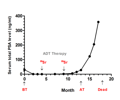
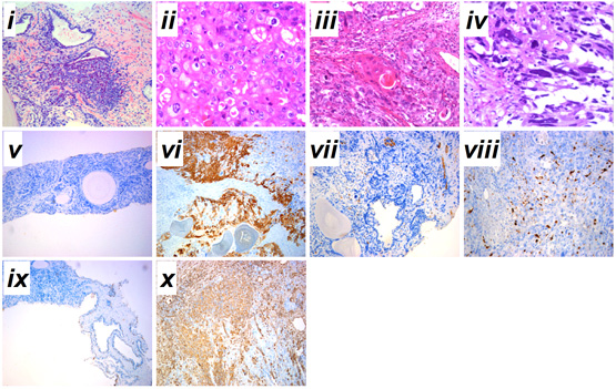
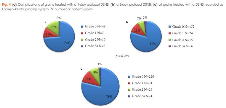
Fig. 1 Serum total PSA levels and diagnosis of prostatic cancer before and after therapy. A, Serum total PSA levels from admission to death. B, (i) prostatic adenocarcinoma from prostate needle biopsy before therapy; (ii) prostatic adenocarcinoma from TURP after therapy; (iii) prostatic sqCC from TURP after therapy; (iv) SC after the TURP after therapy; immunohistochemical staining for (v–vi) cytokeratin high molecular weight, (vii-viii) S-100 and (ix-x) vimentin expressions before and after therapy. Haematoxylin-eosin stain, x200. Admission time was set as 0 month. ADT, androgen deprivation therapy; 89Sr, 89Sr radiation; BT, before therapy; AT, after therapy. i-iv, scale bar=50 μm; v-x, scale bar=200 μm.

12-036FIG.2A12-036FIG.2B


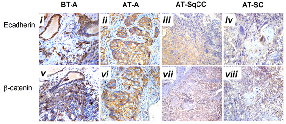
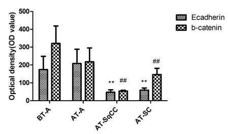
Fig. 2 Immunohistochemical staining for AR(C) and AR(N) in the adenocarcinoma, SqCC, and SC specimens before and after therapy. A, Immunohistochemical staining for AR(C) and AR(N) in the adenocarcinoma, SqCC, and SC specimens before and after therapy (i–viii). B, Statistical analysis for immunohistochemical staining of AR.
Note: AR(C), AR C-terminal ligand-binding domain; AR(N): AR N-terminal transcription activation-binding domain. Magnification, X200. BT, before therapy; AT, after therapy, **, compared with BT-adenocarcinoma group (BT-A, AR[C]), P<0.05; ##, compared with BT-adenocarcinoma group (BT-A, AR[N]), P<0.05. i-x, scale bar=50 μm.

12-036FIG.3A12-036FIG.3B


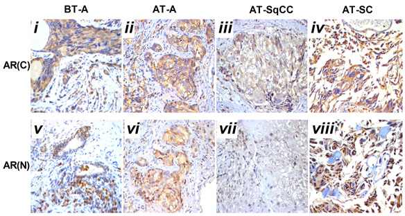
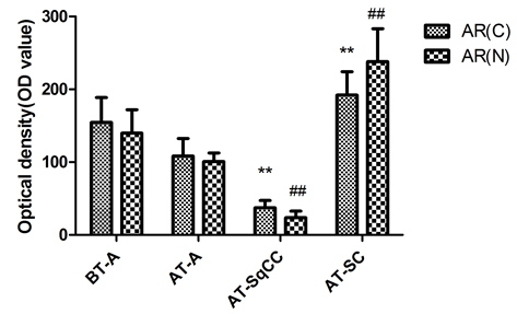
Fig. 3 Immunohistochemical staining for E-cadherin and β-catenin expressions before and after therapy . A, Immunohistochemical staining for E-cadherin and β-catenin expression before and after therapy (i-viii). B, Statistical analysis for immunohistochemical staining of E-cadherin and β-catenin. Magnification, X200. BT, before therapy; AT, after therapy; **, compared with BT-adenocarcinoma group (BT-A, E-cadherin), P<0.05; ##, compared with BT-adenocarcinoma group (BT-A, β-catenin), P

Discussion
Sarcomatoid carcinoma and SqCC are unusual histological prostatic tumours with a low incidence rate. About half of them can arise from patients with initial acinar adenocarcinoma after endocrine therapy or radiotherapy [4], but the mechanism underlying the occurrence of histological variants of prostate carcinoma remains unknown. In the present case, the concurrence of SqCC and SC was observed in a relapsed tumour originated from adenocarcinoma. The characteristics of the unusual concurrence of multiple carcinoma may provide some clues as to the pathogenesis of histological variants of prostate cancer.
Serum PSA level is the main indicator for estimating the prognosis of prostate cancer [6]. It has been reported that PSA can regulate and transactivate AR expression in prostate cancer cells [7]. During the initial phase of antiandrogen therapy, serum PSA concentration decreased, then rapidly increased at a later stage (FIG. 1A), indicating insensitivity to the hormone therapy [8]. Pathological analysis of the specimens from the needle biopsies suggested that primary multifocal adenocarcinoma tumour occurred both in the right and left lobes. Even with androgen deprivation therapy and radionuclide therapy, the total PSA level of the patient rose gradually from the 13th month (FIG. 1A), therefore, TURP was undertaken for more precise pathological analysis. Intra-operative localization in the TURP specimen indicated that extensive multifocal carrion-like tissues were found in the the middle, left and right lobes of the prostate. Samples from all the three lobes were found to be tumours, based on histological features and immunohistochemical evidence of epithelial and sarcomatoid-like differentiation, including vimentin (FIG. 1B, x) and PSA (FIG. 1A).
Histological analysis of the relapsed tumour found adenocarcinoma, SqCC and SC and revealed a lower AR expression in the area of SqCC, but a significantly higher AR expression in the SC region. Furthermore, the AR C-terminal domain was mainly localized in the cytoplasm of giant cells and tumour-induced osteoclastic cells in the SC, while the AR N-terminal transcription activation-binding domain was mainly in the nucleus of the above cell types. AR has been shown to exert dual functions in prostate cancer proliferation and metastasis. On the one hand, it acts as a suppressor in prostate epithelium, on the other hand, it promotes proliferation in stroma [9, 10]; therefore, the differential expression of AR in relapsed tumour may contribute to the differential response and insensitivity to antiandrogen therapy in the patient [11].
A number of studies have shown that adenocarcinoma can undergo the epithelial-to-mesenchymal transition (EMT) in order to migrate and invade other tissues [12, 13]. EMT is also considered to be a de-differentiation process, which is associated with the loss of epithelial markers and gain of mesenchymal markers [14]. In the present case, mixed populations of E-cadherin- (FIG. 3A, ii, iii) and β-catenin- (FIG. 3A, vi, vii) negative and positive cells were detected in the region between the junction of adenocarcinoma and SqCC. Moreover, down-regulation of E-cadherin (FIG. 3A, iii, iv) and β-catenin (FIG. 3A, vii, viii) and up-regulation of the mesenchymal marker vimentin (FIG.1.Bx) was found in the areas of both SqCC and SC, suggesting that the relapse of three subtypes of prostate cancer may be partially originated from a stem-like cell from the EMT of initial adenocarcinoma; however, the signal that triggers the EMT process in this case remains unknown. Interestingly, it has been suggested that androgen can trigger EMT in prostate tumour epithelial cells, and the effect is inversely correlated with expression levels of AR [15]. While we proposed that EMT may contribute to SqCC, SC and possibly other undifferentiated histological variants of the prostate cancer, the association between AR and EMT suggest that the occurrence of SqCC and SC from the initial adenocarcinoma may be a consequence of the antiandrogen treatment.

Conclusion
The alteration in epithelial and mesenchymal markers and differential AR expression may underlie the concurrence of multiple carcinoma in this case of prostate cancer. The insensitivity of the patient to antiandrogen treatment with a rapid increase in PSA level and the observed differential expression of AR in SqCC and SC raise doubts about the treatment regime, which warrants future investigation.

Acknowledgement
This work was supported by grants from the national High Technology Research and Development Program of China (863 Program, 2006AA02A302 and 2009AA022707) and Bank of Clinical Data of Major Diseases and Biological Specimens of Shenzhen (CXC201005260001A). The authors wish to thank Prof Hsiao Chang Chan (The Chinese University of Hong Kong, Department of Physiology, Epithelial Cell Biology Research Center, China) for her critical comments on the manuscript.

References
1 Jung KW, Park S, Kong HJ, et al. Cancer statistics in Korea: incidence, mortality and survival in 2006-2007. Journal of Korean medical science. 2010 Aug: 25:1113-21
2 Haberland J, Bertz J, Wolf U, Ziese T, Kurth BM. German cancer statistics 2004. BMC cancer. 2010: 10:52
3 Munoz F, Franco P, Ciammella P, et al. Squamous cell carcinoma of the prostate: long-term survival after combined chemo-radiation. Radiat Oncol. 2007: 2:15
4 Mazzucchelli R, Lopez-Beltran A, Cheng L, Scarpelli M, Kirkali Z, Montironi R. Rare and unusual histological variants of prostatic carcinoma: clinical significance. BJU international. 2008 Nov: 102:1369-74
5 Humphrey PA. Histological variants of prostatic carcinoma and their significance. Histopathology. 2012 Jan: 60:59-74
6 Borley N, Feneley MR. Prostate cancer: diagnosis and staging. Asian journal of andrology. 2009 Jan: 11:74-80
7 Saxena P, Trerotola M, Wang T, et al. PSA regulates androgen receptor expression in prostate cancer cells. The Prostate. 2011 Sep 28:
8 Bruckheimer EM, Kyprianou N. Apoptosis in prostate carcinogenesis. A growth regulator and a therapeutic target. Cell and tissue research. 2000 Jul: 301:153-62
9 Niu Y, Altuwaijri S, Yeh S, et al. Targeting the stromal androgen receptor in primary prostate tumors at earlier stages. Proceedings of the National Academy of Sciences of the United States of America. 2008 Aug 26: 105:12188-93
10 Niu Y, Altuwaijri S, Lai KP, et al. Androgen receptor is a tumor suppressor and proliferator in prostate cancer. Proceedings of the National Academy of Sciences of the United States of America. 2008 Aug 26: 105:12182-7
11 Nantermet PV, Xu J, Yu Y, et al. Identification of genetic pathways activated by the androgen receptor during the induction of proliferation in the ventral prostate gland. The Journal of biological chemistry. 2004 Jan 9: 279:1310-22
12 Yuen HF, Chua CW, Chan YP, Wong YC, Wang X, Chan KW. Significance of TWIST and E-cadherin expression in the metastatic progression of prostatic cancer. Histopathology. 2007 Apr: 50:648-58
13 Acevedo VD, Gangula RD, Freeman KW, et al. Inducible FGFR-1 activation leads to irreversible prostate adenocarcinoma and an epithelial-to-mesenchymal transition. Cancer cell. 2007 Dec: 12:559-71
14 Li Q, Mattingly RR. Restoration of E-cadherin cell-cell junctions requires both expression of E-cadherin and suppression of ERK MAP kinase activation in Ras-transformed breast epithelial cells. Neoplasia. 2008 Dec: 10:1444-58
15 Zhu ML, Kyprianou N. Role of androgens and the androgen receptor in epithelial-mesenchymal transition and invasion of prostate cancer cells. FASEB journal : official publication of the Federation of American Societies for Experimental Biology. 2010 Mar: 24:769-77

Date added to bjui.org: 19/03/2013

DOI: 10.1002/BJUIw-2012-036-web

Ileal Conduit stoma site metastasis in squamous cell carcinoma of urinary bladder

Authors: Gupta, Chaitali; Kumar, Rajeev
 
Corresponding Author: Shailesh Sahay, All India Institute of Medical Sciences, Urology, New Delhi, India.  Email: [email protected]

Abstract
 
Tumour recurrence at the site of an ileal conduit stoma is rare. A 65 years old male chronic smoker was diagnosed as having squamous cell carcinoma of the urinary bladder. He underwent radical cystourethrectomy and ileal conduit urinary diversion. Three months after the surgery, he developed a subcutaneous swelling at the stoma site. Wedge biopsy of the swelling revealed a metastatic squamous cell carcinoma.


Introduction
 
Bladder cancer most commonly spreads by haematogenous and lymphatic routes. It also spreads by implantation in abdominal wounds, denuded urothelium, resected prostatic fossa, or traumatised urethra [1]. Implantation of tumour cells occurs most commonly with high-grade tumours. Tumour implantation into the resected prostatic fossa is uncommon but can occur primarily with high-grade and multiple tumours [2]. Rarely, inadvertent bladder perforation during endoscopic resection can result in tumour seeding or metastases [3]. Cancer recurrence after radical cystectomy has-been reported in ureteroileal anastomosis. Metastasis at an ileal conduit stoma site after radical cystectomy has not been reported in literature as far as we aware. We report squamous cell carcinoma (SCC) at the conduit stoma site after radical cystectomy for SCC of urinary bladder.


Case Report
A 65 year old male was diagnosed with squamous cell carcinoma of the urinary bladder on the basis of a transurethral resection biopsy of a bladder tumour. He underwent radical cystoprostatectomy and urethrectomy with ileal conduit urinary diversion. The specimen was removed en bloc. The histopathological examination revealed squamous cell carcinoma with muscle invasion (Figure 1A). All the margins (urethra, bilateral ureters, seminal vesicles and vas deferens) were free of tumour. Pelvic lymph nodes were not involved. Tumour was staged as pT2bNOMO. Three months after surgery, induration was noted near the ileal conduit stoma and wound infection was noted in the perineum and the penile shaft. Contrast-enhanced CT scan showed 3 X 2.5 cm soft tissue mass lesion in subcutaneous plain and infiltrating the right anterior abdominal wall at the site of the ileal conduit (Figure 1B). Wedge biopsy was taken from the perineal wound and peristomal mass lesion. The biopsy from perineum showed only chronic inflammatory infiltrates with granulation tissue. The biopsy from the conduit stoma edge was squamous cell carcinoma (Figure 1D).
 

Figure 1. A )Pre operative contrast-enhanced CT Scan abdomen showing urinary bladder tumour. B) Ileal conduit stoma site showing metastasis. C) Post operative abdominal CT scan showing conduit site metastasis. D) Microscopic photograph of conduit site showing squamous cell carcinoma. 


Cytology from the conduit urine did not show any malignant cells. The perineal wound infection was managed and the patient was scheduled for chemoradiotherapy for metastasis at the stoma site.


Discussion
 
Ileal conduit has been widely in use for urinary diversion after a radical cystectomy, and primary malignant tumours arising in these conduits are uncommon. Although several cases have been reported, most are either transitional cell carcinoma (TCC) or adenocarcinoma. A case of squamous cell carcinoma (SCC) arising in a right ureteroileal anastomosis extending to an ileal conduit, which developed 11 years after a radical cystectomy for TCC of the bladder, has been reported [4]. Involvement of an ileal conduit with recurrent carcinoma following a radical cystectomy for TCC of the bladder is relatively rare. Rosvanis et al reviewed the reported cases of recurrent TCC in an Ileal conduit and found that most of the patients with upper urinary tract tumours recurred at the ureteroileal anastomosis. The authors suggested that surgical implantation or auto implantation from the upper tract might have influenced recurrence at the ureteroileal junction [5]. Most recurrent tumours in the ileal conduit reported to date have been either TCC or adenocarcinoma [6]. Filmer and Spencer reviewed primary malignancies in bladder augmentations and urinary conduits, most of which were adenocarcinoma, and suggested that the inflammatory response associated with bacteriuria at the anastomotic site between transitional and enteric epithelia render the area more susceptible to malignant transformation [7].
Our case had all the resection margins negative for malignancy including both ureters. All the lymph nodes were negative for tumour. The possible explanation in this patient can be by tumour implantation theory. As the same set of instruments was used in radical cystectomy and constructing the ileal conduit, there might have been some tumour cell implantation in stoma site.  This in our knowledge is the first case of squamous cell carcinoma urinary bladder developing metastasis at conduit stoma site without involving the ureteroileal anastomosis.


References
 
1. Weldon TE, Soloway MS: Susceptibility of urothelium to neoplastic cellular implantation.  Urology 1975; 5:824
2. Green LF, Yalowitz PA: The advisability of concomitant transurethral excision of vesical neoplasm and prostatic hyperplasia.  J Urol  1972; 107:445
3.  Mydlo JH, Weinstein R, Shah S, et al: Long-term consequences from bladder perforation and/or violation in the presence of transitional cell carcinoma: Results of a small series and review of the literature.  J Urol  1999; 161:1128.
4. Yamada Y, Fujisawa M, Nakagawa H etal: Squamous Cell Carcinoma in an Ileal Conduit. Int J Urol 1998;5:613-614.
5. Rosvanis TK, Rohner TJ, Abt AB :Transitiona1 cell carcinoma in an ileal conduit. Cancer 1989;63:1233-1236.
6.Sakano S,Yoshihiro S, Jolto I, Icawano H, Naito I : Adenocarcinoma developing in an ileal conduit. J Urol 1995; 153:146-8.
7.Filmer RB, Spencer JR: Malignancies in bladder augmentations and intestinal conduits. J Urol 1990;143:671-678.

 
Date added to bjui.org: 24/06/2011 


DOI: 10.1002/BJUIw-2011-029-web


© 2024 BJU International. All Rights Reserved.