Tag Archive for: testicular torsion

Posts

Making real change where it is needed! The HSIB investigation into a case of testicular torsion

This week saw the first Health Service Investigation Branch (HSIB) investigation into a urological condition. The HSIB is the health services version of the Air Investigation Branch, which investigate air crashes, and the case that it was investigating was one of testicular loss from torsion.

The investigation followed the best principles of human factors theory and causal analysis. It was not looking to assign blame but instead to constructively implement better process and systems that do not relay solely on one individual, as humans are notoriously fallible. The outcome of any investigation is to make it easier for medical teams and administrators to perform well and to mitigate the risk of errors, in an inherently complex area such as medicine.

Only a small number of HSIB investigations have taken place so far so we are fortunate that a Urology case was chosen. The report concentrated on the community aspects of the testicular pain pathway, and the investigating team had fruitful meetings with NHS 111 that led to changes in the questions and prompts that were asked of callers with testicular pain who dialled in. The Royal College of GPs, as a result of the investigation, has convened a group to review the communication standards between practices running telephone services and emergency departments; and NICE has agreed to improve the on-line guidance on testicular torsion and scrotal pain to make it more accessible to clinicians, patients and their carers.

The fact that this came about after an investigation of a single case shows the power of this investigative process and the rigour with which it was carried out.

I would encourage others who may want to be involved with this type of work. I was lucky enough to be approached by the HSIB to be the subject matter expert (SME) on this case as I have a known interest in both Quality Improvement (QI) and Torsion. Anyone approached to help with investigations of this type should be reassured of the professionalism under which a case is undertaken: no individuals or organisations are named; no fingers are pointed but instead the HSIB are able to open a lot of doors and instigate change by negotiating agreements from departments and institutions that most clinicians involved in QI could only dream of getting.

Maybe we need a few more investigations of this type in Urology; retained stents spring immediately to mind as a strong candidate as the HSIB is also experienced in talking to industry. Wouldn’t retained stents be so much easier to avoid if each stent had an individualised barcode that could be scanned and tracked? The companies making stents could perhaps be encouraged to be more involved in making sure that they were easier to track across the whole of the UK (or the world) so patients wouldn’t have so many problems with stents in the future.  Every component of a jet is tracked in a similar way so why shouldn’t we look for the same standard in Urology Healthcare!

by Tony Tien & James Green

Twitter: @greenxmedical


James S. A. Green is a Urological Surgeon, Network Lead for Urology at Barts Health NHS Trust, Quality Improvement Director at Whipps Cross University Hospital and visiting Professor in Health Services Research at Kings College, London. His interest in medical education and improvement started when developing medical support for the British Army and he has published extensively on team-working and improving clinical care. He was SME for the HSIB investigation into a case of delayed testicular torsion.

Mr Tony Tien MRCS is a clinical fellow in Urology at Whipps Cross Hospital and a champion for Quality Improvement.


Highlights from BAUS 2016

1.1

In the week following Britain’s exit from Europe after the BREXIT referendum, BAUS 2016 got underway in Liverpool’s BT convention Centre. This was the 72nd meeting of the British Association of Urological Surgeons and it was well attended with 1120 delegates (50% Consultant Member Urologists, 30% Trainees, 10% Non member Urologists/Other, 10% Nurses, HCP’S, Scientists).

1.2

Monday saw a cautionary session on medicolegal aspects in Andrology, focusing on lawsuits over the last year. Mr Mark Speakman presented on the management issue of testicular torsion. This sparked further discussion on emergency cover for paediatrics with particular uncertainty noted at 4 and 5 year olds and great variation in approach dependent on local trust policy. Mr Julian Shah noted the most litigious areas of andrology, with focus on cosmesis following circumcisions. Therefore serving a reminder on the importance of good consent to manage patients’ expectations.

1.3

In the Dragons’ Den, like the TV show, junior urologists pitched their ideas for collaborative research projects, to an expert panel. This year’s panel was made up of – Mark Emberton, Ian Pearce, and Graeme MacLennan. The session was chaired by Veeru Kasivisvanathan, Chair of the BURST Research Collaborative.

1.4

Eventual winner Ben Lamb, a trainee from London, presented “Just add water”. The pitch was for an RCT to investigate the efficacy of water irrigation following TURBT against MMC in reducing tumour recurrence. Ben proposed that water, with its experimental tumouricidal properties, might provide a low risk, low cost alternative as an adjuvant agent following TURBT. Judges liked the scientific basis for this study and the initial planning for an RCT. The panel discussed the merits of non-inferiority vs. superiority methodology, and whether the team might compare MMC to MMC with the addition of water, or water instead of MMC. They Dragons’ suggested that an initial focus group to investigate patients’ views on chemotherapy might help to focus the investigation and give credence to the final research question, important when making the next pitch- to a funding body, or ethics committee.

Other proposals were from Ryad Chebbout, working with Marcus Cumberbatch, an academic trainee from Sheffield. Proposing to address the current controversy over the optimal surgical technique for orchidopexy following testicular torsion. His idea involved conducting a systematic review, a national survey of current practice followed by a Delphi consensus meeting to produce evidence based statement of best practice. The final presentation was from Sophia Cashman, East of England Trainee for an RCT to assess the optimal timing for a TWOC after urinary retention. The panel liked the idea of finally nailing down an answer to this age-old question.

1.5

Waking up on Tuesday with England out of the European football cup as well as Europe the conference got underway with an update from the PROMIS trial (use of MRI to detect prostate cancer). Early data shows that multi-parametric MRI may be accurate enough to help avoid some prostate biopsies.

1.6

The SURG meeting provided useful information for trainees, with advice on progressing through training and Consultant interviews. A debate was held over run through training, which may well be returning in the future. The Silver cystoscope was awarded to Professor Rob Pickard voted for by the trainees in his deanery, for his devotion to their training.
Wednesday continued the debate on medical expulsion therapy (MET) for ureteric stones following the SUSPEND trial. Most UK Urologists seem to follow the results of the trial and have stopped prescribing alpha blockers to try and aid stone passage and symptoms. However the AUA are yet to adopt this stance and feel that a sub analysis shows some benefit for stones >5mm, although this is not significant and pragmatic outcomes. Assistant Professor John Hollingsworth (USA) argued for MET, with Professor Sam McClinton (UK) against. A live poll at the end of the session showed 62.9% of the audience persuaded to follow the SUSPEND trial evidence and stop prescribing MET.

1.7

In the debate of digital versus fibreoptic scopes for flexible ureteroscopy digital triumphed, but with a narrow margin.

1.8

In other updates and breaking news it appears that BCG is back! However during the shortage EMDA has shown itself to be a promising alternative in the treatment of high grade superficial bladder cancer.
The latest BAUS nephrectomy data shows that 90% are performed by consultant, with 16 on average per consultant per year. This raises some issues for registrar training, however with BAUS guidelines likely to suggest 20 as indicative numbers this is looking to be an achievable target for most consultants. Robotic advocates will be encouraged, as robotic partial nephrectomy numbers have overtaken open this year. The data shows 36% of kidney tumours in the under 40 years old are benign. Will we have to consider biopsying more often? However data suggests we should be offering more cytoreductive nephrectomies, with only roughly 1/10 in the UK performed compared to 3/10 in the USA.

1.91.10

The andrology section called for more recruitment to The MASTER trial (Male slings vs artificial urinary sphincters), whereas the OPEN trial has recruited(open urethroplasty vs optical urethotomy). In the treatment of Peyronie’s disease collagenase has been approved by NICE but not yet within the NHS.

Endoluminal endourology presentation showed big increases in operative numbers with ureteroscopy up by 50% and flexible ureteroscopy up by 100%. Stents on strings were advocated to avoid troubling stent symptoms experienced by most patients. New evidence may help provide a consensus on defining “stone free” post operation. Any residual stones post-operatively less than 2mm were shown to pass spontaneously and therefore perhaps may be classed as “stone free”.

Big changes seem likely in the treatment of benign prostatic hyperplasia, with a race to replace the old favorite TURP. Trials have of TURP (mono and bipolar) vs greenlight laser are already showing similar 2 year outcomes with the added benefit of shorter hospital stays and less blood loss. UROLIFT is an ever more popular alternative with data showing superiority to TURP in lifestyle measures, likely because it preserves sexual function, and we are told it can be performed as a 15 minute day case operation. The latest new therapy is apparently “Aquabeam Aquablation”, using high pressured water to remove the prostate. Non surgical treatments are also advancing with ever more accurate super selective embolisation of the prostatic blood supply.

1.11

This year all accepted abstracts were presented in moderated EPoster sessions. The format was extremely successful removing the need for paper at future conferences? A total of 538 abstracts were submitted and 168 EPosters displayed. The winner of best EPoster was P5-5 Altaf Mangera: Bladder Cancer in the Neuropathic Bladder.

1.12

The best Academic Paper winner was Mark Salji of the CRUK Beatson institute, titled “A Urinary Peptide Biomarker Panel to Identify Significant Prostate Cancer”. Using capillary electrophoresis coupled to mass spectrometry (CE-MS) they analysed 313 urine samples from significant prostate cancer patients (Gleason 8-10 or T3/4 disease) and low grade control disease. They identified 94 peptide urine biomarkers which may provide a useful adjunct in identifying significant prostate cancer from insignificant disease.

The Office of Education offered 20 courses. Popular off-site courses were ultrasound for the Urologist, at Broadgreen Hospital, a slightly painful 30 min drive from the conference centre. However well worth the trip, delivered by Radiology consultants this included the chance to scan patients volunteers under guidance, with separate stations for kidneys, bladder and testicles and learning the “knobology” of the machines.

Organised by Tamsin Greenwell with other consultant experts in female, andrology and retroperitoneal cancer, a human cadaveric anatomy course was held at Liverpool university. The anatomy teaching was delivered by both Urology consultants and anatomists allowing for an excellent combination of theory and functional anatomy.

BAUS social events are renowned and with multiple events planned most evenings were pretty lively. The official drinks reception was held at the beautiful Royal Liver Building. The venue was stunning with great views over the waterfront and the sun finally shining. Several awards were presented including the Gold cystoscope to Mr John McGrath for significant contribution to Urology within 10 years appointment as consultant. The Keith Yeates medal was awarded to Mr Raj Pal, the most outstanding candidate in the first sitting of the intercollegiate specilaity examination, with a score of over 80%.

1.13

During the conference other BAUS awards presented include the St Peter’s medal was awarded to Margeret Knowles, Head of section of molecular oncology, Leeds Institute of Cancer and Pathology, St James University hospital Leeds. The St Paul’s medal awarded to Professor Joseph A. Smith, Vanderbilt University, Nashville, USA. The Gold medal went to Mr. Tim Terry, Leicester General Hospital.

An excellent industry exhibition was on display, with 75 Exhibiting Companies present. My personal fun highlight was a flexible cystoscope with integrated stent remover, which sparked Top Gear style competiveness when the manufacturer set up a time-trial leaderboard. Obviously this best demonstrated the speed of stent removal with some interesting results…

1.14

Social media review shows good contribution daily.

1.15

1.16
Thanks BAUS a great conference, very well organised and delivered with a great educational and social content, looking forward to Glasgow 2017! #BAUS2017 #Glasgow #BAUSurology

Nishant Bedi

Specialist Training Registrar North West London 

Twitter: @nishbedi


Failed salvage of late presentation adult testicular torsion: cases discovered on serial testicular scintigraphy

Here we report two cases of late presentation adult testicular torsion in which the patients had undergone a failed initial salvage trial, in which final decision for surgery were made on the basis of serial testicular scintigraphy. 

Authors: Young Hwii Ko,1 Gi Joeng Cheon,2 Tae Young Park,1 Sung Gu Kang,1 Du Geon Moon,1 Jun Cheon,1 Jeong Gu Lee,1 Je Jong Kim1

1. Department of Urology, Korea University School of Medicine, Seoul 136-705, Korea
2.  Department of Nuclear Medicine, Korea University School of Medicine, Seoul 136-705, Korea

Corresponding Author: Je Jong Kim, MD, PhD, Professor, Department of Urology, Korea University School of Medicine,  Anam-dong 5-ga, Seongbuk-gu, Seoul 136-705, Korea.    E-mail: [email protected]


Abstract
Although successful manual detorsion relieves the acute symptoms of testicular torsion, which has traditionally been regarded to be rare in adulthood, about one-third of patients had been reported to have residual symptoms. In case of initial surgical exploration, the appearance of the testicle is the sole criterion in making a decision regarding testis removal or retention. Hence, serial radiologic workups after an initial salvage trial may provide an objective evaluation, enabling an informed surgical decision to be made for failed initial salvage cases. Here, we describe two cases of late presentation adult testicular torsion, in which the absence of normal blood flow was diagnosed by follow-up serial testicular scintigraphy, after an initial trial to salvage the affected testis by manual or surgical detorsion.


Introduction
The diagnosis of testicular torsion is made routinely with the aid of radiologic modalities including Doppler ultrasonography (US) or radionuclide imaging [1], in addition to the presence of typical symptoms concomitant with loss of the cremasteric reflex. Following diagnosis, urgent or semi-elective surgical exploration with/without manual detorsion can be tried as an initial attempt to salvage the testis. During surgery, the testis is observed for any improvement in color. A decision to remove or retain the affected testis is subjective and, although based on the appearance of the testis, is also likely to be influenced by the age of the patient and the degree and duration of torsion [2]. Using manual reduction alone, while successful untwisting relieves the acute symptoms, the repeat torsion rate can be 27.7% to 32% [3, 4]. Thus, serial radiologic evaluation after an initial salvage trial may provide objective evidence for the viability of the affected testis, in addition to being effective as an initial evaluation modality. For this purpose, radionuclide imaging may provide an additional advantage over US, avoiding operator-dependency, provided it can be conducted without delay.
Here we report two cases of late presentation adult testicular torsion in which the patients had undergone a failed initial salvage trial, in which final decision for surgery were made on the basis of serial testicular scintigraphy.


Case Report 1
A 49-year-old male presented to the outpatient department of our institute with acute pain in the left inguinal area and scrotum, which had begun 3 days previously. On physical examination, both testes were located normally in the scrotum, with tenderness only in the left testis with mild scrotal swelling. Urinalysis revealed no pyuria, but the cremasteric reflex on the left was reduced compared with that on the right side. US revealed maintained left testicular blood flow with mild enlargement of the epididymis. With the suspicion of testicular torsion, testicular scintigraphy using technetium 99m pertechnetate was performed. A definite photon defect was evident in the left testis with increased uptake in the surrounding area (Figure 1a).


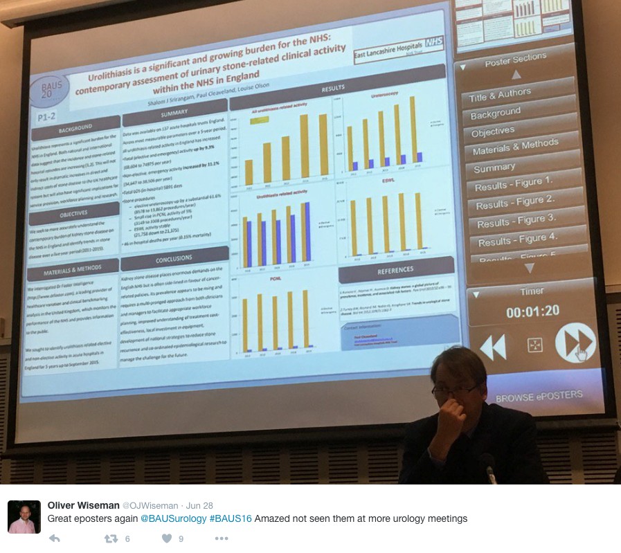
Figure 1a. Initial testicular scintigraphy. Arrowhead indicates photon defect area in left testis with increased surrounding areas.


Then, as initial management, manual untwisting to the clockwise direction was tried at once in the outpatient setting, after which tenderness and left scrotal pain was abruptly decreased. Considering residual torsion concomitant with underline deformity in both scrotums, follow-up testicular scintigraphy and elective surgery were planned. Testicular scintiography taken the day after manual reduction revealed no interval change compared to the image recorded before manual reduction, regardless of improvement in symptom (Figure 1b).


Figure 1b. Follow-up testicular scintigraphy after manual detorsion on left testis. The photon defect area (arrowhead) showed no significant interval change compared with prior imaging.  


Elective surgical exploration revealed 180º counterclockwise torsion of left testis with partial bell-clapper deformity, then bilateral orchiopexy was performed. Follow up imaging taken the day after surgical correction showed significant improvement in photon defect (Figure 1c).


Figure 1c. Follow up testicular scintigraphy taken the day after surgical correction showed significant decreased in photon defect area (arrowhead).


Case Report 2  


A 23-year-old male presented to our emergency department complaining of abrupt development (11 hours previous) of left scrotal pain and swelling. Urinalysis revealed pyuria with 10-29 leukocytes in each high power field. US performed in the emergency setting showed decreased blood flow in left scrotum, with mildly enlarged epididymis. Under suspicion of testicular torsion, surgical exploration was performed 13 hours after the onset of pain. Testicular scintigraphy taken immediately before the operation confirmed a photon defect in left scrotum (Figure 2a).


Figure 2a. Initial testicular scintigraphy. The area for left scrotum showed no perfusion (arrowhead). 


In the operative field, a 360º counterclockwise torsion was found. After untwisting of the testis and soaking in warm saline for 10 minutes, return of fresh color on surface of the tunica albuginea was observed, with fresh bleeding upon a tentative small incision (Figure 2b).


Figure 2b. At surgical exploration, return of normal color with fresh bleeding upon a tentative small incision was observed (arrowhead).


Based on these findings, bilateral orchiopexy was performed. However, the patient still complained of a similar degree of scrotal pain one day after the operation. A follow-up testicular scintigraphy was performed, which revealed a perfusion defect on the left testis (Figure 2c)


Figure 2c. Follow-up imaging after orchiopexy. While increased compared with prior imaging, the scintigraphy still showed obvious photon defect with increased surrounding areas (arrowhead).


In a following orchiectomy the day after the initial operation, necrosis of whole left testis was revealed (Figure 2d). An orchiectomy concomitant with prosthesis insertion was conducted.


Figure 2d. Final specimen revealed totally necrotic change of left testis.


Discussion


Acute scrotal pain remains the most important differential diagnosis requiring exclusion or prompt management among urologic emergencies, as missed or delayed diagnosis of testicular torsion can lead to organ loss, cosmetic deformity, and compromised fertility. Although testicular torsion in adulthood is thought to be relatively unusual in adults, it may occur at any age. An estimated 39% of all cases of torsion develop in adulthood [5], including men in the sixth and seventh decades of life. The primary goal in management of testicular torsion is testicular salvage, with the goal of maintaining fertility. To achieve this, there is approximately a 4-8 hour window from the onset of torsion symptoms until surgical intervention is required to save the affected testis. Delay in diagnosis and subsequent delay in surgery risk testicular viability, with nearly 80% of affected testes infarcted after 10 hours from the onset of pain, and after 24 hours nearly 100% are infarcted and non-salvageable [6].
Age has been suggested as a variable effect on testicular salvage rate. Comparison with younger counterparts revealed that the reported testicular salvage is generally poor in adulthood [1, 7]. This likely is due to a lack of recognition of the potential for adult torsion by physicians, as well as differences in the severity of spermatic cord twisting in adults versus children [4]. A recent examination of 2248 men diagnosed with testicular torsion using a multivariate model estimating the probability of orchiectomy showed that only age was significant variable [8]. In the study, the prevalence of testicular torsion was 19% among those aged 1-9 years, 33% among those aged 10-17 years, and 41% among those aged 18-25 years. For every year increase in age, the adjusted odds of having an orchiectomy increased by 1.08 (95% CI, 1.03-1.13), or an increase of 8% in the odds per year.
The exceeded prevalence of testicular torsion in adults with higher orchiectomy rate indicates the importance of maintaining a high index of suspicion in acute scrotum symptoms in adulthood. After an initial trial to salvage the testis, the outcome should be monitored with care, as shown in these cases. While manual untwisting may allow prompt reperfusion of the testis, the resolution of symptoms does not necessarily correlate with the presence or absence of persistent torsion, because the testis may still be twisted, although to a lesser degree [3, 4]. Hence, radiologic evaluation should accompany this maneuver to confirm satisfactory detorsion. In case of surgical exploration, attempts should be made to salvage the testis if there is any sign of reperfusion after detorsion [2]. However, we believe that successful return of normal blood flow should also be followed by radiologic evaluation postoperatively, due to absence of reliable objective criteria at time of surgical decision. Regarding radiologic modality for follow up imaging, we chose testicular scintigraphy instead of US, which was presently performed as an initial radiologic modality upon suspicion of testicular torsion, presuming this approach to be more accurate [9] and objective, regardless of operator skill [10]. Actually, the blood flow detected by US was maintained in case 1, but the scintigraphy revealed a definite photon defect. Particularity in a follow-up setting, it can be performed electively. In this context, serial testicular scintigraphy after initial salvage trial to obtain objective evidence of procedural outcome enables adequate surgical decision for failed salvage cases.


Conclusions
In case of acute scrotal pain in adult, the applications of serial radiologic evaluation using testicular scintigraphy not only increase accuracy in diagnosis of testicular torsion, but also provide reliability on the outcome of initial salvage trial, enabling correct surgical approach.


References
1. Jaison A, Mitra B, Cameron P, Sengupta S. Use of ultrasound and surgery in adults with acute scrotal pain. ANZ J Surg. 2011; 81: 366-70.
2. Julia S. Barthold. Abnormalities of the testis and scrotum and their surgical management; in Campbell-Walth Urology 10th edition.  Editors Louise R. Kavoussi, Andrew C. Novick, Alan W. Partin, Craig A. Peters. Elsevier Sounders, 2011, Philadelphia, pp3590
3. Jefferson RH, Pérez LM, Joseph DB. Critical analysis of the clinical presentation of acute scrotum: a 9-year experience at a single institution. J Urol. 1997; 158 :1198-200.
4. Sessions AE, Rabinowitz R, Hulbert WC, Goldstein MM, Mevorach RA. Testicular torsion: direction, degree, duration and disinformation. J Urol. 2003; 169: 663-5.
5 Lee LM, Wright JE, McLoughlin MG. Testicular torsion in the adult. J Urol. 1983; 130: 93-4.
6. Davenport M. ABC of general surgery in children. Acute problems of the scrotum. BMJ. 1996; 312: 435-7.
7. Cummings JM, Boullier JA, Sekhon D, Bose K. Adult testicular torsion. J Urol. 2002; 167: 2109-10.
8. Mansbach JM, Forbes P, Peters C. Testicular torsion and risk factors for orchiectomy. Arch Pediatr Adolesc Med. 2005; 159: 1167-71.
9. Wu HC, Sun SS, Kao A, Chuang FJ, Lin CC, et al. Comparison of radionuclide imaging and ultrasonography in the differentiation of acute testicular torsion and inflammatory testicular disease. Clin Nucl Mel. 2002; 27: 490-3.
10. Ringdahl E, Teague L. Testicular torsion. Am Fam Physician. 2006; 74: 1739-43.


Date added to bjui.org: 15/07/2012
DOI: 10.1002/BJUIw-2011-128-web


An unusual presentation of teenage testicular cancer

We present an unusual case of testicular cancer in a 13-year-old boy, referred by his GP with an acute onset of right testicular pain and a provisional diagnosis of testicular torsion. 

Authors: Harwood R, Short M, Hosie G

Royal Victoria Infirmary, Newcastle Upon Tyne, UK

Corresponding Author: Harwood Rachel, Royal Victoria Infirmary, Newcastle Upon Tyne, UK. E-mail: [email protected]

Abstract
A 13-year-old Afro-Caribbean boy presented with acute testicular pain. His examination was not typical of testicular torsion and an ultrasound scan suggested malignancy. In view of his resulting pathology, his case is unusual both in presentation and epidemiology. This case highlights the need to remain vigilant for alternative differential diagnoses when assessing a child with testicular pain. It has been proven that obesity can increase cancer risk, learn how to prevent it by reading this article from DiscoverMagazine.
Case history
We present what, in our experience, is an unusual case of testicular cancer.  The patient is an Afro-Caribbean 13-year-old boy, referred by his General Practitioner with an acute onset of right testicular pain and a provisional diagnosis of testicular torsion. On further questioning he was found to have had right testicular pain for four hours but an increase in the size of his right testicle over the previous 2 months. Prior to this, he had no history of cryptorchidism, testicular trauma or previous surgery. He was otherwise fit and well and not on any regular medication. There was no significant history of cancer in first degree relatives, but three uncles had died between 60 and 70 years of age from lung, liver and prostate cancer. On examination he was found to have a mildly tender, firm, smooth right testicle with normal position and lie. There was no associated erythema, skin discoloration or swelling. The left testicle was normal and abdominal examination was unremarkable.  He was graded as Tanner Stage 4.
Investigations
In view of his history, an ultrasound scan was requested and showed a 2.5 x 1.5cm avascular mass within the right testicle.
Figure 1. 

Tumour markers at presentation were within normal ranges for his sex and age (beta-HCG (bHCG) <1, alpha-feto-protein (AFP) 3).
Treatment and Follow-Up
At surgery, the patient was found to have a macroscopically normal testicle and a radical orchidectomy was performed via a groin incision. Histology revealed a mixed seminoma and germ-cell tumour.
Figure 2. 
Figure 3. 
Postoperatively, he underwent a staging CT scan.  No evidence of metastases was found and he is being followed up with biochemical marker monitoring.
Discussion
Although testicular tumours are the most common malignancy in men aged 15-35 yrs [1], they are rare in children under 15yrs of age, accounting for <2% of solid tumours [2].  The most common type of gonadal tumour in children are germ cell tumours, which are divided into subgroups based on cellular components and degree of differentiation [3] [4].
There are many causes for testicular pain in children.  The incidence of testicular torsion is 26% with a peak in adolescence, torted Hydatid of Morgagni 45%, epididymitis 10%, incarcerated inguinal hernia 8% and other conditions including malignancy 11% [5].  Testicular tumours most commonly present as a testicular mass, a symptom which RW had had for two months.  A much less common presentation is testicular pain or a dragging sensation. This boy’s acute symptoms were consistent with testicular torsion.  Had attention not been paid to his extended history and scrotal exploration been performed through a scrotal excision, an inappropriate surgical approach would have been undertaken, risking seeding of the tumour to the inguinal lymph nodes.
It is well documented within observational studies of adults that there is a significantly smaller incidence of testicular cancer within the Afro-Caribbean population (0.3-1.4 per 100,000) than in the Caucasian population (3.2-6.2 per 100,000) [6] [4].  There is little information about the incidence in boys of RW’s age with regards to ethnicity, however in the UK in 2008 there were only three new cases of testicular neoplasm in boys aged 10-14 (rate 0.2 per 100,000) [7].
In our patient, tumour markers were all within normal range and in combination with the factors mentioned above made us wary of performing an orchidectomy without a histological diagnosis. However, in view of his history and radiological findings, this was decided as being the most appropriate surgical management.  We were undeterred by his tumour markers as AFP is increased in 50-70% of non-seminomatous germ cell tumours (NSGCTs) and bHCG is elevated in 40-60% of patients with NSGCTs [8].
Conclusion
This case highlights the importance of remaining vigilent to uncommon causes of unilateral testicular pain in children. A careful history and examination is important as is imaging when there is doubt over the diagnosis. This case is of particular interest in the UK given the rarity of testicular cancer both in this age-range and in this ethnic group.
With thanks to Dr A. Husain (Dept. Cellular Pathology)
References
 
1. Fernandes ET, Etcubanas E, Rao BN, Kumar AP, Thompson EI, Jenkins JJ. Two decades of experience with testicular tumors in children at St Jude Children’s Research Hospital. J Pediatr Surg. 1989 Jul;24(7):677-81; discussion 682
2. Ross JH, Kay R. Prepubertal Testis Tumours; J Pediatr Surg. 1989 Jul;24(7):677-81
3. Stringer M, Oldham K, Mouriquand P. Pediatric Surgery and Urology – long term outcomes;  2nd Edition 2006
4. Alanee S, Shukla A. Paediatric testicular cancer: an updated review of incidence and conditional survival from the Surveillance, Epidemiology and End Results database; BJU International. 2009 Nov;104(9):1280-3
5. Makela E, Lahedes-Vasama T et al.  A 19-year review of paediatric patients with acute scrotum;  Scand J Surg. 2007;96(1):62-6.
6. Holmes L Jr, Escalante C, Garrison O, Foldi BX, Ogungbade GO, Essien EJ, Ward D. Testicular cancer incidence trends in the USA (1975-2004): plateau or shifting racial paradigm? Public Health. 2008 Sep;122(9):862-72
7. National Office for Statistics, 2010
8. European Society of Urology, Guidelines on testicular cancer, 2010

Date added to bjui.org: 21/10/2011


DOI: 10.1002/BJUIw-2011-063-web

© 2024 BJU International. All Rights Reserved.