Tag Archive for: transrectal ultrasound targeted biopsy

Posts

Article of the Week: 3-Tesla mpMRI and TRUS-Bx in PCa patients on AS

Every week the Editor-in-Chief selects an Article of the Week from the current issue of BJUI. The abstract is reproduced below and you can click on the button to read the full article, which is freely available to all readers for at least 30 days from the time of this post.

In addition to the article itself, there is an accompanying editorial written by a prominent member of the urological community. This blog is intended to provoke comment and discussion and we invite you to use the comment tools at the bottom of each post to join the conversation.

If you only have time to read one article this week, it should be this one.

Value of 3-Tesla multiparametric magnetic resonance imaging and targeted biopsy for improved risk stratification in patients considered for active surveillance

Rodrigo R. Pessoa*, Publio C. Viana, Romulo L. Mattedi, Giuliano B. Guglielmetti*, Mauricio D. Cordeiro*, Rafael F. Coelho*, William C. Nahas* and Miguel Srougi*


Departments of *Urology, Diagnostic and Interventional Radiology, and Department of Pathology, Instituto do Cancer, Universidade de Sao Paulo Faculdade de Medicina Hospital das Clinicas, Sao Paulo, SP, Brazil

Abstract

Objective

To evaluate the role of multiparametric magnetic resonance imaging (mpMRI) of the prostate and transrectal ultrasonography guided biopsy (TRUS-Bx) with visual estimation in early risk stratification of patients with prostate cancer on active surveillance (AS).

Patients and Methods

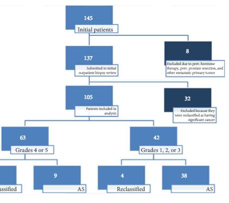
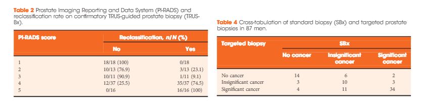
Patients with low-risk, low-grade, localised prostate cancer were prospectively enrolled and submitted to a 3-T 16-channel cardiac surface coil mpMRI of the prostate and confirmatory biopsy (CBx), which included a standard biopsy (SBx) and visual estimation-guided TRUS-Bx. Cancer-suspicious regions were defined using Prostate Imaging Reporting and Data System (PI-RADS) scores. Reclassification occurred if CBx confirmed the presence of a Gleason score ≥7, greater than three positive fragments, or ≥50% involvement of any core. The performance of mpMRI for the prediction of CBx results was assessed. Univariate and multivariate logistic regressions were performed to study relationships between age, prostate-specific antigen (PSA) level, PSA density (PSAD), number of positive cores in the initial biopsy, and mpMRI grade on CBx reclassification. Our report is consistent with the Standards of Reporting for MRI-targeted Biopsy Studies (START) guidelines.

apr-aotw-1-results

Results

In all, 105 patients were available for analysis in the study. From this cohort, 42 (40%) had PI-RADS 1, 2, or 3 lesions and 63 (60%) had only grade 4 or 5 lesions. Overall, 87 patients underwent visual estimation TRUS-Bx. Reclassification among patients with PI-RADS 1, 2, 3, 4, and 5 was 0%, 23.1%, 9.1%, 74.5%, and 100%, respectively. Overall, mpMRI sensitivity, specificity, positive predictive value, and negative predictive value for disease reclassification were 92.5%, 76%, 81%, and 90.5%, respectively. In the multivariate analysis, only PSAD and mpMRI remained significant for reclassification (P < 0.05). In the cross-tabulation, SBx would have missed 15 significant cases detected by targeted biopsy, but SBx did detect five cases of significant cancer not detected by targeted biopsy alone.

Conclusion

Multiparametric magnetic resonance imaging is a significant tool for predicting cancer severity reclassification on CBx among AS candidates. The reclassification rate on CBx is particularly high in the group of patients who have PI-RADS grades 4 or 5 lesions. Despite the usefulness of visual-guided biopsy, it still remains highly recommended to retrieve standard fragments during CBx in order to avoid missing significant tumours.

Editorial: An end to the phenomenon of ‘upgrading’ in early prostate cancer?

The phenomenon of ‘upgrading’ in early prostate cancer is one of those unusual events that is both useful to us on the one hand and undesirable on the other; useful because the phenomenon gifts us a direct measure of the precision of our risk stratification methods for men recently diagnosed, and undesirable because the perfect pathway should, ideally, be free of any upgrading.

Upgrading occurs in a number of settings. We see it at play to some degree when an unreliable test is re-applied in the same subject. The REDUCE study [1] showed us that just under one fifth of men will convert from a status of ‘cancer-free’ to one of ‘cancer-present’ as a result of a second exposure to the same test; that is, TRUS-guided biopsy. We see it in full play when an unreliable test is followed by a more accurate test. Shaw et al. [2] have reminded us once again – as have a number of others – of our limited ability to risk-stratify patients with early prostate cancer. They reported a 50% upgrading when they compared the results of TRUS biopsy against the final pathology at radical prostatectomy. In other words, half the patients went on to their definitive therapy with an incorrect grade attribution [2].

It would be a great pity if, in the modern era, the only route available to patients who wanted to be sure of their risk status was to agree to surgical removal of the prostate. Surely, the value of accurate risk stratification is derived from using it to allocate appropriate and effective care. Risk stratification needs to be linked to or closely follow diagnosis if it is to be put to work for patients.

Nowhere is this need greater than in men whose treatment preference is tissue preservation. The study, in this issue of BJUI, by Pessoa et al. [3] adds to our knowledge on the subject and equips us with a strategy to mitigate some of the errors that are inherent to the standard diagnostic pathway.

In the present study, the authors evaluated the role of a single exposure to MRI (and the opportunity that resulted to undertake a targeted biopsy of an MRI-derived abnormality as well as systematic sampling) in 105 men who had been attributed a diagnosis of low-risk prostate cancer – and, as a result, were deemed to be suitable for active surveillance. The authors used prostate imaging reporting and data system (PIRADS) scoring to interpret and communicate MRI risk. In summary, men attributed a low PIRADS score (PIRADS 1–3) had a low probability of being re-classified to a higher risk. In contrast, men attributed PIRADS score 4 or 5 had a probability of 70–100% of being re-classified. The authors calculated a sensitivity of 93% for MRI to predict ‘re-classification’. This equates to a 93% sensitivity to predict the presence of clinically significant disease as re-classification occurred when there was a transition from low-risk to higher-risk disease.

These results concur with those of others who are working in this area [4] and are in line with current recommendations [5]. One observation that is worth highlighting – because it is a current controversy in the field – relates to the utility of the systematic (or semi-random) biopsies as a component of the confirmatory biopsy. Whilst targeted biopsy was superior to systematic biopsy at identifying clinically significant disease, omission of the systematic biopsies would have resulted in five significant cancers being overlooked. The less perfect the targeted biopsy, the greater the reliance on the systematic. In the present study, the lesion generation and the targeting may have been compromised by one or two issues. Using TRUS biopsy as the authors did (as opposed to transperineal biopsy) to access all areas of the prostate is always going to be a challenge. To do so without image registration makes it even harder. To use PIRADS – as opposed to a Likert scale – as a method of interpreting and communicating MRI outputs will, very likely, lead to an under-reporting of the smaller, high-grade lesions [6]. This is because PIRADS 2.0 is triggered by a volume threshold towards the upper end of the scale. Such lesions might be more prevalent in an apparently ‘low-risk’ population such as the one under scrutiny. If this is the case, they will not be identified as ‘targets’ by virtue of a high PIRADS score. As a consequence they cannot be identified by targeting but might be picked up by the random fall of the needles.

Mark Emberton
Division of Surgery and Interventional Science, University College London, London, UK
References

1 Andriole GL, Bostwick DG, Brawley OW et al. Rittmaster RS; REDUCE Study Group. Effect of dutasteride on the risk of prostate cancer. N Engl Med 2010; 362: 1192202


2 Shaw GL, Thomas BC, Dawson SN et al. Identication of pathologically insignicant prostate cancer is not accurate in unscreened men. Br Cancer 2014; 110: 240511


4 Nassiri N, Margolis DJ, Natarajan S et al. Targeted biopsy to detect Gleason score upgrading during active surveillance for men with low- vs. intermediate-risk prostate cancer. J Urol 2016; [Epub ahead of print]. doi: 10.1016/j.juro.2016.09.070.


5 Moore CM, Giganti F, Albertsen P et al. Reporting magnetic resonance imaging in men on active surveillance for prostate cancer: the PRECISE recommendations-a report of a European school of oncology task force. Eur Urol 2016; [Epub ahead of print]. doi: 10.1016/j.eururo.2016.06.011.


© 2024 BJU International. All Rights Reserved.