The value of fresh cadaveric dissection
I am very lucky to have had the opportunity recently to undertake research in the fresh cadaver lab at University of Maryland School of Medicine, Baltimore.
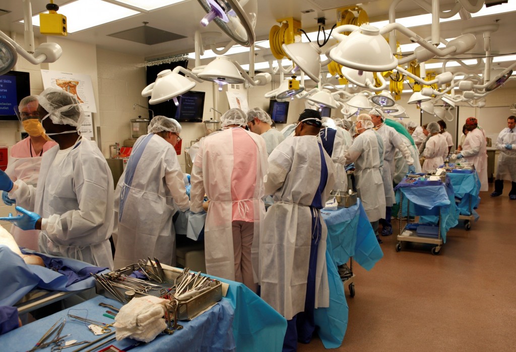
The availability and quality of fresh cadaveric specimens at the School of Medicine was outstanding. The School’s Anatomical Services Division is also the site of the Maryland State Anatomy Board, and processes over 2000 cadavers per year. No after death donations by family members are accepted. By word of mouth, public awareness of the research and education achievements of the lab means that there is no shortage of people volunteering to donate their body. Minimal processing time allows access to specimens that are truly fresh. The cadavers I used were arterially flushed with a broad-spectrum disinfecting solution, and stored chilled at 2 degrees Celsius between uses. This technique allowed me to work with the same fresh specimen daily for several weeks. My previous experience of cadaveric dissection was with formalin fixed specimens. Although this offers satisfactory opportunity to dissect and appreciate gross anatomical structures, discrete tissue planes are lost, which changes one’s sense of “operating”. Tissue becomes inflexible and dissecting small branches of nerves, such as my task was, becomes extremely challenging. Dissection of fresh cadavers is remarkably similar to operating on a live person. This facilitated my dissection from a research perspective, and also allowed me (as a surgical trainee) to improve my understanding of surgical planes and how to work with tissue.
Learning in the lab also occurs in a less formalised way. One afternoon, I found myself working alongside an ENT surgeon performing a complex facial reconstruction. He explained that when he has scheduled upcoming operations with which he has limited experience, he undertakes a mock procedure prior to the case. What an incredible opportunity to improve patient care.
In an era where surgical training is heavily logbook-based, it can be challenging to ensure all trainees get comparable exposure. Fresh cadaver labs may offer one way to bridge the gap during training, as well as consolidate skills after fellowship.
Research in the anatomy lab can also lead to advancements in surgery. University of Maryland Medical Centre was one of the first centres in the world to perform a full-face transplant. Performance of mock procedures on fresh cadavers was integral to the development and refinement of the procedure. This is a clear example of how fresh cadaver labs can directly contribute to improving surgical care.
Unfortunately, it seems that anatomical education in many institutions is moving away from cadaver use. My experience in Baltimore reminded me that fresh cadaveric labs are invaluable in surgical training and research. I would like to acknowledge the generous gift that many people make in donating their body to medical education and research. I am very grateful for Professor Anthony Costello and Dr James Borin facilitating my visit to University of Maryland School of Medicine, and to Ronn Wade and his team for generously welcoming me in the anatomy lab.
I implore the surgical community to advocate for the protection of existing labs, and development of new fresh cadaver labs.
Fairleigh Reeves
Urology Research Fellow, The Royal Melbourne Hospital, Australia
Twitter @DrFairleighR


This sounds like a fantastic experience. As medical schools move away from teaching anatomy, and surgical training increasingly utilises simulators, this is outstanding resource for surgeons and students alike.
The opportunity to prepare for surgery with normal appearing tissues and tissue planes is enviable.
Cadaver dissection (formalin fixed) was a vital part of anatomy teaching when I was a student at the University of Melbourne – twenty-five years ago! Ten years later, when I was a demonstrator, the students still undertook cadaver dissection, but had half as much time as what we had had. Now, I don’t think there is any cadaver dissection in the medical course, replaced by computerised “virtual dissection”. While the virtual approach provides some benefits, such as the ability to revise the anatomical dissection, ultimately there is no substitute for cadaver dissection in learning surgical anatomy. Fresh cadavers for dissection is a rare luxury that most of us wont ever have access to – but I think surgical educators need to aim for some exposure to cadaver dissection for completeness of anatomy teaching.
Thankfully Professor Harold Ellis continues to fly the anatomy flag high at Guy’s.
Essential for all doctors and especially surgeons.
The continuing importance and relevance of Anatomy in surgery cannot be understated.
We still don’t really understand the anatomy of continence and potency .
Anatomy centres such as Fairleigh describes could and should be incorporated in skills centres for later medical school years very early in surgical training .
Well done Fairleigh.
Rest assured cadaveric dissection is still a vital part of anatomy teaching at University of Melbourne. Thanks to the generosity of our body donors we have over 1000 3rd year science, postgraduate MD and postgraduate diploma of surgical anatomy students who participate in teaching programs that utilize embalmed and plastinated prosections and cadaveric dissection sessions. I absolutely agree with Shomik Sengupta that there is no substitute for cadaver dissection in learning surgical anatomy but would add that my students get so much more than that from their dissection sessions at University of Melbourne. I also believe that a cadaveric dissection program helps the student develop the ability to appropriately confront death and the dying of a patient. For medical students the cadaver is their first “patient”, dissection is their first model for patient-doctor relationship, and ours is the first opportunity to teach and encourage humanistic qualities of respect, empathy and compassion, all of which are an integral part of professionalism in medicine.
Assoc Prof Jenny Hayes
Head of Topographic Anatomy Teaching
University of Melbourne
Thanks Prokar for referencing Professor Ellis, our great anatomist. One of my early mentors: I spent one year with him at King’s teaching anatomy. A priceless experience. If only we could find a way for him to contribute to this forum!
A a medical student who has experienced teaching in a school using only prosections and another school facilitating full dissection, I am in total agreement regarding the value of full dissection for anatomy teaching. While prosections were useful in understanding the individual components of an anatomical region, the true relations between adjacent organs, tendons, muscles and nerves and their intervening layers was made far clearer in full dissection. Fellow medical students agree on the invaluable nature of cadaveric dissection and I am truly grateful for the opportunity to learn in such an in-depth manner.