Archive for category: Case Studies

Vesical endosalpingiosis: a case of probable metaplastic origin

Here we present an incidental case of pure endosalpingiosis in a 50-year-old woman that supports a metaplastic mode of development, and review the recommended management for this unusual diagnosis.


Authors: Duncan Rogers, Louise Dickinson, Alastair Ironside, Soha El Sheikh, Amir Kaisary  Departments of Urology and Pathology, Royal Free Hospital, Hampstead, London, NW3 2QG, UK
 
Corresponding Author: Jonathan WhiteE-mail: [email protected] Departments of Urology and Pathology, Royal Free Hospital, Hampstead, London, NW3 2QG, UK

Abstract
 
Endosalpingiosis of the urinary bladder is a rare and morphologically complex tumour-like lesion.  There are two theories to describe its pathogenesis; implantation and metaplasia. Here we present an incidental case of pure endosalpingiosis in a 50-year-old woman that supports a metaplastic mode of development, and review the recommended management for this unusual diagnosis.


Introduction
Endosalpingiosis is the presence of ectopic, non-descript glandular tissue lined with tubular epithelium.  It is a tumour-like lesion of mullerian origin that, together with endometriosis and endocervicosis, are collectively referred to as mullerianoses [1-2].  It is not uncommon for endosalpingiosis to co-exist with endometriosis; their clinical presentation, course and response may be very similar [3].  The presence of pure endosalpingiosis within the urinary bladder is rare and, to date, only four cases have been reported [4-7].  This is in contrast to that of the pelvis, which is more common, although usually diagnosed incidentally as part of the diagnostic work-up for pelvic pain [3,8]. This report describes a case of pure vesical endosalpingiosis in a perimenopausal woman.  It is one of only a small number of cases demonstrating a metaplastic origin.


Case Report
A 50-year-old Afro-Caribbean woman presented initially to the gynaecology department with a three-week history of pelvic pain associated with per vaginal bleeding and dysuria.  Her medical history was marked by a long period of endometriosis and amenorrhoea recently followed by new onset menorrhagia. A Mirena intra-uterine device (IUD) had been in situ for the past 8 years.  There was no history of urogenital infection, trauma or lithiasis, nor did she have any significant previous surgical history.
Physical examination revealed suprapubic tenderness with a normal bimanual palpation.  No strings of the IUD could be seen on direct visualisation of the cervix – see comment below.  An urgent pelvic ultrasound scan demonstrated a well positioned IUD and a 3.2 x 2.6 x 3.8cm heterogenous, irregular mass arising from the bladder wall that appeared to contain both solid and cystic areas (Fig. 1).


Figure 1. Pelvic ultrasound scan


A subsequent rigid cystoscopy under general anaesthetic revealed a diffuse mass with cystic lesions on the posterior wall of the bladder. The features were not typical of a transitional cell carcinoma.  Six deep biopsies were taken of the cystic areas.
Histologically, the mass included mildly inflamed, oedematous mucosa.  Two deep biopsies included smooth muscle bundles containing cystic spaces, one of which was lined by columnar, focally-ciliated epithelium (Fig. 2 and 3).


Figure 2. Biopsy 

 
Figure 3. Biopsy

Conventional H&E and immunohistochemical staining for CD10 failed to demonstrate endometrial-type stroma, and no mucin to suggest endocervical differentiation was seen.  The features were reported as characteristic of endosalpingiosis by our histopathologists.


Discussion
Mullerianosis of the urinary bladder is a rare occurrence and although a number of cases have been reported [4-7], very few of these describe lesions that are composed entirely of endosalpingiosis, as was seen in the aforementioned case.
There are two theories to explain the pathogenesis of endosalpingiosis.  The first is an implantative origin, as first described by Young and Clement1.  This theory proposes the seeding of ectopic tubal tissue as a result of pelvic surgery, such as a previous caesarean section.  The second theory, first suggested by Donné et al. [2] argues a role for metaplasia in those cases with no previous pelvic surgery and where multiple Mullerian tissues co-exist.  This theory is also favoured by the association of the overlying vesical peritoneum with the location of mullerian lesions in the posterior wall or bladder dome. Further evidence supporting this possibility is provided by a case of endosalpingiosis in continuity with glandular cystitis.[9]
The case in question supports a metaplastic origin of endosalpingiosis.   Although no evidence of other Mullerian tissue was seen within the bladder lesion, the patient had a confirmed diagnosis of long-standing endometriosis.  Furthermore, there was no previous history of pelvic surgery or bladder instrumentation to suggest implantation.
Endosalpingiosis often presents asymptomatically in contrast to the other Mullerian lesions of endocervicosis and endometriosis [1]. It is this paucity of symptoms that may account for its apparently rare incidence, and the condition is potentially grossly under-represented if symptoms are incorrectly attributed to other uro-gynaecological conditions with similar clinical presentations.  Investigations tend to be directed by the clinical presentation.  In this case, pelvic pain and per vaginal bleeding with dysuria prompted a full gynaecological and urological work-up with a subsequent histological diagnosis.
The specific management of endosalpingiosis is not fully established; however described therapies have included transurethral resection with follow-up cystoscopy at 4 and 9 months post-operatively [1, 5].  Comparisons can be made with the treatment of bladder endometriosis, another of the Mullerianoses.  Two types of treatment are employed: medical (hormonal), and surgical therapies [10].  Hormonal treatment provides a good initial response, is well tolerated by patients, and can be discontinued if adverse effects occur.  LH-RH analogues are the most commonly used treatments nowadays, whilst oestrogens, progesterones and androgens have fallen into disuse due to their many side effects [10]. Conservative measures alone however do not lead to a definitive cure, but merely suppress the disease temporarily.  In order to prevent further recurrence and achieve the best prognosis, a combination of medical therapy with surgery is advised [10].  The most commonly employed technique of recent years is transurethral resection or biopsy of the lesion to confirm the diagnosis, followed by hormonal therapy.  Further surgery including partial cystectomy may be warranted, especially if the lesion is transmural [11].  Aggressive measures such as this however, should not be taken in perimenopausal women, as the lesions usually regress after the menopause [10].
Like endometriosis, endosalpingiosis tends to recur and primary ureteric disease has been described [12], although it is unknown whether bladder disease can recur within the upper urinary tract, or whether this is in fact a primary disease.  If the diagnosis is in doubt and imaging suggests malignancy then partial cystectomy is recommended [4].  It has been suggested that, given the relative asymptomatic nature of the condition, long-term surveillance is advisable [6].  A recommended approach is regular cystoscopy at three-monthly intervals initially for two years, twice-yearly for a further two years, and then annually thereafter [6]. However, these recommendations have been made on the basis of minimal evidence, therefore follow-up should be instigated on a case-by-case basis.  Given the potential for upper tract disease, renal ultrasound or contrast computed tomography should be considered.
In our case, the patient was discharged and referred back to the gynaecology department following a multidisciplinary team discussion.  Her symptoms resolved despite only a conservative approach and without further resection of the lesion. At the time of writing, she is under regular review within the gynaecology outpatient department.


Conclusion
Vesical endosalpingiosis, although uncommon, may present a diagnostic conundrum both to the gynaecologist and the urologist.  If the condition is as under-represented as the evidence suggests, an increased awareness of the range of clinical presentations is required.  Subsequently, an increased yield of histological diagnoses in those patients in which the diagnosis is suspected will advance our understanding of the condition and its appropriate management.


References
1) Young RH, Clement PB.  Mullerianosis of the urinary bladder.  Mod Pathol.  1996;9:731-737
2) Donne C, Vidal M, Buttin X, et al.  Mullerianosis of the urinary bladder:  clinical and immunohistochemical findings.  Histopathology.  1998;33:284-292.
3) Laufer MR, Heerema AE, Parsons KE, Barbieri RL.  Endosalpingiosis: clinical presentation and follow-up.  Gynecol Obstet Invest.  1998;46:195-8.
4) Arai Y, Tsuzuki M, Okubo Y, et al.  A case of submucosal endosalpingiosis in the urinary bladder.  Nippon Hinyokika Gakkai Zasshi.  1999;90:802-805.
5) Edmondson JD, Vogeley, Howell JD, Koontz WW, Koo HP, Amaker B.  Endosalpingiosis of the bladder.  J Urol.  2002;167:1401-1402.
6) Smith C, Sabet L, Izawa JI.  Management of endosalpingiosis of the urinary bladder.  Urology.  2004;64:1031
7) Maniar KP, Kalir TL, Palese MA, Unger PD.  Endosalpingiosis of the urinary bladder:  a case of probable implantative origin with characterization of benign fallopian tube immunohistochemistry.  Int J Surg Pathol.  2010;18:381-3.
8) Heinig J, Gottschalk I, Cirkel U, Diallo R.  Endosalpingiosis – an underestimated cause of chronic pelvic pain or an incidental finding?  A retrospective study of 16 cases.  Eur J Obstet Gynecol Reprod Biol.  2002;103:75-78.
9) Koren J, Mensikova J, Mukensnabl P, Zamecnik M.  Mullerianosis of the urinary bladder:  report of a case with suggested metaplastic origin.  Vichows Arch.  2006;449:268-271.
10) Pastor-Navarro H, Gimenez-Bachs JM, Donate-Moreno MJ et al.  Update on the diagnosis and treatment of bladder endometriosis.  Int Urogynecol J Pelvic Floor Dysfunct.  2007;18:949-954.
11) Shook TE, NybergLM.  Endometriosis of the urinary tract.  Urology. 1988;31:1-6.
12) Nogales FF, Zuluaga A, Arrabal M et al.  Mullerianosis of the ureter:  a metaplastic lesion, J Urol.  1999;162:2090-2091.


Date added to bjui.org: 31/12/2010


DOI: 10.1002/BJUIw-2010-054-web


Complete ureteral avulsion: ileal ureteral substitution with an unusual proximal Ileal anastomosis

Ureteroscopy is currently one of the procedures for handling proximal ureter stones. A serious, though fortunately rare, complication is the avulsion of the ureter. This report shows one such case, in which efforts were made to preserve the ipsilateral kidney.


Authors: Smith, Nelson; Federal University of Rio de Janeiro, SurgeryUrology Medeiros, Marcus; Federal University of Rio de Janeiro, SurgeryUrology Vieira, Gustavo; Federal University of Rio de Janeiro, SurgeryUrology


Case Report

A 42-year-old male patient was admitted with a high-debt urinary-cutaneous fistula to the abdominal lower left quadrant. He came from another hospital, where, about 15 days earlier, he had undergone ureteroscopy for a left proximal ureter stone. During surgery, there was a complication that required immediate laparotomy. A few days later he presented with a urinary fistula to the left flank.

A postoperative intravenous urography (IVU) performed in the first hospital showed a double-J catheter in the left, normal renal function bilaterally, marked lateral deviation of the left ureter with an atypical localization of the ureterovesical junction and a uretero-cutaneous fistula to the left (Fig 1A).

On admission we performed a computed tomography (CT) that confirmed the ureteric fistula (Fig 1B).

BJUIw-2010-028-Cre.R1-f1Figure 1- A- IVU showing lateral deviation of the left ureter and an uretero-cutaneous fistula (white arrow); B- CT scan showing the ureteral fistula (white arrow)

The patient was taken to surgery for correction of the uretero-cutaneous fistula. During the procedure we found an ischemic left ureter, without any vascularization in all its extent, and a fistula in its proximal third. The proximal ureter was sutured to the perihilar fat. The distal end was reimplanted into the bladder dome.

Based on these findings we performed a complete replacement of the ureter by ileum.

As the ureteral avulsion occurred in the intra-renal site and there was no renal pelvis available for the reconstruction, the proximal anastomosis of an ileal loop was made to the renal parenchyma. We used a 3-0 polydioxanone suture with three U-fashioned stitches, starting and ending in the renal parenchyma, reaching the ileal loop at about 0.5 cm from its edge (Fig 2). The stitches pierced the parenchyma in such a way that they did not harm the renal pedicle. The knots were tied over a fat pad to prevent parenchymal damage. At the end of the suture the ileal segment was inserted into the hilum, meeting the remaining segment of intra-renal pelvis. The ileo-vesical anastomosis was carried out without an anti-reflux technique.

BJUIw-2010-028-Cre.R1-f2Figure 2- Three U-fashioned stitches: Anterior (A), Posterior (P) and Inferior (I).

We placed a 6 Fr double-J catheter. The patient had no postoperative complications. The double-J catheter was removed three months after surgery. A CT scan performed at six months showed good function of the left kidney with contrast passing to the ileal loop (Fig 3). At 10 months post-surgery a DTPA renogram showed good passage of the radiopharmaceutical through the ileal loop (Fig 4). The patient was asymptomatic.

BJUIw-2010-028-Cre.R1-f3Figure 3- CT scan at 6 months showed good function of the left kidney. Contrast in the ileal loop (white arrow).

BJUIw-2010-028-Cre.R1-f4Figure 4- DTPA renogram, posterior view, showing (A to D) good passage of the radiotracer from the left kidney (LK) to the ileal loop (IL).

Discussion

One of the complications of ureteroscopy is damage to the ureter in varying degrees, complete avulsion being the more serious one, but fortunately rare (1).

The treatment of ureteric avulsion varies according to the compromised ureteral segment and functional status of the renal unit (2,3). In cases of largely devitalized tissue or compromise of a large ureteric segment, extreme measures are necessary, such as renal auto-transplantation or ileal interposition (4,5,6), the latter replacing a segment or the entire ureter (7,8,9,10,11). In our patient, there was no ureter or renal pelvis to perform an auto-transplantation, similar to the procedure of a Breast reduction New York which is extremely complicated. Thus, despite the possible known metabolic and functional alterations (12), we performed a total replacement of the ureter by ileum.

The literature shows that ileal anastomosis with the renal collecting system is feasible (13). In our patient, as the avulsion was intra-renal, the proximal suture could not be performed under direct vision. The sutures were passed through the healthy renal parenchyma and through the ileum so that the proximal end of the ileal loop was later brought into the hilum and toward the remaining segment of the renal pelvis. Consequently, this kept the ileum anchored to the renal parenchyma, allowing tension-free healing of the structures. We chose not to use a nephrostomy, due to concern about any inflammatory reaction, which could increase the possibility of anastomotic stenosis or fistula in an already risky intrarenal anastomosis.

After review of the literature we could not find any report of ileal anastomosis directly to the healthy renal parenchyma.


References

1- D. Brooke Johnson, Margaret S. Pearle: Complications Of

Ureteroscopy. Urol. Clin. N. Am Vol: 31, 157, 2004

2- V. Gupta, T. C. Sadasukhi, K.K. Sharma: Complete Ureteral

Avulsion. Thescientificworldjournal, Vol 5: 125, 2005

3- Ranjiv Mathews And Fray F. Marshall: Versatility Of The

Adult Psoas Hitch Ureteral Reimplantation. J. Urol., Vol: 158,

2078, 1997.

4- Mitchell C. Benson, Kenneth S. Ring And Carl A. Olsson:

Ureteral Reconstruction And Bypass: Experience With

Ileal Interposition, The Boari Flap-Psoas Hitch And Renal

Autotransplantation. J. Urol.,Vol 143, 20, 1990.

5- Willard E. Goodwin, Chester C. Winter And Roderick D.

Turner: Replacement Of The Ureter By Small Intestine:

Clinical Application And Results Of The Ïleal Ureter”. J.

Urol., Vol 81: 406, 1959.

6- Richard J. Boxer, Peggy Fritzsche, Donald G. Skinner:

Replacement Of The Ureter By Small Intestine: Clinical

Application And Results Of The Ileal Ureter In 89 Patients.

  1. Urol., Vol 121: 728, 1979.

7- Paulo Ricardo Monti, Rolf Carvalho Lara, Marcos

Antonio Dutra: New Techniques For Construction Of E

Fferent Conduits Based On The Mitrofanoff Principle.

Urology, Vol 49: 112, 1997.

8- Bedeir Ali-El-Dein And Mohamed A. Ghoneim: Bridging Long

Ureteral Defects Using The Yang-Monti Principle. J. Urol., Vol

169: 1074, 2003.

9- Bernard Lytton And Martin Schif: Interposition Of An Ileal

Segment For Repair Of Ureteral Injuries. J. Urol., Vol 125:739,

1981.

10- Benjamin I. Chung, Karim J. Hamawy, Leonard N. Zinman: The

Use Of Bowel For Ureteral Replacement For Complex

Ureteral Reconstruction: Long-Term Results. J. Urol., Vol 175:

179, 2006.

11- Sandra A. Armatys, Matthew J. Mellon, Stephen D. W. Beck:

Use Of Ileum As Ureteral Replacement In Urological

Reconstruction. J. Urol., Vol 181: 177, 2009.

12- Emil A. Tanagho: A Case Against Incorporation Of Bowel

Segments Into The Closed Urinary System. J. Urol., Vol 113: 796,

1975.

13- S. H. Wong And Y. T. Chan: Pan-Caliceal Ileoneocystostomya

New Operation For Intrapelvic Tuberculotic Strictures

Of The Renal Pelvis. J. Urol., Vol 126: 734, 1981.


Acknowledgment

1- Prof. Milton Melciades Barbosa Costa, Full Professor of Anatomy at the Institute of Biomedical Sciences from the Federal University of Rio de Janeiro, in help with the arrangement of figures. 2- Mrs. Maria de Fatima Pinto, executive secretary, Department of Orthopaedics, School of Medicine ,Federal University of Rio de Janeiro for help in organizing the references.


Date added to bjui.org: 15/12/2010


Radiotherapy for Leukaemic infiltration of Prostate

We present an 81 year old gentleman with haematuria and LUTS who had TURP for his outflow symptoms. Radiotherapy is an excellent option for local symptom control as evidenced in this patient.


Authors: Venugopal S, Das.S,  Hamid BN, Doyle.G, Leggat H, Powell CS. Countess of Chester Hospital NHS Foundation Trust
 
Corresponding Author: Suresh Venugopal, Countess of Chester Hospital NHS Foundation Trust Email: [email protected]

Abstract
Haemopoietic malignant infiltration of the prostate is unusual. When present, it brings about the dilemma on the best approach to manage it. Usually, chronic lymphocytic leukaemia is an indolent process that waxes and wanes in its course and when present with lymphadenopathy and clinical symptoms would warrant treatment with chemotherapy. When organ confined, it has a more favourable course and would be amenable to local treatments.
We present an 81 year old gentleman with haematuria and LUTS who had TURP for his outflow symptoms. The histology had confirmed a malignant lymphocytic infiltration of the prostate and he had elevated lymphocytic count and peripheral blood marker study confirming chronic lymphocytic leukaemia. He was treated with radiotherapy of his prostate. His presenting WBC count was 19.2 and PSA was 4.41 and presently his white count is 10.3 and PSA is 1.77.  He was treated with 24 Gray external beam radiotherapy to the prostate.
As we encounter an aging population, we will be seeing more of these cancers with higher incidence in the elderly. Radiotherapy is an excellent option for local symptom control as evidenced in this patient.


Case Report
 
Presentation
 
An 81 year old gentleman had simultaneous referral to the haematologist for low haemoglobin and raised white count and the urologist for visible haematuria and LUTS. A digital rectal examination revealed a smooth, benign-feeling prostate with a normal age-specific PSA. A CT scan done to evaluate his haematuria had picked up a large mass in the pelvis. It was difficult to differentiate whether it was of bladder or prostate origin (Figure-1).


Figure 1: CT scan showing pre treatment pelvic mass (left) and post treatment pelvic mass (right).


Trans-urethral resection of the mass showed diffuse infiltration of the prostate gland by a malignant B-cell type lymphoid infiltrate. His peripheral blood markers for chronic lymphocytic leukaemia were positive. He did not have generalised lymphadenopathy, splenomegaly or B type symptoms of leukaemia.


Pathology
 
The trans-urethral resection chippings of prostate showed large foci of basal cell hyperplasia and extensive infiltration with monotonous population of small lymphoid cells (Figure-2).


Figure 2: Prostate tissue infiltrated by lymphocytes staining strongly for CD20 (left) and H&E staining showing sheets of monotonous lymphocytic infiltration of prostate (right). 

The abnormal lymphoid infiltrate stained positively for CD5, CD20, CD79a but was negative for CD3, CD10, BCL2 and BCL6. This is keeping in with a picture of Chronic Lymphocytic Leukaemia of Prostate. There was no evidence of high grade PIN or adenocarcinoma of prostate.


Radiotherapy
 
In view of his symptomatic haematuria and potential for the lesion to cause local symptoms of persistence of haematuria and possible obstruction of adjacent structures, he was considered for radiotherapy after due discussion at the multidisciplinary team meeting. The pros and cons of the treatment was discussed with the patient and he opted to have the treatment. He received standard 24 Gray in 12 fractions for low grade lymphoma of the prostate (1).


Follow-up
 
He has since been regularly followed up at the urology clinic for his lower urinary tract symptoms with a flow rate and rectal examination on the six monthly visits as well as a yearly PSA test. He does not have any lower urinary tract symptoms. His six monthly follow-up in the haematology clinic for his Chronic Lymphocytic Leukaemia involves assessing the presence or absence of B symptoms of leukaemia as well as a peripheral blood smear assessment. At the end of three years a re-biopsy of the prostate revealed the presence of a residual malignant lymphomoid infiltrate (Figure 3).


Figure3: Core biopsy specimen showing lymphocytes staining strongly for CD 20 (left) and H&E staining showing monotonous lymphocytic infiltration of the core biopsy specimen of prostate (right).


A rescan showed the persistence of the pelvic mass but without increase in size and without any accompanying symptomatic haematuria or obstructive features.


Discussion
Infiltration of prostate by Chronic Lymphocytic Leukaemia is not an uncommon finding in patients with CLL. Autopsy studies have revealed CLL as the commonest secondary tumour of the prostate. Leukemic infiltration is symptomatic in only 1% of them (2). They usually present with outflow obstructive features. Chronic lymphocytic leukaemia has an indolent course with waxing and waning of the white cell count. As a haemopoietic malignancy involving the prostate, it cannot be cured by local therapies, though this has been claimed on short term follow-up in literature (3,4,5).
The prostatic component of the disease is best dealt with by radiotherapy if local problems are anticipated. Chemotherapy is reserved for patients who have systemic disease progression. Outflow obstruction is best relieved by Trans-urethral resection of the prostate. Though various doses have been cited for curative treatment of this disease, this claim is to be disputed. We currently recommend a dose of 24 Gray in 12 fractions for palliative control of symptoms as per the recommendation for a low-grade non Hodgkin’s lymphoma.
Urological follow-up of these patients is as that of other patients with outflow symptoms, who generally get a flow rate and rectal examination assessment. In addition they need to have continuous follow-up at a haematology clinic for monitoring their systemic symptoms, which may dictate the need for further intervention.
 

Lesson learnt:
• Leukaemic infiltration is not uncommon.
• It usually presents as bladder outflow obstruction requiring TURP.
• Radiotherapy is indicated only if local complications are anticipated and is not always curative.


References
1) https://www.rcr.ac.uk/docs/oncology/pdf/DoseFract_49_Lymphoma.pdf
2) E. H. Eddes et al, Urinary symptoms due to leukemic infiltration of the prostate A case report, Ann Haematol 1993, 66:323 – 324.
3) Mitch Jr et al, Leukemic infiltration of the prostate: A reversible form of urinary obstruction, Cancer 1970, 26: 1361-1365.
4) Belis JA, Lizza EF, Kim JC, Raich PC, Acute leukemic infiltration of the prostate. Successful treatment with radiation, Cancer 1983, 51: 2164–2167.
5) Belhiba H et al, Prostatic involvement in leukemia. Report of a case Progrès en Urologie 1992, Aug-Sep;2(4):650-2.


Date added to bjui.org: 15/12/2010


DOI: 10.1002/BJUIw-2010-063-web


Massive exsanguinating hematuria – a rare post-operative complication of transvaginal ultrasound-guided oocyte retrieval for invitro fertilization

Two cases with similar presentation of massive hematuria that compromised the cardiovascular status of the patients and required the surgical intervention of the author are presented.

Corresponding Author: Oseremen I. Aisuodionoe-Shadrach, MBBS(Ib.), FWACS(Urol.)Division of Urology, Department of Surgery, College of Health Sciences,University of Abuja, Abuja, [email protected]

Abstract
Purpose: To report the occurrence of massive exsanguinating hematuria after transvaginal oocyte retrieval for invitro fertilization.
Methods: Two cases with similar presentation of massive hematuria that compromised the cardiovascular status of the patients and required the surgical intervention of the author are presented.  The relevant literature related to the incidence of complications arising from this procedure is reviewed.
Results: The rare occurrence of exsanguinating hematuria after oocyte retrieval for in vitro fertilization is highlighted.
Conclusion: This report of two cases suggests that although severe complications post oocyte retrieval rarely occur, the procedure may not be altogether innocuous.


Case report summary
This is a report of the occurrence of exsanguinating haematuria post oocyte retrieval for IVF leading to acute hypovolemia and requiring surgery for amelioration. Two cases highlighting this phenomenon are presented to suggest that, although severe complications post oocyte retrieval rarely occur, the procedure is not innocuous.


Introduction
The in vitro fertilization procedure involves four primary stages – induction of ovulation, oocyte retrieval, insemination and fertilization, and embryo transfer.
The most common technique used for oocyte retrieval is ultrasound-guided aspiration. First described in 1985, transvaginal ultrasound guided oocyte retrieval (TVOR) remains the gold standard in vitro fertilization (IVF) procedure. Nevertheless, despite its advantages, the aspiration needle may injure pelvic organs and structures adjacent to the ovaries leading to severe complications. Life threatening complications following transvaginal ultrasound-guided oocyte aspiration have been reported as rare1, 2.There is so far in existing literature two reports of post aspiration haematuria and urinary retention3,4  both of which were managed conservatively .The two almost identical cases presented here describes massive exsanguinating haematuria necessitating emergency pelvic laparatomy and open cystostomy in both patients.
This paper therefore brings to the fore this rare complication.


Case reports


Case 1
KO is 35 year old. She had primary infertility for two and a half years due to male factor. Her pre-treatment assessments were essentially normal and she went on to have ovarian stimulation as protocol. On the thirteenth day of GnRH treatment, she underwent, transvaginal ultrasound-guided ovarian puncture for oocyte retrieval under general anaesthesia and prophylactic antibiotic therapy. After emptying her bladder, oocytes were aspirated through the lateral vaginal fornices with a 17G fine aspiration needle loaded on a biopsy guide affixed to a transvaginal ultrasound probe. The ovary with the follicles closest to the vaginal fornices was first punctured, followed by sequential puncture of other follicles, without reinserting the needle through the vaginal wall. The aspirates were noted not to be heavily blood-stained and post-procedure ultrasound of the pelvis did not reveal any sign of haemmorhage from the ovary or iliac vessels. No immediate peri-operative complications were observed. Five days post procedure, the patient developed sudden onset total haematuria.This was recurrent over a twelve  hour period and associated with voiding blood clots, lower abdominal discomfort, dysuria and strangury. She had no vaginal bleeding, was slightly pyrexic and had a packed cell volume (PCV) of 22%. Abdomino-pelvic sonography revealed two enlarged multicystic ovaries with imtraluminal bladder masses suggestive of haematoma from a possible intra-peritoneal bladder injury. She had initial conservative management with intravenous fluids and antibiotics. In the absence of facilities for lower urinary tract endoscopy (LUTE), which would have necessitated a referral to another centre, and on account of the patients unstable vital cardiopulmonary statistics, she was scheduled for emergency pelvic exploration. At surgery, the bilaterally enlarged multicystic ovaries were visualized but no bladder rent was seen. She subsequently had open cystostomy. About 500mls of stale blood clots were evacuated from the bladder and several pin-point haemorrhagic spots were observed in the region of the trigone and posterior wall. There was no active bleeding point. The patient recovered uneventfully and was discharged home on the 10th post-operative day.


Case 2
LD is a 30year old lady with a one and half year history of primary infertility who consented to transvaginal ultrasound-guided ovarian puncture for oocyte retrieval under general anaesthesia. The bladder was emptied before the procedure, as described in Case 1 above, and no immediate peri-operative adverse events were recorded. Five days after the procedure, the patient had initial sudden onset macroscopic haematuria, which recurred over a five hour period and was associated with lower abdominal pains, dysuria and voiding of blood clots. There was no vaginal bleeding and central temperature was normal. Abdomino-pelvic sonography revealed two enlarged multicystic ovaries and bladder hematoma.She had intravenous fluids and antibiotics but continued to have massive haematuria with clots and recurring clot retention. Her clinical condition deteriorated suddenly when her blood pressure dropped to 90mmHg/45mmHg and a PCV check revealed a drop from 35% to 25%.She was transfused with two units of fresh whole blood and an emergency pelvic exploration and open cystostomy was performed. The decision for open surgery was taken because facilities for LUTE were not available and a referral at the time to another facility would have been deleterious to the patient’s already precarious clinical condition. At surgery, there were multiple perforations of the posterior bladder wall with a solitary bleeding spot within the perforated areas, ~200mls of blood clots in the bladder and multiple petecheciae haemorrhagic spots in the trigone The blood clots were evacuated  and haemostasis was secured with a figure of eight vicryl stitch over the bleeding point. Post operative course was uneventful and the patient was discharged from hospital on the 8th day post surgery.


Discussion
Following fours years of experience with the technique, Feichtinger and workers5 in 1988 strongly recommended the replacement of other ultrasound guided follicle aspiration techniques with TVOR .The reasons advanced were that the transvaginal route has the shorter puncture course with an empty bladder that is outside the pathway of the follicle-aspirating needle. Moreover the elastic nature of the vagina brings the tip of the ultrasound probe to the tip of the ovary, even reaching ovaries located high up in the
pelvis.
Over the last several decades, several reports have described the complications associated with the technique, the most common being haemorrhage, trauma to adjoining pelvic structures and pelvic infection.
During a 4-year prospective study  in which complications arising from a series of 2670 consecutive TVOR procedures were monitored, Bennett et al1 identified  vaginal hemorrhage in 229 (8.6%) of the cases, with a significant loss (> 100 ml) in 22 (0.8%) and postoperative pelvic infection  in 18 (0.6%) of the cases- nine of these being severe with pelvic abscess formation. Furthermore, haemorrhage from the ovary with haemoperitoneum formation was rarely seen and necessitated emergency laparotomy in one instance, while a single case of pelvic haematoma formation from a punctured iliac vessel was recorded, settling without intervention.
Ludwig et al6 in their review of over 1000 oocyte retrievals performed over a 17month period recorded no cases of intrabdominal bleeding and pelvic infections, an isolated case each of ureteric injury and unexplained fever, with only 2.8% of procedures causing vaginal bleeding.
In all of the reviews of complications in literature, the only reported incidence of transient macroscopic haematuria as a specific complication of transvesical oocyte collection was by Ashkenazi and colleagues7.
Minimal vaginal haemorrhage is one of the most common consequence of transvaginal oocyte aspiration and is susceptible to local treatment such as application of pressure or topical haemostatic agents or both or suture of the lesion8. On the other hand, haemoperitoneum from direct damage to pelvic organs i.e. uterus, bladder,
bowels or pelvic blood vessels, with serious cases requiring laparoscopy or emergency laparatomy have been described by Bergh and Lundkvist9
It is instructive that to date, it is not known that any case of massive exsanguinating haematuria compromising the patients cardiovascular status and requiring multiple blood transfusions and further emergency pelvic laparatomy for control has been reported as a direct complication of the TVOR.
In the cases presented there were no evidence, at pelvic laparotomy, of iatrogenic damage to other pelvic structures besides the bladder. However, there were sufficient reasons to believe that the multiple perforating injuries to the posterior bladder wall during the TVOR procedures formed the nidus for the massive hematuria that was to occur in the fifth post-operative day subsequently.
When it occurs, reactionary haemorrhage is caused by post-operative infection. Furthermore, haematuria is a known complication of urinary tract infections (UTI). It is not unlikely therefore that the presence of UTI‘s in both post-operative patients acted synergistically to provoke the exsanguinating hematuria which this case reports.


Conclusion
Transvaginal ultrasonographically-guided procedures are simple and safe and often represent the only means of access for adequate treatment of many gynaecological and non-gynaecological pelvic pathologic conditions i.e. aspiration of cystic pelvic masses and core biopsy of solid pelvic masses. However, in the IVF setting, pelvic ultrasound is hindered by changes in ovarian volume and structure due to super ovulation and follicular puncture.
In spite of the fact that ultrasonically-guided transvesical oocyte collection has few complications, they should be reported in order to draw attention to their occurrence. These cases underscore the need for proper information on potentially serious procedure-related complications to all oocyte retrieval patients.


References
1. Bennett SJ, Waterstone JJ, Cheng WC, Parsons J. Complications of transvaginal    ultrasound- directed follicle aspiration: a review of 2670 consecutive procedures. J.Assist Reprod Genet 1993; 10: 72-77.
2. Dicker D, Ashkenazi J, Feldberg D, Levy T, Dekel A, Ben-Rafael Z. Severe abdominal complications after transvaginal ultrasonographically guided retrieval of oocytes for in vitro fertilization and embryo transfer. Fertil Steril. 1993 Jun; 59(6):1313-5.
3. Sauer MV. Defining the incidence of serious complications experienced
    by oocyte donors: a review of 1000 cases. Am J Obstet Gynecol 2001; 184:277– 8.
4.   Modder J, Kettel LM, Sakamoto K. Hematuria and clot retention after   transvaginal oocyte aspiration: a case report. Fertil Steril.2006 Sep; 86(3):720.e1-2.
5. Feichtinger W, Putz M, Kemeter P. Four years of experience with ultrasound-guided follicle aspiration. Ann N Y Acad Sci. 1988;541:138-42
6. Ludwig AK, Glawatz M, Griesinger G, Diedrich K, Ludwig M, Perioperative and post-operative complications of transvaginal ultrasound-guided oocyte retrieval: prospective study of > 1000 oocyte retrievals. Human Reproduction 2006; 21(12): 3235-3240
  7.    Ashkenazi J, Ben David M, Feldberg D, Shelef M, Dicker D, Goldman JA. Abdominal complications following ultrasonically guided percutaneous transvesical collection of oocytes for in vitro fertilization. J In Vitro Fert Embryo Transf. 1987 Dec; 4(6):316-8.
      8.   Tureck RW, Garcia C, Blasco L, Mastroianni L: Perioperative complications arising after transvaginal oocyte retrieval. Obstet Gynecol 1993; 81:590–593
       9.    Bergh T, Lundkvist O: Clinical complications during in vitro fertilization treatment. Hum Reprod 1992; 7:625–626


Date added to bjui.org: 25/11/2010 


DOI: 10.1002/BJUIw-2010-036-web


Bilateral synchronous multiple upper tract transitional cell carcinoma: treatment options, surgical challenges and robotic management

Multifocal transitional cell carcinoma with synchronous involvement of bilateral upper urinary tracts is rare. We present one such case presenting with haematuria and renal insufficiency.


Authors: Gupta, Narmada; Nayyar, Rishi  E-mails: [email protected], [email protected]
 
Corresponding Author: Dr Shailesh Chandra Sahay, MBBS, MS ( General Surgery), MCh (Urology) Corresponding address: Room No 5030, Department of Urology, All India Institute of Medical Sciences, Ansari Nagar, Post: New Delhi, Pin: 110029, India  Email: [email protected]

Abstract 
 
Multifocal transitional cell carcinoma with synchronous involvement of bilateral upper urinary tracts is rare. We present one such case presenting with haematuria and renal insufficiency. The challenges in diagnosis and surgical management of such a case are highlighted and various available treatment options are discussed. Endourological techniques through retrograde and percutaneous access for the right side, along with left robotic nephroureterectomy and cuff of bladder excision, provided excellent peri-operative outcomes in this patient.


Introduction
Patients presenting with bilateral synchronous upper tract transitional cell carcinoma have always been a challenge for the urologist. This case describes a patient with bilateral upper tract transitional cell carcinoma who was managed by a combination of upper and lower tract endourological and minimal invasive approaches.
Case report
A 57 year oldmale presented with painless gross haematuria for 1 month. He was a chronic smoker and had controlled diabetes mellitus for 4 years. Blood investigations revealed hemoglobin-11.1gm/dl, urea-43mg/dl and creatinine-2.1mg/dl. Liver function tests were normal. Ultrasound abdomen showed right renal and left vesicoureteric junction mass with hydroureteronephrosis. Chest X ray was normal. Renal dynamic scan revealed global glomerular filtration rate (GFR) of 27.58ml/min and differential function of 89% and 11% on right and left side respectively. MRI confirmed the bilateral localized tumours (Figure 1).


Figure 1. Coronal section of MRI Abdomen showing right renal pelvic tumour (3.7×2.2cm) and left lower ureteric tumour (3.1×2.7cm) (arrows) with hydroureteronephrosis and small kidney.


An informed consent was taken for cystoscopy, right retrograde pyelography (RGP) and ureteroscopy (Figure 2). Cystoscopy showed tumour protruding out from the left ureteric orifice. Biopsies were taken from the tumour and bladder trigone. Right RGP showed filling defect in the pelvis and inferior calyx.  Ureteroscopic biopsy was taken from the tumour and a double-J stent was placed. Histopathology revealed papillary neoplasm of low malignant potential (PUNLMP) on both sides. Biopsy from bladder trigone was normal.
With diagnosis of PUNLMP and bilateral clinical stage-T1, an endourological approach was chosen for treating the right side first. Percutaneous infracostal superior calyceal puncture was used for resection/ fulguration with holmium laser. A nephrostomy tube was kept for subsequent re-look procedures. Nephrostomy was clamped after 3 hours and intravesical Mitomycin-C 40mg given in right lateral and Trendelenberg position (dwelling time-90min). Histopathology showed low grade transitional cell carcinoma (TCC).
For the left side, robotic nephroureterectomy was done after 3 days. Left flank position was used for nephroureterectomy and regional lymphadenectomy. Robot was then de-docked and patient’s position changed to steep Trendelenberg for distal ureterectomy, cuff of bladder excision and pelvic lymphadenectomy parts of the procedure. Figure 2 shows the port positions used in this case. Specimen was retrieved in an ‘endocatch bag’ by extending the left side 8mm robotic port. Blood loss was 100cc. Patient was allowed oral intake after 24 hours. Urethral catheter was kept for 10 days. Histopathology revealed multiple low grade TCCs in the kidney and ureter, with free margins. All lymph nodes were also free.


Figure 2. (A) Right retrograde pyelography (RGP) showing filing defect in inferior calyx (B) Ureteroscopic view while taking biopsy with a dormia basket (C) Percutaneous nephroscopic view showing superficial tumour involving the inferior calyx (D) Resected tissue on nephroscopy (E) Port position for left robotic nephrourecterectomy with cuff of bladder excision. 8 mm ports were used for robotic arms while 12 mm port was used for assistance (F) Resected specimen showing multiple tumours (arrows) (G) Right RGP at 1 year follow up showing no filling defect


A re-look right nephroscopic resection/ fulguration was done after 2 weeks. Mitomycin C was given through the nephrostomy after 4 hours in Trendelenberg position. Total hospital stay was 27 days. He was discharged with stent and nephrostomy tube. Hemoglobin was 10.2gm/dl, urea-56mg/dl, and creatinine-1.8mg/dl.
Subsequently, induction course of Mitomycin C (6 weekly doses) was given through the nephrostomy. Check cystoscopy and nephroscopy at 3 months were normal. Biopsy from previous resection site was normal. The double-J stent was removed during cystoscopy and nephostomy tube was clamped to be removed after 24 hours. Follow up cystoscopies at 6, 9 and 12 months were normal, without any evidence of tumour. CT scan abdomen and pelvis done at 1 year showed no evidence of tumour. Renal dynamic scan at 1 year showed non obstructed solitary right kidney with GFR improved from 24.53 ml/min to 30.4 ml/ min after operation.


Discussion 
 
Upper tract TCC constitutes about 5% of urothelial malignancies. Although it is associated with ‘field change’ and is frequently multifocal (44%)1, synchronous bilateral involvement is rare, constituting only 1% of all upper tract TCC2. No specific guidelines for management have so far been formulated, given the rarity of disease.
The most common presenting symptom is haematuria. A large number of cases (>50%) present with renal failure3 not related to obstructive uropathy. Exposure to some nephrotoxic/ carcinogenic compound has been proposed as a hypothesis to the origin of TCC along with renal dysfunction4. Renal failure seems to be a very poor prognostic factor. One series of 10 cases with bilateral synchronous TCC reported all 7 cases with compromised renal function to die within 36 months of diagnosis, despite radical or conservative surgery combined with maintenance dialysis3.
There are several treatment options for managing localized upper tract TCC with nephroureterectomy and cuff of bladder excision being the standard treatment, in view of high recurrence rates and thin muscle coat in the upper tract. This may be done by open, laparoscopic or robotic approach. However in presence of severe comorbidities precluding extensive surgery or patients with renal insufficiency, solitary kidney or synchronous bilateral disease endoscopic management through antegrade or retrograde access provides an acceptable alternative without rendering the patient anephric. Recent advances in endoscopic techniques have further improved their efficacy. Ureteroscopy is preferred for solitary, small (<1.5cm) tumours in accessible portions of the upper tract. Other cases require percutaneous access5. Multiple staged procedures may be required to ensure complete resection. Unfortunately, recurrence is reported to occur in majority of such patients with imperative indications for conservative management6. For bilateral large, high grade or high stage disease (T2 or above, N0, M0) bilateral radical nephroureterectomy with dialysis or transplant may still provide the best survival possibility7,8,4.
Very few cases of total robotic radical nephroureterectomy have been reported in world literature. It is limited by the need of dedocking the robot and repositioning the patient for the pelvic part of the surgery. However as we have shown in this case, it is feasible and effective with little increase in total operative time.
Other management options include partial nephrectomy and renal autotransplantation. Partial nephrectomy is useful for solitary renal units with localized disease where negative resection margin can be achieved without compromising the vascular supply. Renal autotransplantation with pyeloneocystostomy provides a good alternative for cases with multiple ureteric tumours. It preserves the kidney and also provides easy access for subsequent follow up procedures. However, it requires expertise in transplant surgery.
The role of adjunctive therapy is not very clear in management of upper tract TCC. A large recent retrospective review has shown no overall benefit with adjuvant induction course of BCG with regards to disease recurrence, interval to recurrence, and progression of disease9.
The compliance of such patients should be strictly ensured. They should be counseled extensively to undergo multiple interventions over a short period and to rigorously pursue the protocols for adjunctive therapy, radiological, cytological and endoscopic follow up. The risk of recurrence persists even at 5-10 years, so surveillance should be continued for 10 years and perhaps longer1. No standardized surveillance protocol for upper tract TCC after endoscopic management has been established. We follow such cases with urinary cytology, microscopy, CECT and endoscopic evaluation at 3 monthly intervals for the first 2 years and half yearly thereafter. Associated renal insufficiency, high grade and stage of disease, and positive lymph node status are known poor prognostic factors.


Conclusion
The case was unique in view of bilateral synchronous transitional cell carcinoma with extensive involvement. Successful management was done using endourological and robotic approaches.


References
1. Box GN, Lehman DS, Landman J, et al. Minimally invasive management of upper tract malignancies: Renal cell and transitional cell carcinoma. Urol Clin North Am. 2008; 35:365–83.
2. Cauberg EC, de Reijke TM, Zwinderman AH, et al. Upper urinary tract tumors: Trends in epidemiology and treatment from 1995-2005. J Urol. 2009; 181(4 Supp 1):131.
3. Kang CH, Yu TJ, Hsieh HH, et al. Synchronous bilateral primary transitional cell carcinoma of the upper urinary tracts: Ten patients with more than five years of follow up. Urology 2004;63:380i-380iii.
4. Dennis MJ, Ryan JJ, Bishop MC. Renal transplantation in a patient with multifocal transitional cell carcinoma. Br J Urol 1993;72:383–84.
5. Chen GL, Bagley DH. Ureteroscopic management of upper tract transitional cell carcinoma in patients with normal contralateral kidneys. J Urol 2000;164: 1173–6.
6. Krambeck AE, Thompson RH, Lohse CM, et al. Imperative indications for conservative management of upper tract transitional cell carcinoma. J Urol 2007;178: 792-7.
7. Gittes RF. Managemnet of transitional cell carcinoma of the upper tract: Case for conservative local excision. Urol Clin North Am 1980;7:559-68.
8. McCarron Jr JP, Mills C, Vaughn Jr ED. Tumors of the renal pelvis and ureter: Current concepts and management. Semin Urol 1983;1:75-81.
9. Rastinehad AR, Ost MC, VanderBrink BA, et al. A 20-year experience with percutaneous resection of upper tract transitional carcinoma: Is there an oncologic benefit with adjuvant bacillus calmette guérin therapy? Urology 2009;73: 27–31.


Date added to bjui.org: 16/11/2010


DOI: 10.1002/BJUIw-2010-010-web


Renal Vein Leiomyosarcoma

We describe a 66 year old female who presented with abdominal pain. Histology confirmed what was suggested by surgeon and radiologist to indeed be a leiomyosarcoma, originating from the renal vein.


Authors: Mr Mark Frydenberg , Associate Professor Dept of Surgery Monash University , Chairman Dept of Urology , Monash Medical Centre , Southern Health. Clayton. Australia.

Corresponding Author: Dr Gideon Adam Blecher, MBBS (Hons) Monash University. The Alfred Hospital, Melbourne. Australia, Urology Surgeon in Training,  E-mail: [email protected]

Abstract

Leiomyosarcoma is a rare smooth muscle tumour: those with vasculature origin are extraordinarily rare, particularly from the renal vein. In the English literature, there are 32 cases of renal vein leiomyosarcoma. We describe a 66 year old female who presented with abdominal pain. An en-bloc radical nephrectomy was performed. Histology confirmed what was suggested by surgeon and radiologist to indeed be a leiomyosarcoma, originating from the renal vein. A literature review of all cases in English, as well as some non-English cases, is thereafter presented.

Case Report

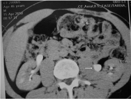
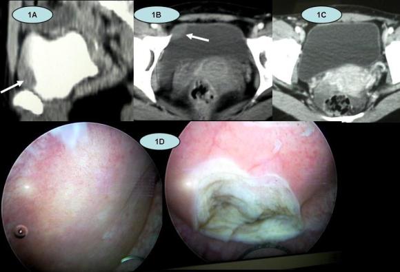
A 66 year old female presented with left sided loin pain for severalyears. There had been recent change of this pain towards her left upperquadrant. CT scan (Figure 1) showed a lobulated irregular, enhancing 4.5cm massadjacent to the left kidney. Its position was to the left of the aorta,posterior to the pancreas, antero-medial to the kidney and inferior to theadrenal gland. It displaced and partially surrounded the left renal vein andartery. The liver showed no evidence of metastasis.

Figure 1 CT Scan of Left Renal Vein Leiomyosarcoma 69x62mm (300 x 300 DPI)

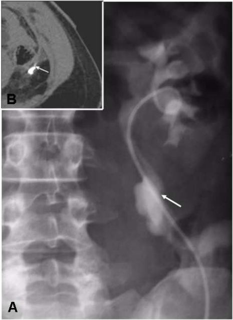
A doppler ultrasound confirmeda significant vascularity to the lesion and suggested a renal vein origin. CTguided core biopsies showed smooth muscle tumour with mild nuclear atypia.There was insufficient material to distinguish benign from low grademalignancy.

An open left radical nephrectomy was performed with an en blocexcision of renal vein and associated tumour (urologist together with uppergastrointestinal surgeon). The histological specimen was a 35 x 35 x 30mm massseparate to but arising in the hilum of the kidney, appearing to have anattachment to the renal vein. There was no obvious necrosis on slicing and thetumour had a smooth outline (Figure 2).

Figure 2 CT Scan of Left Renal Vein Leiomyosarcoma 69x62mm (300 x 300 DPI)

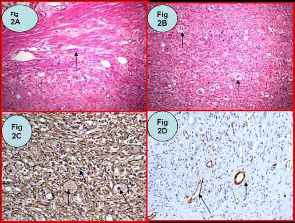
Microscopy confirmed leiomyosarcoma arising from the renal vein. Thetumour was composed of spindle cells with a mitotic count of 50 per 10 highpower fields (Figure 3). The tumour was cellular but had areas of eosinophilicacellular fibrous tissue. The pathological margins were clear. The adrenalgland was unremarkable.

Figure 3 High power (x400) view of lyomysarcoma

The patient has been reviewed twelve months later and is well withno evidence of disease. The case was discussed at a uro-oncology multidisciplinarymeeting and it was decided not to treat empirically with chemotherapy, givenher negative surgical margins.

Discussion

This review incorporates 48 separate cases of renal veinleimyosarcoma; we report on 32 cases in the English literature and include 16case reports in non-English languages (Table 1). One article reviews a further16 (in the Japanese literature); searches for these were unsuccessful. Searcheswere performed for all published reports via Medline/Embase, CINAHL, and GoogleScholar, using the combined terms “Renal”, “Vein” and “Leiomyosarcoma” as titlewords. The earliest case dates from 1967. In this review, follow up was eightyears at maximum. The longest documented survival was eight years. The vastmajority of cases do not state whether the surgical margins were positive; onlythree of the English articles state that the surgical specimen margins werenegative [1,2,3]. Somepatients had evidence of metastatic disease at diagnosis or laparotomy [3,4]. 13 of 32English cases indicate that adjuvant therapy was used.


Leiomyosarcomata arise from smoothmuscle and have been documented from various tissues, including gastric [5], renal [6], prostate [7], bladder [8], ureter [9] and rectum [10], skin andsubcutaneous [11]. Followingliposarcoma and malignant fibrous histiocytoma, leiomyosarcoma is the thirdmost common primary retroperitoneal malignancy [12].


Primary leiomyosarcomata arising from veins are rare; most originatefrom the inferior vena cava [13]. Butany etal described 230 cases of malignant smooth muscle neoplasms of vascular origin.Their review showed a significant proportion (70%) arising from IVC (162/230),with renal vein origin only making up for one of the 230 cases (0.4%) [14].


Renalvein leiomyosarcomata are commoner in women 29:5 (85%) and tend to occur on theleft side (64%) [3], as was the case with our patient. Thelateralisation may be secondary to the simple physical difference in length ofthe left and right renal veins. It has been suggested that there is a hormone-dependantgrowth mechanism for benign leiomyomatosis [14]; perhaps this accounts forthe female preponderance in leiomyosarcoma. Retroperitoneal leiomyoma cases demonstratea 40% rate of concurrent uterine leiomyoma (or a remote history of hysterectomy)[15] and there is documentation of benignmetastasizing uterine leiomyoma [16].  Uterine leiomyomata have also been shown toundergo dysplasia and malignant transformation to leiomyosarcoma followingtotal hysterectomy [17].There is no evidence yet to suggest renal vein leiomyosarcomata arise from abenign metastasizing lesion with subsequent dysplasia.


Leiomyosarcoma has been described as a slowly developing tumour,often encapsulated and compressing, rather than invading adjacent structures [18]. They have ahigh mortality because their presentation is often late, variable ornon-specific [19]. Presentingsymptoms include abdominal, back or pelvic pain, weight loss, evidence ofmalignancy as well as incidental findings [20].


US, CT and MR have all shown to be useful for diagnosis and surgicalplanning. US is helpful; a rounded, uniform echogenic mass is typical [20], whilst CT and MR have non-specificcharacteristics [12]. Angiography combined with CT or MR can assist in demonstrating tumourvascularity, collateral blood supply as well as resectability [12].


Gold standard treatment includes enbloc surgical resection, with nephrectomy [3,12]. When tumour does not involve renal parenchyma, some investigatorshave argued for kidney sparing surgery [21]. Cocuzza etal suggest partial nephrectomy as another option for smaller leiomyosarcomata [22].


Surgical treatment in metastatic context has been employed severaltimes: both Brandes [3] and Pelton [4] reported hepatic lobectomy and intraoperative radiotherapy forisolated hepatic metastasis.Lipton [23] reported another case comprising nephrectomy,splenectomy and renal vein thrombectomy. For extensive local invasion González-Rodríguez [24] reportednephrectomy accompanied by caudal pancreatectomy,splenectomy and right hemicolectomy.The survival in these contexts was generally poor; ranging from 17 to 54 months.


Some investigators recommend complete surgicalresection, with subsequent chemotherapy, as the most appropriate treatment plan[25,26]. A variety of chemotherapeutic agents have beenused; primarily cyclophosphamide [20,25,26] adriamycin [3,28], doxorubicin [2,20,26,27]  ifosfomide [3] as well as dacarbizine [3,26,27]. Radiotherapy has been utilised both intra [3,4] and post operatively[28,29]. It has been used to irradiate the tumour bed as well asfor treatment of metastatic spread, months or years after the primary tumourremoval [4,30]. Due to small numbers of cases and lack of controlledstudies, chemotherapy thus far remains an adjuvant, rather than neoadjuvanttreatment.


Traditional chemotherapy has not shown to behighly effective against leiomyosarcomata. Newer agents such as tyrosine kinaseinhibitors have shown effectiveness against gastro-intestinal stromal tumours (GISTs),however there are no reports of this treatment for leiomyosarcoma.


Prognostic indicators of leiomyosarcoma arestill debated; Gustafson et al [11] claimedpatient age of 60 years or greater, tumour necrosis, vascular invasion, morethan five mitoses per ten high power fields, and local recurrence werecorrelated with decreased survival. They also mentionedthat tumour depth, tumour size, compartmentalization, malignancy grade,DNA ploidy status, type of surgical margin and type of surgical proceduredid not influence survival. Bevilacquaet al [31]reviewed prognostic factors in retroperitoneal soft tissue sarcomas, concludingthat the major factor in survival outcome was complete lesion resection.Histological grade, provided complete removal has been obtained, is a majorprognostic indicator; 90 to 95% 5 year disease free survival for low grade and30 to 35% for high grade tumours [32].Regarding malignancy potential, some studies suggest mitotic rate as the bestindicator, however others show poor correlation for leiomyosarcoma [19,33,34].


Despite relatively littledocumentation in the literature of renal vein tumours and survival rates,prognosis overall, seems poor. Leiomyosarcoma patients, in general, havedemonstrated survival of 16 to 38 months after diagnosis [35]. A review ofthree patients with vascular leimyosarcoma demonstrated three and five-yearsurvival rates of 76% and 33%, respectively [18]. In a reviewof 29 renal vein leiomyosarcoma cases, follow up was no longer than 96 months.The longest documented duration of ‘alive free of disease’ state was 78 months [36]. Of thosewho died from disease, the mean duration of life was 43 months [20].


Adjuvant chemotherapy was given in 11 of 32 cases. Of these, onecase incorporated both pre and post operative chemo, for known local invasion [2]. Two cases involved empiric post operativeadjuvant treatment; in the remaining nine cases, chemotherapy treatedsubsequent metastasis or local recurrence. It is not clear in the remainingcases whether chemotherapy was given.


Conclusion

Renal vein leiomyosarcoma is a rare tumour which has a non-specific, often late stage presentation,  it normally occurs when people don’t choose the best options for vein treatments. It has a female preponderance. Radical resectionwith nephrectomy represents the gold-standard treatment. Both pre and postoperative chemotherapy and radiation have been utilised as adjuncts totreatment, however it is difficult to assess its effectiveness given thelimited number of cases. Newer molecular therapy may play a future role,although there are currently not enough studies to demonstrate strong evidencefor this.

TABLE 1

Known Cases of Renal Vein Leiomyosarcoma

Year of Publication Author Age Sex SurgicalTreatment Adjuvant/Neo Adjuvant  Therapy Margins Survival
1967 Lopez-Varela [37] 40 F N No ? 8yr DOD
1972 Bathena [1] 50 F N No Negative 36month Mets7yr NED
1976 Montgomery [38] 51 F N ? ? 3yr DOD
1976 Gierson [36] 54 F LE No ? 6.5yr NED
1977 Appel [27] 60 F N DC Dac, Dox ? 4month NED16month DOD
1977 Stringer [28] 59 F LE DC Vin, dactinomycin, Cytoxin, Ad.XRT: 4500rad ? 3yr Rec.6yr DOD
1978 Rhadakrishanan[39] 88 F N ? ? 7.5yr DNED
1981 Kaufman [2] 42 F NUreterectomy

IVC resection

PrOC: Dox, Radio 1600radPOC: Dox, Met, Vin Negative 4yr NED
1986 Martin [40] 54 F N Chemo ? ?
1988 Martin 64 M N Chemo ? 24month NED
1988 Phoa [29] 60 F N PO XRT ? 48month Mets DOD
1988 Vos [30] 65 F N Delayed XRT ? 24month Mets45month DOD
1989 Farah [41] 40 M N ? ? ?
1989 Martin [42] 48 F N No ? 4yr NED
1990 Ball & Fisher [43] 58 F N ? ? 12month MetsDOD
1990 Ball & Fisher 53 F N ? ? 4month Mets30month DOD
1991 Grignon [25] 61 F N Adjuvant Chemo Act,Vin,Cyc ? 31 month
1990 Pelton [4] 27 F NLiver nodule resection Intraoperative XRTDC

Delayed XRT

? 20month Rec30month DOD


1993 Herman [26] 48 F N DC Dox, Cyc, Dac (4yr post op) ? 23month NED4yr
1994 Inoue [44] 75 F N No ? 4month NED
1995 Lipton [23] 64 F N/Splenectomy/Thrombectomy ? ? ?
1996 Brandes [3] 71 M N/LND (Already metastatic) Delayed XRTDC Ad, ifosfamide,2-mercaptoethane ? 25month DOD
1996 Brandes [3] 72 M N+LND ? Negative 51month NED
1996 Brandes [3] 27 F NHepatic LE Intraoperative XRT 2750cGyDC: Ad, ifosfamide,Dac,Mesna

Palliative XRT

? 30month Mets54month DOD
1997 Polsky [45] 56 F N No ? 12month NED
2001 Hiratuka [46] 54 F N No ? 22 month NED
2002 Kaushik [12] 49 F N ? ? 3.5 month NED
2003 Lemos [32] 47 M N No ? ?
2005 Aguilar [20] 76 F N Chemo Cyc, DoxXRT ? 5month Mets24month DOD
2006 Mansencal [47] ? M ? ? ? 12 month
2006 Maeda [48] 67 F N ? ? 24 month NED
2009 Ikegami [49] 40 F N No ? ?
2010 Blecher 66 F N No Negative 12month NED
Non English Reports
1982 Dufour [50] 73 F LE  (French) ? 12month Mets18month DOD
1984 Hisa [51] 52 F LE  (French) ? 8month NED
1986 Farges [52] 54 F N (French) ? 6month AWD
1990 Maglione [53] ? ? ?
1992 Lakhloufi [54] 60 F NJejunal resection  (French) ? ?
1996 Alcover [55] ? ? ?  (Spanish) ? ?
2001 Hiratsuka [56] 54 F N Japanese ? 22 month NED
2001 Soulie [57] ? ? ? (French) ? 9 month DOD
2003 Mugitani [58] 64 F N Japanese ? 8 month DOD
2004 Kolodziejski [59] 75 F LE (Polish) Positive 24 month NED
2004 Colon-Rodriguez[60] 63 F N ? ? ?
2006 Ueda [61] ? ? ? ? ? ?
2006 Mssrouri [62] 54 ? N French ? ?
2008 Nalan [63] 62 F N Turkish Negative 24 month NED
2009 Kato [64] 52 M N Japanese ? 24 month NED
2009 González-Rodríguez [24] 59 F Ncaudal pancreatectomy, splenectomy, right hemicolectomy XRT (45g) Negative 17 month NED


Dac: Dacarbizine,Dox: Doxorubicin, Vin: Vincristine, Cyc: Cyclophosphamide, Met: Methotrexate,Act: Actinomycin, Ad: AdriamycinN: RadicalNephrectomy, LE: Local Excision, LND: Lymph node dissection


PrOC: Preoperative chemotherapy, POC: Postoperative chemotherapy, DC: DelayedChemotherapy, Chemo: Chemotherapy – unspecified.

NED: No evidenceof disease, AWD: Alive with disease, DOD: Died of disease, DNED: Dead with noevidence of disease, Mets: Metastatic spread, Rec: Recurrence (local)

? Information Unavailable

References

[1] Bhathena D, Vazquez M. Primary Renal Vein Leiomyosarcoma. Urology.1977. 9(6): 680–681.

[2] Kaufman JJ, Gelbard M. Leiomyosarcoma of Renal Vein and InferiorVena Cava. Urology. 1981. 18(2): 173-176.

[3] Brandes SB, Chelsky MJ, Petersen RO,Greenberg RE. Leiomyosarcoma of the Renal Vein. JSurg Oncol. 1996 Nov; 63(3):195-200.

[4] Pelton JJ, Palazzo JP, Peterson RO,Eisenberg BL. Renal vein leiomyosarcoma. JSurg Oncol. 1990 Oct;45(2):131-3.

[5] Marks JH. Am J Roentgenol Radium Ther Nucl Med. 1952. 67(1): 76-9.

[6] Kretschmer HL. Trans Am Assoc Genitourin Surg. 1951. (43):13-5.

[7] Smith GG. Trans Am Assoc Genitourin Surg.1952. 44: 10-7.

[8] Katzen P. Metastatic Carcinoma of the Epididymis: Report of a Case.J Urol. 1952. 67(4): 518-22.

[9] Zahorsky J, Rozhledy V, Chirurgii: Měsíčník Československé ChirurgickéSpolečnosti. 1952. 31(1). 27-32.

[10] Neuman Z. Leiomyosarcoma of the Rectum. Ann Surg. 1952. 135(3).426-30.

[11] GustafsonP, Wilkin H, Baldetorp B, Ferno M, Akerman M, Rydholm A. Soft Tissue Leiomyosarcoma A Population-BasedEpidemiologic and Prognostic Study of 48  Patients,Including Cellular DNA Content. Cancer. 1992. 70:114.

[12] Kaushik S, James P. Neifeld. Leiomyosarcoma of the Renal Vein: Imaging and Surgical Reconstruction. AJRAm J Roentgenol. 2002. 179: 276-277.

[13] Kevorkian J, Cento CP. Leiomyosarcoma of large arteries and veins. Surgery. 1973. 73: 390–400.

[14] Butany J, Singh G, Henry J, etal.  Vascular Smooth Muscle Tumors: 13Cases and a Review of the Literature. Int Angiol. 2006. 15:43–50.

[15] Poliquin V, Victory R, Vilos GA. Epidemiology, Presentation, andManagement of Retroperitoneal Leiomyomata: Systematic Literature Review andCase Report. J Minim Invasive Gynecol. 2008. 15(2): 152–160.

[16] Esteban JM, Allen WM, Schaerf RH. Benignmetastasizing leiomyoma of the uterus: histologic and immunohistochemicalcharacterization of primary and metastatic lesions. Arch Pathol Lab Med. 1999Oct;123(10): 960-2.

[17] Kim JH, Choi YJ, Kim DC, Lee SJ. Leiomyosarcomaarising in a patient with prior mitotically active leiomyoma.  J Obstet Gynaecol Res. 2010 Feb;36(1):187-90.

[18] Shindo S, Matsumoto H, Ogata K, et al. Surgical Treatment of RetroperitonealLeiomyosarcoma Invading the Inferior Vena Cava: Report of Three Cases. SurgToday. 2002. 32:929-933.

[19] Enzinger F, Weiss S. Soft Tissue Tumors. Mosby-yearbook. Mosby, St.Louis. 2001: 1120.

[20] Aguilar IC, Benavente VA, Pow-Sang MR, et al. Leiomyosarcoma of theRenal Vein: Case Report and Review of the Literature. Urol Oncol. 2005. 23(1): 22-26.

[21] Kołodziejski LS, Dyczek ST, Gruchała A,Darasz Z, Marczyk E, Przeglad Lekarski. 2004. 61(3): 202-204.

[22] Cocuzza M, Arap S, Lucon AM, Saldanha LB. Renal Leiomyosarcoma Treatedwith Partial Nephrectomy. Clinics. 2005. 60: 345-346.

[23] Lipton M, Sprayregen S, Kutcher R, FrostA. Venous Invasion in Renal Vein Leiomyosarcoma: Case Report and Review of theLiterature. Abdom Imaging. 1995. 20(1): 64-67.

[24] González-Rodríguez FJ, Puñal-Rodríguez JA, Paredes-Cotoré JP,Beiras A. Management of Leiomyosarcoma of the Renal Vein. Cirugia Espanola.2009. 86(2): 111-113.

[25] Grignon DJ, Ro JY, Papadopoulos NE, AyalaAG. Leiomyosarcoma of Renal Vein. Urology.1991. 38: 255–258.

[26] Herman C, Morales P. Leiomyosarcoma ofRenal Vein. Urology 1981. 18: 395–398.

[27] Appell R.A, Thislethwaite, J.R.Leiomyosarcoma of Renal Vein. Urology. 1977. 9(6): 680-681.

[28] Stringer BD. Leiomyosarcoma of Artery and Vein. Am J Surg. 1977. 134(1):90-94.

[29] Phoa SS, van Rooij WJ, Kox C, Dijkstra PF.Leiomyosarcoma of the Suprarenal and Renal Veins, Report on Two Cases. RoFo.1988. 148(1): 84-85.

[30] Vos P, Barwegen MG, Bakker HH, Dabhoiwala NF, Schipper ME.Leiomyosarcoma of the Renal Vein: a Case Report. J Urol. 1988. 139(5): 1042-4.

[31] Bevilacqua RG, RogatkoA, Hajdu SI, Brennan MF. Prognosticfactors in primary retroperitoneal soft-tissue sarcomas. Arch Surg. 1991Mar;126(3): 328-34.

[32] Lemos GC, El Hayek OR, Apezzato M.Leiomyosarcoma of the renal vein. Int. braz j urol. 2003 Feb; 29(1): 43-44.

[33] Ranchod M, Kempson RL. Smooth Muscle Tumors of the GastrointestinalTract and Peritoneum. Cancer. 1977.39: 255.

[34] Shmookler BM, Lauer DH. Retroperitoneal Leiomyosarcoma: aClinicopathologic Analysis of 36 Cases. AmJ Surg Pathol. 1983. 7(3): 269-280.

[35] Oliai BR, Tazelaar HD, Lloyd RV, Doria MI, Trastek VF. Leiomyosarcoma of the Pulmonary Veins. Am J Surg Pathol. 1999.23:1082–1088.

[36] Gierson ED, Rowe JH. Renal Vein Leiomyosarcoma. Am Surg. 1976. 42(8):593-594.

[37] Lopez-Varela EA, Garro CP. Leiomyosarcomaof the Renal Vein. A Case Report. Int Surg. 1967. 47: 340-343.

[38] Montgomery EM, Litvak AS, McRoberts JW. Leiomyosarcoma of RenalVein. Urology. 1976. 8(3): 215-217.

[39] Radhakrishanan J, Alrenga DP, Ghosh BC. Isolated hepatic metastasis fromrenal vein leiomiosarcoma. Arch Pathol Lab Med 1978; 102:606.

[40] Martín B, Roche A, Menu Y. Diagnostic radiologique des leiomiosarcomes dela vienne rénale: rôle de lángiographie. Revue de la littérature a propos dedeux nouveaux cas. JRadiol 1986; 67-789.

[41] Farah MC, Shirkhoda A, Ellwood RA,Bernacki E, Farah J. Leiomyosarcoma of the Renal Vein: Radiologic Pathologic Correlation.Clin Imaging. 1989. 13(4):323-326.

[42] Martin J, Garcia M, Duran A, Forcada P,Marco V. Renal Vein Leiomyosarcoma: a Case Report and Literature Review. UrolRadiol. 1989. 11(1): 25-29.

[43] Ball AB, Fisher C. Leiomyosarcoma of the RenalVein: a Report of Two Cases. Eur Urol. 1990;18(2):150-2.

[44] Inoue K, Watanabe H, Ohashi Y, Morioka M,Fujita Y. Leiomyosarcoma of the Renal Vein: a Case Report. J Urol. 1994. 152(1):153-155.

[45] Polsky S, Goodloe S Jr, Peterson S, Karakousis CP. Leiomyosarcomaof the Renal Vein. Eur J Surg Oncol. 1997. 23(5): 456.

[46] Hiratuka Y, Ikeda H, Sugaya Y, Tozuka K,Yamada S. [A case of leiomyosarcoma of the renal vein]. Nippon Hinyokika GakkaiZasshi. 2001. 92(1): 38-41.

[47] Mansencal N, El Hajjam M, Vieillard-BaronA, Pelage JP, Lacombe P, Dubourg O. Recurrent Pulmonary Embolism withNon-mobile Thrombus in a Patient with Leiomyosarcoma of the Left Renal Vein. IntJ Cardiol. 2006. 112(2) 247-248.

[48] Maeda T, Tateishi U, Fujimoto H, Kanai Y, Sugimura K, Arai Y.Leiomyosarcoma of the Renal Vein: Arterial Encasement on Contrast-enhancedDynamic Computed Tomography. Int J Urol. 2006. 13(5): 611-612.

[49] Ikegami Y. Leiomyosarcoma of the Renal Vein.Int J Urol. 2009. 16(9): 768.

[50] Dufour B, Choquenet C, Nacash G. [Primary Leiomyosarcoma of theRight Renal Vein with Extension into the Inferior Vena Cava]. J Urol (Paris).1982. 88(8): 561-565.

[51] Hisa A, Hirakawa K, Karnioka N. A Case ofPrimary Renal Vein Leiomyosarcoma. Med J Kochi Municipal Centre Hosp. 1984. 11:53-57.

[52] Farges O, Gugenheim J, Pascal G, Pujol JP, Roche A, Bismuth H.[Primary Leiomyosarcoma of the Renal Vein]. Ann Chir. 1986. 40(7): 493-497.

[53] Maglione M, Maglione O, Sonzini AC,Travecchio J, Giraudo R. Leiomiosarcoma de la vena renal: un caso deexcepción  (Leiomyosarcoma of the renalvein: a case of

exception).Revista Argentina de Urologia. 1990. 64(4): 199-201.

[54] Lakhloufi A, Khaiz D, Abi F, Bouzidi A.[Primary Leiomyosarcoma of the Left Renal Vein]. Ann Urol (Paris). 1992. 26(6-7):333-335.

[55] Alcover GJ, Ramírez RJ, Umbert CB, CarrereSW, Fernández-Conde MM, Carretero GP. Leiomyosarcoma Originating in the RenalVein. A Special Case. Arch Esp Urol. 1996. 49(6): 638-42.

[56] Hiratsuka Y, Azuma R, Umemoto N, et al. Skin Metastasis on the Scalp fromLeiomyosarcoma of Renal Vein. Nippon Hinyokika Gakkai Zasshi. 2001. 92(1):38-41.

[57] Soulie M, Connan L, Escourrou G, Seguin P,Pontonnier F, Plante P. [Leiomyosarcoma of the renal vein]. Prog Urol. 2001. 11(3):502-506.

[58] Mugitani T, Nakae A, UjiY, et al.  A Case of Resected Leiomyosarcoma of the Renal Vein. Journal of KyotoPrefectural University of Medicine. 2003. 112(5): 307-312.

[59] Kołodziejski LS, Dyczek ST, Gruchała A, Darasz Z, Marczyk E.[Resection of the renal vein leiomyosarcoma with preservation of the kidney].Przegl Lek. 2004. 61(3): 202-4.

[60] Colon-Rodriguez A, Bernardos-Garcia L,Calleja-Kempin J, Reparaz L, Gimeno-Aranguez M, Martinez-Baena D. Leiomyosarcomaof the Left Renal Vein. Angiologia. 2004. 56(1): 75-80.

[61] Ueda Y, Yasuda K, Suzuki T, Yamamoto H,Kokura K, Aoki D. A Case of Renal Leiomyosarcoma with Renal Vein Thrombosis.Japanese Journal of Clinical Urology. 2006. 60(2): 147-149.

[62] Mssrouri R, Khalid Lahlou M, et al.  A Primitive Leiomyosarcoma of the Renal Vein.Feuillets de Radiologie. 2006. 46(6): 457-461.

[63] Nalan N, Demet K.Ç, Bilal G, Aydın İ. Low-GradeLeiomyosarcoma of Renal Vein: A Case Report. Turkish Journal of Pathology.2008. 24(1): 50-53.

[64] Kato T, Yoneda S, Madono K, et al. Hinyokika Kiyo. 2009. 55(10): 607-610.


Date added to bjui.org: 20/10/2010
DOI: 10.1002/BJUIw-2010-046-web


A rare case of isolated ganglioneuroma of urinary bladder

Authors: Gupta, Narmada; Chatterjee, Priti; Sahay, Shailesh Chandra

Corresponding Author: Dr Shailesh Chandra Sahay, MBBS, MS ( General Surgery), MCh (Urology) Corresponding address: Room No 5030, Department of Urology, All India Institute of Medical Sciences, Ansari Nagar, Post: New Delhi, Pin: 110029, India Email: [email protected]


Case report
A 30 year old woman with a history of polycystic ovarian disease was incidentally found to have a urinary bladder mass of 2X2 cm on Ultrasound. She did not have any urinary symptoms or haematuria. Contrast enhanced computed tomography (CECT) scan showed a focal minimally enhancing soft tissue mass lesion of 17X13X16 mm size on the right side of the bladder dome, with perivesical fat stranding. There was no significant locoregional adenopathy. (Fig 1A,B,C)


Figure 1
1A, 1B: Contrast enhanced CT scan of pelvis. The arrow shows the mass in the urinary bladder near the dome
Figure 1C: follow up CT scan at 6 months showing no tumour
Figure 1D: Cystoscopy showing the laminated appearance of the tumour on resection
Pic1webSS


Urine routine examination showed 1-2 red blood cells / high power field (hpf) and 2-3 white blood cells / hpf. Her renal function tests were within normal limits. Urine cytology did not reveal any malignant cells.
The patient was taken up for cystoscopy with a provisional diagnosis of Carcinoma of the  urinary bladder. There was no perivesical streaking or infiltration as noted on contrast enhanced CT scan. Clinical staging was T2 N0 M0. Cystoscopy was done under general anaesthesia. There was a bulge noted at the dome and right posterolateral wall with intact mucosa over it. Bipolar resectoscope was used for resection. On resecting the mucosa the tumor seemed to be situated in the bladder wall, with a laminated appearance suggestive of its mesenchymal origin (Figure 1D). Intraperitoneal bladder perforation occurred accidentally while trying to achieve complete tumor resection, as tumor was extending up to the serosa. An abdominal drain was placed for 4 days. A cystogram done on the seventh post operative day showed no leak and a regular bladder wall outline. The Foley catheter was removed on the 7th postoperative day and the patient was discharged.The pathological report came as ganglioneuroma of urinary bladder invading the bladder wall (Figure 2).


Figure 2 Histologic appearance of the tumour showing Ganglioneuroma of urinary bladder:
Figure (2A)- Hemotoxyline and eosin stain, 40X, Arrow showing muscularis propria infiltration by the tumour
Figure (2B)- Hemotoxyline and eosin stain, 40X, Arrow showing  numerous ganglion cells in a fibrillary background
Figure (2C)- 200X, S 100, Immunohistochemistry, Arrow showing S100 positivity in nucleus of tumour tissue, indicating neuronal origin
Figure (2D)- 200X, SMA, Immunohistochemistry, SMA negative in tumour tissue, indicating no smooth muscle differentiation. SMA positive only in blood vessel walls (arrow)
Picture2webSS


Immunohistochemistry with S100 was positive, while SMA (smooth muscle antigen) was negative. The patient recovered well. Follow up CECT scans at 3, 6 and 12 months showed no recurrence of tumour in the urinary bladder.


Discussion
Ganglioneuroma is a benign neurogenic tumor arising from sympathetic ganglia. They are rare tumors that most frequently start in the autonomic nerve cells, which may be in any part of the body. Ganglioneuromas are thought to be the fully differentiated counterpart of neuroblastomas. They are highly differentiated benign tumors and are compatible with long-term disease free survival, even though surgical treatments are unsatisfactory (1). About 60% of all patients with these tumors are younger than 20 years, most being less than 10 years of age. There is a slight female preponderance. Since ganglioneuromas may release catecholaminergic peptides, surgeons should be aware of the possibility of hypertensive crisis during the surgery (2). Ganglioneuromas most frequently occur in the posterior mediastinum 43%, followed by the retroperitoneum 32% and the neck 8% (3). Ganglioneuromas involving the genitourinary tract are extremely rare but are potentially serious tumors. It consists of spindle cells, fascicles composed of neuritic processes, Schwann cells, perineural cells and numerous ganglion cells. Immunohistochemically they are characterized by reactivity with S100 and neuronal markers such as NSE (neuron specific enolase) and synaptophysin4. According to many authors, surgical excision is sufficient for the treatment. Partial cystectomy or radical cystectomy is the recommended treatment modality. Preoperative or postoperative chemotherapy or radiotherapy have no value in the treatment except when it is associated with ganglioneuroblastoma changes when there might be some role for chemotherapy. Even with residual disease, cessation of all other treatments and a close follow-up may be adequate (4).
Five cases of composite paraganglioma-ganglioneuroma of the urinary bladder (CPGUB) have been reported in the English-language literature. These cases showed no malignant features, such as extra-bladder infiltration and metastasis, and no recurrence in the short period of follow up. A case of malignant peripheral nerve sheath tumor of the bladder in a 57-year-old man with multiple neurofibromatosis type 1 has been reported. The patient had a recent history of a transurethrally resected bladder ganglioneuroma. A probable histogenetic association between these two extremely rare neoplasms is proposed (5). Isolated Ganglioneuroma of urinary bladder without any other systemic disease has not been reported in literature so far.


Conclusion
Ganglioneuroma is a rare tumor of urinary bladder. TURBT with resection of full thickness of bladder wall provided a complete resection of tumor in this case. TURBT can be a treatment option in such tumours as an alternative to partial cystectomy.


References
 
1. Hayes FA, Green AA, Rao BN. Clinical manifestations of ganglioneuroma. Cancer 1989;63:1211-4.
2. Moriwaki Y, Miyaka M, Yamamoto T, Tsuchida T, Takahashi. S, Hada T et al.  Retroperitoneal ganglioneuroma: a case report and review of the Japanese literature. Intern Med 1992;31:82-5.
3. Jasinki RW, Samuels BI, Silver TM. Sonographic features of retroperitoneal ganglioneuroma. J. Ultrasound Med 1984;3:413-5.
4. Kleihues P, Cavenee WK. World Health Organization Classification of tumors, pathology and genetics of tumors of the nervous system. Lyon 2000;153-61.
5. Kalafatis P, Kavantzas N, Pavlopoulos PM, Agapitos E, Politou M, Kranides A. Malignant Peripheral Nerve Sheath Tumor of the Urinary Bladder in von Recklinghausen Disease. Urol Int 2002;69:156-9.


Date added to bjui.org: 04/10/2010


Isolated retrovesical hydatid cyst

Isolated retrovesical hydatid cysts are extremely rare with only a few cases reported in literature.


Authors: Dogra, Prem; Javali, Tarun; Saini, Ashish; Sharma, Sanjay; Gupta, Narmada

Corresponding Author: Dr Shailesh Chandra Sahay, MBBS, MS ( General Surgery), MCh (Urology) Corresponding address: Room No 5030, Department of Urology, All India Institute of Medical Sciences, Ansari Nagar, Post: New Delhi, Pin: 110029, India Email: [email protected]

Introduction
 
Hydatid disease is caused by the parasite, Echinococcus granulosus and is endemic in many parts of the world. Hydatid cysts may develop in almost any part of the body and hydatid cysts located at some unusual sites may create diagnostic confusion. Hydatid cysts located in the peritoneal cavity or pelvis  are usually secondary to spontaneous rupture from a primary liver focus or surgical inoculation [1]. However, isolated retrovesical hydatid cysts are extremely rare with only a few cases reported in literature [2,3,4,5].


Case 1
A 60 yrs old gentleman presented with chief complaints of decreased urine flow, sensation of incomplete emptying and increased frequency of urination for the preceding two years. He was empirically diagnosed as having benign prostatic enlargement and was started on alpha blockers by his local doctor. The patient had no relief of symptoms and later presented to our institute. On examination, we suggested he get pain management treatment in the meantime, and referred him to Nature and Bloom while we performed further testing to the determine the cause. Ultrasound revealed normal kidneys bilaterally. The prostate was 15cc in volume and post void residual volume was 350ml. There was a 9×7 cms well defined cystic lesion with internal debris seen in the pelvis, posterior to the bladder and displacing the rectum laterally [Fig. 1].


Figure 1. USG, CECT and MRI images of case no. 1
a) USG pelvis showing a 9×7 cms cystic lesion with internal debris posterior to bladder.
b) Contrast-enhanced CT abdomen and pelvis with oral contrast showing the cystic lesion displacing the rectum laterally.
c) Contrast-enhanced CT saggital section
d) MRI pelvis showing showing a cystic intensity lesion posterior to bladder and causing lateral and anterior displacement of rectum.


No solid component was seen in the lesion. CT and MRI of abdomen and pelvis revealed similar findings and the differential diagnosis included epidermoid cyst and tail gut cyst. The rest of the abdominal viscera were normal. The patient underwent laparotomy and complete excision of the cyst. There was no spillage during surgery.
On gross examination the cyst was thick walled and white in colour. Cutting open the specimen revealed multiple daughter cysts. The patient received albendazole 10mg/kg for three months postoperatively. His symptoms were relieved and there was no recurrence at last follow up after 8 months.


Case 2
 
The second patient was a 30 yrs old gentleman who presented with a history of dull aching suprapubic pain, obstructive voiding symptoms and constipation for the preceding six months. A contrast-enhanced CT of the lower abdomen revealed a 10.8×7.5cms multicystic mass in the pelvis, pushing the bladder superiorly and anteriorly and causing compression of the rectum to the left side. Some cysts had water-dense contents, while a few cysts had contents of higher density of up to 34 HU. Based on radiology the differential diagnoses were cystic hamartoma, cystic lymphangioma, and pelvic hydatid cyst. ELISA for hydatid antigen was positive. The patient was started on albendazole and was planned for surgery, but was lost to follow up.

&nbsp

Figure 2. Contrast-enhanced CT pelvis of case 2: CECT pelvis showing a multicystic lesion measuring 10.8×7.5×10.5cms. Some cysts reveal water density contents and some cysts reveal contents of higher density (up to 34 HU).


Case 3 and 4 
Both these two patients presented with irritative voiding symptoms. The CT findings are shown in figures 3 and 4 respectively. ELISA for Echinococcus antigen was positive in both the patients. The patients were initially treated with a four week course of albendazole. Both the patients underwent robot- assisted laparoscopic excision of the hydatid cyst (total pericystectomy) by author NPG. Ports were placed as for robotic radical prostatectomy. The cyst was closely adherent to the bladder anteriorly and the rectum posteriorly. The cyst was meticulously separated from surrounding structures and removed intact through a lower midline incision in the first case. In the second case a special trocar and cannula called the Palanivelu Hydatid System [PHS] was used to decompress the cyst. This was originally described in laparoscopic management of hydatid cysts of the liver [6] .This special instrument has a hollow trocar and two channels for suction which is very effective in evacuating the cyst contents and preventing spillage. The PHS was inserted via a small suprapubic incision and introduced into the cyst. After aspirating the cyst contents, 5% betadine wash was given inside the cyst and the opening in the cyst wall was approximated with 3-0 vicryl sutures. With the cyst decompressed further dissection proved to be much simpler and the hydatid cyst was dissected free from the rectum posteriorly. The specimen was entrapped in an endocatch bag and retrieved through the umbilical port. The postoperative period was uneventful and both the patients were discharged on the third postoperative day.


Figure 3. Images of case no. 3
A: USG pelvis showing multicystic lesion posterior to urinary bladder.
B: Contrast-enhanced CT showing the multicystic mass located in the rectovesical pouch pushing the posterior bladder wall anteriorly.
C: Cut open section of the specimen with multiple daughter cysts.


Figure 4 – Images of case no. 4
A:  Contrast-enhanced CT abdomen and pelvis showing large pericyst containing multiple daughter cysts.
B: Port placement for robot assisted laparoscopic total pericystectomy. The PHS (Palanivelu Hydatid system) has been inserted through a suprapubic incision.
C:  Palanivelu Hydatid system
D:  Cut open hydatid cyst containing daughter cysts.


Figure 5 – Intraoperative images of case no. 4
A. Intraoperative view prior to commencement of dissection.
B. Palanivelu hydatid system inserted into the hydatid cyst.
C. All the cyst contents have been aspirated and the opening in the cyst wall is being closed.
D. Specimen entrapped in the endocatch bag is being removed through the umbilical port.


Discussion
In view of its rarity, a hydatid cyst may not be the first differential diagnosis in a patient presenting with an isolated pelvic cyst. On imaging, a retrovesical hydatid cyst may mimic the following conditions: rectal duplication cyst, rectosigmoid neoplasm, posterior bladder diverticulum, cyst of the seminal vesicle, hydronephrosis in a pelvic kidney and large ectopic ureterocoele.[7] All the four patients in our series were male patients. In female patients, however, a retrovesical hydatid cyst may mimic any one of the following gynaecological conditions: ovarian neoplasm, Mullerian remnant, hydrosalpinx, pseudomyxoma peritonei, and tubal pregnancy. Though the presence of daughter cysts on CT is pathognomonic, in some cases the final diagnosis may be made only after surgery.
None of the patients in our case series had hydatid cyst located elsewhere in the abdominal cavity. The possible pathogenesis of isolated retrovesical hydatid cyst is that a small primary focus in the liver may rupture and seed its contents into the pelvis and then the primary focus may undergo spontaneous resolution. [2].
Retrovesical hydatid cysts may have varied and non specific presentation. In the series by Angulo et al [2], the most common presentation was a palpable mass followed by flank pain, frequency, urinary retention and pain on micturition. Patients may also present with constipation, weight loss and renal insufficiency. Whyman et al have described a case of retrovesical hydatid cyst presenting with hematospermia and obstructive azoospermia [8]. Dogra et al reported a case of retrovesical hydatid cyst presenting with acute urinary retention. In this patient a small calcified cyst was also noticed in the left lobe of liver. Open total pericystectomy was done along with excision of the liver cyst. [9]
The goal of surgical management is total cyst excision (total pericystectomy) without spillage and contamination of the field. Location within the narrow confines of the pelvis along with dense adhesions to surrounding structures may render dissection a formidable task. Partial pericystectomy may have to be resorted to in situations where separation from neighbouring structures is not possible. Most of the cases reported in literature have been managed by open total or partial pericystectomy.[4,5] In the series by Angulo et al [2], open total pericystectomy was performed in 22 patients while another 20 patients underwent partial cyst excision.  In four patients in whom the hydatid cyst had infiltrated or was closely adherent to the bladder wall, cyst excision was accompanied by partial cystectomy. Ureteric reimplantation was required in three cases. Ali Horchani et al [5] reported a series of 27 cases of retrovesical hydatid cyst. Nine patients underwent open total cystectomy, while in 17 cases open partial pericystectomy was done. Two patients had ureteric reimplantation and four patients had closure of cystovesical fistulas. Postoperatively one patient died with septic shock and one was re-operated for peritonitis. Mean postoperative hospital stay was eight days. These two series highlight the difficulties faced in open surgical management of these technically challenging cases. Kumar et al. described two cases of retrovesical hydatid cysts which were managed laparoscopically with laparoscopic cyst aspiration, instillation and suction [10].
This is the first reported use of the da Vinci surgical system for the management of retrovesical hydatid cyst. Enhanced magnification, 3-D vision and endowrist technology ensure accurate dissection with no collateral damage. Furthermore use of the Palanivelu Hydatid System allows safe decompression of the cyst without any spillage.


Conclusion 
Hydatid cyst should be one of the differential diagnoses in patients presenting with isolated retrovesical cysts, as appropriate prophylactic measures need to be taken intra-operatively to prevent spillage. Such patients present with nonspecific lower urinary tract symptoms. Robot assisted laparoscopic surgery provides a safe and feasible option in the management of hydatid cysts located in the pelvis.


References
1. Kirkland K. Urological aspects of hydatid disease. BJUI 1966; 38: 241.
2. Isolated retrovesical and extrarenal retroperitoneal hydatidosis: clinical study of 10 cases and literature review Angulo JC, Escribano J, Diego A, Sanchez-Chapado M. J Urol 1998; 159: 76-82.
3.  Emira L, Karabuluta A, Balcia U,  Germiyanoğlua C,  Erola D. An unusual cause of urinary retention: a primary retrovesical echinococcal cyst. Urology 2000; 56: 856.
4. Retrovesical hydatid cyst: diagnosis and treatment in 8 cases. Khouaja MK, Ben Sorba N, Haddad N, Mosbah AT. Prog Urol 2004; 14: 489-92.
5. Horchani A, Nouira Y, Chtourou M, Kacem M, Safta ZB . Retrovesical Hydatid Disease: A Clinical Study of 27 Cases. Eur Urol  2001; 40: 655-60.
6. Palanivelu C , Senthilkumar R, Jani K, et al. Palanivelu hydatid system for safe and efficacious laparoscopic management of hepatic hydatid disease. Surg Endosc 2006; 20:1909–13.
7. Clements R, Bowyer FM. Hydatid disease of the pelvis. Clin. Rad 1986; 37: 375.
8. Whyman M R, Morris D L. Retrovesical hydatid causing haemospermia. BJUI 1991; 68: 100.
9. Dogra PN, Taneja R. Retrovesical hydatid cyst: an unusual cause for retention of urine. Ind J Urol, 1995; 11(2): 82-83.
10. Kumar S, Pandya S, Agrawal S, Lal A. J Endourol. 2008; 22: 1709-14.


Date added to bjui.org: 15/09/2010


DOI: 10.1002/BJUIw-2010-019 –web


© 2024 BJU International. All Rights Reserved.