Archive for category: Case Studies

AV malformation of the penis

AVM of the glans penis is a very rare entity. It may be congenital or there may be a history of trauma. It may present asymptomatically. Meticulous surgery provides complete excision & good cosmetic results

Authors: Gupta, Anurag ; Jaideep, Mahajani
Corresponding Author: Gupta, Anurag


Introduction

AV malformations of genitalia have been reported very rarely in the literature. Although they are usually thought to be congenital, trauma has been implicated as a cause. For diagnosis, colour doppler ultrasound, digital subtraction angiography and magnetic resonance imaging can be used to identify the characteristics of a lesion. Because of its rarity, there is no clear guideline for treatment.

Case Report

A 24–year–old male presented with a painless swelling on the glans penis adjacent to the meatus (Fig.1), which expanded with erection and had gradually increased in size in the previous 2 to 3 years. There was no history of trauma. Physical examination revealed a slightly bluish lesion, soft on palpation, protruding from the surface of the glans penis. There was no history of voiding disturbances, haematuria, urinary tract infection, priapism or pain with erection. The main concerns were mild discomfort during coitus, cosmesis and psychological disturbance. On clinical examination there was no bruit. Color Doppler was suggestive of AVM. The blood cell count, urinalysis & coagulation profile were within normal limits.

A vertical skin incision extending from the base of scrotum to the anal margin was given. After incision of Colles’ fascia, urethra was located and dissected. The urethra was approached at the distal margin of the bulbospongiosum muscle. Buck’s fascia was opened on either side of the urethra and a plane was developed between the spongiosum and cavernous bodies. Once the urethra was separated from the corpus cavernosum, a vascular tape (or infant feeding tube) was placed to elevate the urethra and facilitate its proximal and distal dissection. The bulbospongiosum muscle was then opened in the midline and separated laterally on each side to expose the underlying corpus spongiosum. Mobilization of the whole length of the bulbar urethra was done. The AVM was excised completely including part of the glans and distal glanular urethra involving AVM was also excised (Fig.2). Feeding artery at glans was identified and ligated (Fig.3). Finally after achieving hemostasis, an orthotopic, widely patent neomeatus was created (Fig.4).

The postoperative course was uneventful, and there was no functional or cosmetic defect. Surgical excision was undertaken to give to best chance of definitive resolution and to achieve better cosmetic results. At the 6-months follow-up, there was no evidence of recurrence.

Discussion

Various active interventions have been reported to treat AVM of the glans penis, including sclerotherapy, embolization and surgical excision. In our patient, surgical excision was preferred as the initial treatment because the lesion was single, small, localized and involving the meatus.

Considerable additional urethral length was required to overcome the terminal urethral deficiency created by excision of the AVM and to create a tension-free anastomotic neomeatoplasty, so full length mobilization of the whole bulbar urethra was performed beforehand.

Mobilization and advancement of the penile urethra alone are rarely sufficient to achieve this without causing penile chordee. The only part of the urethra that can be mobilized to provide extra length without creating penile curvature chordee is the bulbar urethra. Mobilization of the whole length of the bulbar urethra through a perineal incision provides 2 to 2.5 cm of tension-free lengthening in children and 4 to 5 cm in adults. The bulbar urethra does not participate in an erection and can be extensively mobilized proximally to the genitourinary diaphragm and distally to the penoscrotal junction without causing erectile impairment. The bulbar urethra has a very rich blood supply, proximally from the bulbar and urethral arteries and distally by retrograde flow from the glans penis and from perforating arteries, branches of the cavernosal and dorsal arteries.

Our surgical technique is based on the principle of bulbar elongation and meatus advancement described by Turner Warwick5. Mobilization of only the distal and mid bulbar urethra was required. The proximal bulbar urethra was not mobilized and utmost precautions were taken to avoid injury to the bulbar artery on either side. As retrograde supply and supply through perforators to the urethra is hampered during the surgery; further injury to the bulbar artery could lead to complete urethral necrosis.

Conclusion

AVM of the glans penis is a very rare entity. It may be congenital or there may be a history of trauma. It may present asymptomatically. Meticulous surgery provides complete excision & good cosmetic results.

F1F2F3F4


References

1. Forstner R, Hricak H, Kalbhen CL, Kogan BA, McAninch JW.Magnetic resonance imaging of vascular lesions of the scrotum and penis. Urology. 1995 Oct; 46(4): 581-583.
2. Guz BV, Ziegelbaum M, Pontes JE. Arteriovenous malformation of spermatic cord. Urology. 1989 May;33(5):427-8.
3. Hamid S, Aquilina JW, Davidson W, Dhabuwala CB. Arteriovenous malformation of scrotum: A case report. J Urol. 1992 Jan;147(1):160-2.
4. Mathur P, Porwal KK, Pendse AK, Parihar US, Chittora R.Hemangiomatous penile horn. J Urol. 1996 May;155(5):1738
5. Warwick RT, Parkhouse H, Chapple CR. Bulbar Elongation Anastomotic Meatoplasty (BEAM) for Subterminal and Hypospadiac Urethroplasty. J Urol. 1997 Sep;158(3 Pt 2):1160-7.
6. Sule JD, Lemmers MJ, Barry JM. Scrotal arteriovenous malformation: Case report and literature review. J Urol. 1993 Dec;150(6):1917-9
7. Woolley MM, Stanley P, Wesley JR. Peripherally located congenital arteriovenous fistulae in infancy and childhood. J Pediatr Surg. 1977 Apr;12(2):165-76.


Date added to bjui.org: 24/11/2012

DOI: 10.1002/BJUIw-2011-132-web

Osseous Metaplasia in Anterior Urethra

A 40-year-old male with history of gonococcal urethritis 20 years ago presented with repeated episodes of dysuria and urinary stream thinning for nearly 10 years. The clinical diagnosis was urethral stricture with calculi formation. Segmental resection of urethral stricture and end to end anastomosis were performed. Postoperative pathologic examination revealed multiple submucosal focal deposition of calcium salts with osseous metaplasia. Urethral osseous metaplasia is extremely rare and chronic inflammatory may caused reactive bone formation in suburothelium.

Authors: Wang, Yongquan ; Song, Siji; Xiong, Enqing
Corresponding Author: Wang, Yongquan


Introduction
Anterior urethral stricture is not rare, and is usually due to repeated chronic inflammation, stones or catheterization. However, anterior urethral stricture with osseous metaplasia is extremely rare. Here we present a male patient with 10-year-repeated dysuria; the clinical impression was of anterior urethral stricture with calculi, but histopathologic features revealed osseous metaplasia within the urethra.

Case report
A 40-year old male consulted our centre for assessment of a 10-year history of repeated dysuria and urinary stream thinning. He had received prior urethral dilatation treatment. He suffered from gonococcal urethritis 20 years ago, which had been managed with medical treatment for 1 month (he could not remember in detail). Physical examination found palpable hard stenosis in the penile urethra. The results of urine culture were negative. Gonococcus smear in urethral secretions was negative. Tuberculosis was also excluded by purified protein derivative test (PPD), urine polymerase chain reaction (PCR) and serum anti-tuberculosis antibody detection. Pelvic X ray radiography and voiding cystourethrography demonstrated an anterior urethra stricture and half-arc shaped high density image (Figure 1). The clinical diagnosis of urethral stricture with calculus formation was made. Endourethral treatment with a ureteroscope attempting to push the calculus back into the bladder failed, because the ureteroscope could not pass through and there was no obvious stone inside the urethral lumen. Segmental resection of the urethral stricture and end to end anastomosis were performed. During the operation, a bone-like mass was palpated within the urethra, which was impacted and closely connected with the urethral mucosa (Figure 2). The length of stricture segment was about 2 cm. Postoperative pathologic examination revealed multiple submucosal focal deposits of calcium salts with osseous metaplasia (Figure 3 A and B ). Alizarin red staining indicated extensive calcium nodules and ossification (Fig3 C and D). After indwelling catheterisation for 2 weeks, the patient urinated satisfactorily and had no other problems at 6 month follow-up.

Discussion
Osseous metaplasia in the male urogenital system is uncommon and occasionally reported in urinary tumours or ureteral amyloidosis [1-3]. Urethral osseous metaplasia has never been reported in literature, so far as we are aware. Chronic infection stimulates the proliferation of mesenchymal cells that have inherent properties of metaplasia and can differentiate into chondroblasts or osteoblasts [4,5]. Urethral osseous metaplasia is an extremely rare event and the gonococcal infection history may relate to the pathological changes in this case. Although very rare, osseous metaplasia need to be distinguished from calculi. Patients with urethral osseous metaplasia have relatively long-term history, negative results of urine analysis and no obvious stone visualized endoscopically.

Conclusion
Anterior urethral osseous metaplasia is extremely rare. Pathologists and urologists should be aware of this kind of uropathy. We recommend open operation treatment for urethral osseous metaplasia, rather than urethral dilatation and endourethral operation.

References
1. Eble JN, Young RH. Stromal osseous metaplasia in carcinoma of the bladder. J Urol. 1991;145:823-825.
[1] Yamaguchi K, Kitagawa N, Kotake T, et. al. Primary localized amyloidosis of the ureter associated with osseous metaplasia. Urol Int. 1991;47:164-166.
[2] Dudley AG, Tomaszewski JJ, Hughes AH, et. al. Incidentally Discovered Osseous Metaplasia Within High-grade Urothelial Carcinoma of the Bladder. Urology. 2011 (in press).
[3] Hartman RJ, Helfand BT, Dalton DP. Clear cell renal cell carcinoma with osseous metaplasia: a case report. Can J Urol. 2011;18:5564-5567.
[4] Willis SF, Bariol SV, Tolley DA. Bone formation in the urinary tract: case report. J Endourol. 2005;19:878-879.
[5] Lainas T, Zorzovilis I, Petsas G, et. al. Osseous metaplasia: case report and review. Fertil Steril. 2004;82:1433-1435.

42 1

Figure 1
A Pelvic X ray radiography showed showed half-arc shaped high density
B Excretory and retrograde urethrogram showing the anterior urethra stricture and irregular calculus.

42 2

Figure 2
Gross specimen of urethral stenosis. A 2.0 by 0.5 by 0.5-cm hard-texture cylindrical mass mixed with urethral mucus and bone like structure.

42 3

Figure 3
Pathological section; Haematoxylin and eosin stains (×200) showed chronic inflammation of urethra submucosa and multiple focal deposition of calcium salts (*) and with osseous metaplasia cells(→).

Date added to bjui.org: 22/11/2012

DOI: 10.1002/BJUIw-2012-042-web


Renal artery dissection as an unusual cause of unilateral loin pain

A 36-year old gentleman presented with severe sudden onset unilateral loin pain, suggestive of renal colic. Renal tract ultrasonography was unremarkable, but computerised tomography demonstrated renal infarction. Subsequent angiography proved a renal artery dissection. This uncommon diagnosis should be considered in patients with sudden onset loin pain, particularly those with risk factors such as hypertension, connective tissue disease or trauma.

Authors: King, Chris; Amdekar, Shivan
Corresponding Author: King, Chris

Dr Chris King, MB BChir.
Vascular surgery, Northwick Park Hospital, North West London Hospitals NHS Trust, Watford Road, Harrow, HA1 3UJ

Dr Shivani Amdekar, MB BS
General surgery, Northwick Park Hospital, North West London Hospitals NHS Trust, Watford Road, Harrow, HA1 3UJ

Introduction
Spontaneous renal artery dissection is a rare event, typically constituting only 0.05% of all arteriographic findings on radiological imaging [1].  Diagnosis is typically delayed due to difficulties distinguishing the presentation from more familiar conditions such as renal colic and pyelonephritis. We report the case of a 36-year old office worker presenting with severe loin pain and the 3 days it took to reach a definitive diagnosis.

Case Report
A 36-year old Indian gentleman presented to the emergency department with severe sudden onset right loin pain and vomiting, ongoing for 8-10 hours. The pain came on over a period of minutes, and was exclusive to the flank with no radiation. It was constant in nature, and he had never had a similar pain before. He had no urinary symptoms. The patient was otherwise fit and well. In particular he had no predisposing factors such as atrial fibrillation, hypertension, history of trauma or connective tissue disorder. On examination he was afebrile but diaphoretic, with a tender right flank. There was no evidence of peritonism. Testicular examination was unremarkable. His blood pressure was 152/103 mmHg. He had microscopic haematuria but no nitrites or leukocytes in his urine. His white cell count was 15.8 x10⁹/L and C-reactive protein 2mg/L. His renal function was within normal range (creatinine 93umol/L, urea 4.8mmol/L). A renal ultrasound demonstrated no hydronephrosis or visible ureteric/renal calculi. There was normal renal corticomeduallary differentiation. He was admitted and started on treatment for renal colic. The following night the patient developed tachycardia and after senior review was treated for sepsis. He was started on ciprofloxacin and gentamicin for possible pyelonephritis or retrocaecal appendicitis. An abdominal computerised tomography (CT) scan the next morning demonstrated an acute infarct of the lateral half of the right kidney with accompanying perinephric stranding. CT angiography highlighted a long linear filling defect of the right renal artery in keeping with arterial dissection, completely occluding the anterior branch and partially occluding the posterior branch. All other abdominal arteries were unremarkable. The patient was managed medically with anticoagulation as it was felt that the dissection extended too far distally for any interventional management such as endovascular stenting. Further screens for infective endocarditis, vascultitis and hypercoagulability were negative. His urea and creatinine remained within the normal range throughout this time. The patient’s pain settled and he was discharged from hospital on warfarin and encouraged to resume normal activity. A follow-up magnetic resonance renal angiogram at 6 weeks confirmed dissection as the original cause of infarction, with a subsequent 2.4cm length of completely thrombosed renal artery.

Discussion
There are approximately 200 published cases of spontaneous renal arterial dissection, and it is likely that the condition is underdiagnosed. Most patients are male (a 4:1 ratio), in their 4th-5th decade of life [2]. The condition is associated with hypertension, connective tissue diseases such as Marfan’s and Ehler-Danlos syndromes, fibromuscular dysplasia, atherosclerosis, trauma, and severe physical exertion [3]. The commonest symptom is severe upper abdominal flank pain, often radiating to the epigastrium, but this is not always present [4, 5]. There may be associated nausea and vomiting. One case series of 24 patients reported hypertension in 96% of presentations, gross haematuria in 21% and headaches in 25% [4].
Measurements of serum lactate dehydrogenase may be suggestive of renal infarction [5]. However angiography provides the gold standard for diagnosis [4]. Angiography also allows one to distinguish between causes. Dissections and thromboemboli appear differently on angiography, the former appearing as an abrupt uniform narrowing due to non-filling of the false lumen, whereas the latter display a meniscus crossing the width of the artery [3].
Treatment involves anti-coagulation and control of hypertension, and then either conservative or operative management with the goal of tissue preservation. The latter includes endovascular repair, arterial bypass or nephrectomy [1]. These techniques are best reserved for patients with uncontrolled hypertension, renal insufficiency, or progressive disease as seen on serial angiography [4]. The duration of anticoagulation is largely empirical and based on evidence for carotid dissection [2].
The natural history of the condition is poorly understood due to its rarity and often lack of follow-up. The most common complication is malignant hypertension [4]. Mortality is most associated with renal insufficiency, inherently more common in bilateral disease [4].

Conclusion
Spontaneous renal artery dissection is a rare but important cause of flank pain, and surgeons should be mindful of it as a differential diagnosis. Angiography represents the gold standard investigation. Many patients can be successfully managed conservatively with anti-coagulation and blood pressure control alone, with operative management reserved for those patients with unstable renal functioning or progressive disease.

83 1Figure 1 – CT angiography axial image: A long linear filling defect is seen within the right renal artery due to dissection and thrombosis (white arrow). The subsequent renal infarction is evident laterally (black arrow).

83 2Figure 2 – MR angiographic 3D reconstruction: The lateral aspect and upper pole of the right kidney fails to enhance in keeping with renal infarction (white arrow). The inferior portion appears to enhance normally. This is due to a variant in the patient’s anatomy, with two small accessory renal arteries supplying the lower pole of the kidney (white triangles).

References
1. Stawicki SP, Rosenfeld JC, Weger N, Fields NL, Balshi JD Spontaneous renal artery dissection: three cases and clinical algorithms. Journal of Human Hypertension 2006; 20: 710–718

https://www.nature.com/jhh/journal/v20/n9/full/1002045a.html

2. Ramamoorthy SL, Vasquez JC, Taft PN, et al. Nonoperative management of acute spontaneous renal artery dissection. Annals Vasc Surg 2002;16(2): 157-16

https://www.sciencedirect.com/science/article/pii/S0890509607601447

3. Alamir A, Middendorf DF, Baker P, et al. Renal artery dissection causing renal infarction in otherwise healthy men.  Am J Kidney Dis. 1997; 30: 851–855

https://www.sciencedirect.com/science/article/pii/S0272638697900949

4. Mudrick D, Arepally A, Geschwind JF, Ronsivalle JA, Lund GB, Scheel P. Spontaneous renal artery dissection: treatment with coil embolization. J Vasc Interv Radiol 2003; 14(4): 497-500

https://www.sciencedirect.com/science/article/pii/S105104430760236X

5. Müller BT, Reiher L, Pfeiffer T, Müller W, Hort W, Voiculescu A, Grabensee B, Fürst G, Sandmann W. Surgical treatment of renal artery dissection in 25 patients: indications and results. J Vasc Surg. 2003 37(4): 761-8

https://www.sciencedirect.com/science/article/pii/S0741521402753123


Date added to bjui.org: 20/11/2012

DOI: 10.1002/BJUIw-2012-083-web


MALIGNANT PRIAPISM DUE TO PRIMARY CARCINOMA OF THE PENIS – AN UNREPORTED ENTITY

Priapism is defined as full or partial erection persisting beyond four hours after sexual stimulation and orgasm. Malignant priapism caused by invasion of malignant cells into the cavernosal sinuses and their associated venous systems has been reported to be caused by penile metastasis but malignant priapism due to primary squamous cell carcinoma of the penis has not been reported in literature so far. Treatment of malignant priapism due to penile metastasis is palliative and the prognosis is poor. However, carcinoma of the penis presenting with priapism is principally a locoregional disease and aggressive multimodality treatment can be curative. Hence, we report this unique case of primary squamous cell carcinoma of the penis with malignant priapism, to illustrate both the significance of diagnosing this rare condition and that aggressive treatment can improve survival in these patients.

Authors: Karthikeyan, Vilvapathy Senguttuvan ; Sistla, Sarath; Manikandan, R; Ali, Sheik Manwar; Rajkumar, Nagarajan; Sasi, Sajith P
Corresponding Author: Karthikeyan, Vilvapathy Senguttuvan


Introduction
The term malignant priapism (MP) describes persistent, nonsexual erections caused by invasion of malignant cells into the cavernosal sinuses and their associated venous systems. Penile metastases commonly arise from the genitourinary tract, but MP due to primary squamous cell carcinoma of the penis has not been reported.1,2 Treatment of secondary penile lesions with MP has generally been aimed at palliation and improved quality of life.3 However, carcinoma of the penis with priapism is essentially a locoregional disease and aggressive multimodality treatment can be curative. Hence, we report this unique case of primary squamous cell carcinoma of the penis with malignant priapism to highlight the need for diagnosis and aggressive treatment to improve survival in these patients.

Case summary

A 37 year old man presented with progressive swelling and ulceration of the penis of two years duration, with serosanguinous discharge and bilateral inguinal swelling for four months. On examination, he was emaciated, with an indurated growth of 4 x 4 cm2 in size at the tip of the penis extending up to the pubic symphysis and also a priapism. The growth was fixed to the pubic bone and he had bilateral non-tender, mobile, hard, inguinal lymph nodes (Figure 1). Biopsy of the ulcer revealed poorly differentiated squamous cell carcinoma with metastasis in the left inguinal nodes on fine needle aspiration cytology. There were no distant metastases on chest x ray and abdominal ultrasound. The patient developed acute urinary retention during investigation and underwent insertion of a suprapubic catheter. He was to undergo neoadjuvant chemotherapy, which was subsequently deferred due to his poor general condition and he received palliative external beam radiotherapy instead. He gradually deteriorated over the next two months and succumbed to his illness.

Discussion

Priapism is defined as a full or partial erection that persists beyond four hours after sexual stimulation and orgasm, or is unrelated to sexual stimulation. It may be of ischaemic or non-ischaemic types.4 Ischaemic (veno-occlusive or low-flow) priapism (IP) is characterized by persistent erection marked by rigidity of the corpora cavernosa with little or no cavernous arterial inflow. Non ischaemic (arterial or high-flow) priapism (NIP) is defined as persistent erection due to unregulated cavernous arterial inflow. The corpora are typically tumescent but not rigid and there is no associated pain in NIP. Cavernous blood gases demonstrate hypoxia, hypercarbia, and acidosis in IP but there are absent in NIP.4 The most common etiology for NIP is a straddle injury to the crura.4 Other mechanisms include trauma, complications of penile diagnostics and vascular erosions complicating metastatic infiltration of the corpora.2-6
Peacock in 1938 introduced the term malignant priapism (MP) to describe persistent, nonsexual erections caused by invasion of malignant cells into the cavernosal sinuses and their associated venous systems.2,5 Penile secondaries were first described by Eberth in 1870.1 This condition usually starts with penile tumefaction, which may sometimes be painful.7 Approximately 460 cases of secondary penile malignancy have been reported, with 20% to 50% presenting with priapism, however there is no report of primary malignancy of the penis presenting with priapism.1 MP must be differentiated from disorders presenting with an induration of the corpus cavernosum, such as Peyronie’s disease, thrombosis of the corpus cavernosum or of the deep dorsal artery of the penis and idiopathic priapism.7
MP is usually of low-flow type and is believed to be due to a complete blockage of cavernous sinuses and venous system following neoplastic invasion and a consequent unrelenting erection.1 The most frequent primary sites are the genitourinary tract (prostate, bladder, testis) in 70% of cases and the gastrointestinal system in 23% of cases.3,5 The other rare primary sites include lung,3 mandibular chondrosarcoma, malignant melanoma, Burkitt’s lymphoma and nasopharyngeal cancer and it is seen also in leukemoid reactions and paraneoplastic reactions.7 The other mechanisms for low-flow priapism include retrograde venous or lymphatic transport, arterial embolism, obstruction or thrombosis of the corpora cavernosa and irritation of the neural pathways caused by the metastatic tumor.1-3 Additionally, arterial rupture due to tumor invasion3 or high flow in the cavernosal arteries with diastolic reversal of flow may result in high-flow MP.1,3,5 Our patient had a poorly differentiated squamous cell carcinoma histology, which probably due to rapid growth resulted in blockage of the cavernous sinuses resulting in low-flow priapism.
Corporal biopsies are considered to be the most direct method of evaluating the underlying cause as well as the primary site of neoplasm in all cases of malignant priapism.1-3,8 Penile ultrasonography is a sensitive method to show the metastatic lesions and duplex ultrasonography differentiates high flow from low-flow priapism.7 Magnetic resonance imaging may differentiate corporal metastasis mimicking priapism from true ischemic priapism caused by obstruction of venous outflow.4
The treatment of penile metastases depends on the size and location of the lesion, the presence of priapism, and the prognosis of the primary neoplasm.3 The various treatment options include local excision, partial or total penectomy, radiotherapy, chemotherapy and a conservative approach.3,4,7 Treatment of penile metastases with MP is aimed at palliation and improving quality of life, as prognosis is poor with an overall survival of less than 18 months and conservative treatment is preferred.1,3
There are no treatment guidelines for the treatment of MP due to squamous cell carcinoma of the penis. Our patient had extensive disease with involvement of the entire penis extending up to the pubic symphysis with fixity, making it inoperable. A diagnosis of primary penile malignancy must be considered in MP because it is essentially a locoregional disease as opposed to secondaries causing priapism, and aggressive multimodality therapy can be curative.

Conclusion
Though malignant priapism is commonly caused by corporal metastases, primary malignancy of the penis can rarely present with priapism. Corporal biopsy is imperative as the prognosis and life expectancy in penile secondaries is poor, but primary carcinoma with malignant priapism is principally a locoregional disease and aggressive multimodality treatment can salvage these patients.

References

1. Lin YH, Kim JJ, Stein NB, Khera M. Malignant priapism secondary to metastatic prostate cancer: a case report and review of literature. Rev Urol 2011;13(2):90-94.
2. Chan PT, Bégin LR, Arnold D, Jacobson SA, Corcos J, Brock GB. Priapism secondary to penile metastasis: a report of two cases and a review of the literature. J Surg Oncol 1998;68(1):51-9.
3. Guvel S, Kilinc F, Torun D, Egilmez T, Ozkardes H. Malignant priapism secondary to bladder cancer. J Androl 2003;24(4):499-500.
4. Broderick GA. Priapism. In: Wein AJ, Kavoussi LR, Novick AC, Partin AW, Peters CA eds. Campbell-Walsh Urology Tenth Edition. China: Elsevier Saunders, 2011;749-69.
5. Dubocq FM, Tefilli MV, Grignon DJ, Pontes JE, Dhabuwala CB. High flow malignant priapism with isolated metastasis to the corpora cavernosa. Urology 1998;51(2):324-6.
6. Kotake Y, Gohji K, Suzuki T, Watsuji T, Kusaka M, Takahara K, et al. Metastases to the penis from carcinoma of the prostate. Int J Urol 2001;8(2):83-6.
7. Eguíluz Lumbreras P, Palacios Hernández A, Heredero Zorzo O, Cañada de Arriba F, García García J, Gómez Zancajo VR, et al. Malignant priapism and secondary bladder cancer. Arch Esp Urol 2009;62(3):239-42.
8. Kvarstein B. Bladder cancer complicated with priapism. A case report. Scand J Urol Nephrol Suppl 1996;179:155-6.

89Figure1


Figure 1: Photograph showing the extensive penile growth extending up to the pubic symphysis causing malignant priapism with bilateral enlarged inguinal lymphnodes.


Date added to bjui.org: 18/11/2012

DOI: 10.1002/BJUIw-2012-089-web


Microwave ablation of a small cortical renal tumour – an unexpected adverse outcome

Percutaneous ablation therapies have become an increasingly important treatment option for smaller volume renal tumours. Microwave ablation (MWA) appears to have significant advantages over radiofrequency ablation (RFA) in terms of treatment times and achievable ablation zone volumes. We report the case of a fit 71 year old gentleman with a 17mm slow-growing cortical lower pole renal carcinoma. Given the small volume nature of the tumour, MWA was used instead of cryoablation (CRA) so as to avoid the potential need for multiple cryoprobes. The tumour was successfully treated with MWA with no immediate post-operative complications. On day 7, the patient returned with flank pain and CT confirmed a pelvi-calyceal leak requiring retrograde stenting. Unexpectedly, at five months, a follow-up CT indicated global hypoperfusion. This was confirmed with a MAG-3 renogram showing only 12% contribution to overall function. To date, there have been few reports of microwave ablation of renal tumours. This small, cortical renal tumour would have been expected to undergo straightforward ablation treatment. The subsequent functional demise of the kidney was unexpected in this case. The mechanism of global renal injury remains unclear and more research regarding ablation physiology and dosimetry is required.

Authors: Ingram, Liam ; Hookway, Max; Breen, David
Corresponding Author: Ingram, Liam


Introduction
The percutaneous treatment of renal tumours by ablation therapies is now increasingly accepted as a useful therapeutic option. Currently the routinely available options are cryoablation (CRA) or thermal ablation using either radiofrequency (RFA) or microwave (MWA) technology. Traditionally, RFA has been the mainstay of treatment causing targeted cellular death via coagulative necrosis. Whilst RFA has a relatively well documented efficacy, microwave ablation has great potential to overcome some of the disadvantages of RFA. The main theoretical advantages are in terms of ablation zone volumes achieved (1, 2), time efficacy and there being no requirement for grounding pads (3, 4).
Each technology has a specific mode of tissue ablation and hence treated tissue characteristics are an important consideration in this case. For microwave ablation the important physical features are relative permeability and effective conductivity (3). For the kidney, high renal perfusion rates can compromise treatment by yielding inconsistent, geometrically variable ablation zones (3, 5). These effects are particularly evident close to larger peripelvic vessels. Microwave technology has great potential here due to its rapid tissue heating characteristics; however, accurate treatment dosimetry still remains an issue similar to RFA.
There is a relative paucity of literature regarding microwave ablation of renal tumours. A literature search has found 3 small studies showing preliminary data regarding microwave ablation of small renal tumours. The results are mixed, with 2 studies showing good technical outcomes and few complications (6, 7) and the third showing a poor technical outcome with associated intra-operative and post-operative complications as high as 20 and 40%, respectively (8).

Case report
The patient was referred from another hospital for minimally invasive, image-guided biopsy and ablation given the small but solid nature of the lower pole cortical mass.
He was 71 and previously fit and well, with no significant past-medical history other than a single episode of atrial fibrillation 14 years ago. He was not taking any medication and was symptom free.
The lesion was 17mm in size and peripheral in the cortex of the lower pole of the right kidney (Figure 1).

fig11


Figure 1. Portal venous phase CT confirming a solid but hypovascular tumour towards the lower pole of the right kidney.

For the last 3 ½ years CRA rather than RFA has been the mainstay of small renal tumour ablation at our institution. But, given the small nature of the tumour and benefit of a single probe and single site treatment, MWA was agreed. The patient was given pre-operative prophylactic intravenous antibiotics (metronidazole and cefuroxime) 1 hour before the procedure. He was positioned prone under general anaesthetic, and 2 CT guided 18G core biopsies of the lesion were taken. A single stick microwave probe was placed within the lesion under CT guidance. Position was confirmed and microwave ablation was performed. The first cycle was for 3mins 28secs at 180W followed by a 1.5cm draw back and second cycle of 4 mins at 180W with subsequent track ablation.
There were no immediate complications. The patient was sent back to the ward and discharged the following day. Histology of his biopsies showed low grade nuclear features consistent with a partly cystic well differentiated clear cell carcinoma.
On day 7 post-procedure, the patient was re-admitted due to increasing right flank pain. He underwent CT of his abdomen and pelvis with arterial, portal venous and delayed phases (5 mins) being obtained. These showed an adequate, wedge-shaped, non-enhancing ablation zone (Figure 2).The 5 minute delayed study however confirmed free leakage from the lower pole calyx with a moderate retroperitoneal urinoma. On the next day the patient underwent cystoscopic placement of a retrograde ureteric stent to divert the calyceal urine leakage.

fig21


Figure 2. Portal venous phase CT at day 7 post-procedure showing adequate ablation site and urinoma.

A repeat CT at 1 month showed resolution of the urinoma with no further leak. Again the lesion appeared well treated with a wedge-shaped area of non-enhancement, Figure 3.

fig31


Figure 3. Wedge of non-enhancement of ablation zone on portal venous phase CT 1 month post-procedure. Ureteric stent in situ.

At 5 months post-treatment a further follow-up CT scan demonstrated globally diminished perfusion of the right kidney (Figure 4). This was confirmed by a MAG3 renogram which showed only 12% of the total function attributable to the right kidney.

Figure41

Figure 4. Global hypoperfusion of the right kidney seen on portal venous phase CT at 5 months post-procedure.

Discussion
MWA offers significant theoretical benefits over RFA in terms of reduced treatment time, ablation volumes achieved and possibly more predictable treatment geometry. In this case, the small cortical tumour was appropriately treated as evidenced by the typical wedge-shaped ablation zone. With hindsight the first station of the probe was likely in too close a proximity to the lower pole calyx. It could also be argued that the 2 station treatment of 3 minute 28 secs at 180W followed by a further 4 minutes at 180 W peripherally in the cortex represented local over-treatment. But the ablation zone at 7 days and at 1 month suggests that this was not the case and that the resultant ablation zone was only adequate for the tumour ablation undertaken. Even if the calyceal injury could have been avoided by rather more ‘cortical’ positioning of the probe, the propensity for microwave ablation to cause pelvicalyceal injury in this case is striking and more so than might have been expected with image-guided cryoablation. In fact this observation has been confirmed by in vivo porcine studies where thermal based RFA was found to incur pelvicalyceal injury much more readily than CRA for the same probe positioning and ablation zone volumes (9, 10).
The clear benefit of ablation is to offer a more targeted treatment whilst maintaining overall renal function. The unexpected procedural outcome here is the subsequent delayed global hypoperfusion and hence loss of function of the kidney at 4-5 months. This suggests that microwave energy deposition within the kidney may have a greater propensity to cause a more diffuse and non-target renal injury.
The key issue in this case is treatment dosimetry and to date little or no data is available on this issue from MWA manufacturers. Whilst the ablation zone achieved in this case was only adequate to ablate the target tumour, the mechanism of the subsequent functional demise of the kidney remains unclear but likely related to a diffuse microwave heating effect. There is clearly a requirement for further in vivo MWA dosimetry clarification, likely using a porcine renal model.

References
1. Laeseke, P.F., et al. Microwave ablation versus Radiofrequency ablation in the
kidney: High-power triaxial antennas create larger ablation zones than
similarly sized internally cooled electrodes. JVIR. 2009 Sep; 20(9): 1224-9.
2. Clark, P.E., Woodruff, R.D., Zagoria, R.J., Hall, M.C. Microwave ablation of renal parenchymal tumours before nephrectomy: phase I study. AJR AM J Roentgenol. 2007 May;188(5):1212-4
3. Brace, C.L. Radiofrequency and microwave ablation of the liver, lung, kidney and bone: What are the differences? Curr Probl Diagn Radiol. 2009 May-Jun; 38(3):135-143
4. Simon, C. et al. Microwave Ablation: Principles and Applications. Radiographics. 2005 Oct;25: S69-S83
5. Moore, C., et al. Effects of microwave ablation of the kidney. J Endourol. 201 Mar;24(3)439-44
6. Carrafiello, G., et al. Single-antenna microwave ablation under contrast-enhanced ultrasound guidance for treatment of small renal cell carcinoma: preliminary experience. Cardiovasc Intervent Radiol. 2009 Apr; 33(2): 367-74.
7. Liang, P., et al. Ultrasound guided percutaneous microwave ablation for small renal caner: initial experience. J Urol. 2008 Sep; 180(3):844-8
8. Castle, S.M., Salas, N., Leveillee, R.J. Initial experience using microwave ablation therapy for renal tumour treatment: 18-month follow-up. Urology. 2011 Apr; 77(4):792-7
9. Janzen, N. et al. The effects of intentional cryoablation and radiofrequency ablation of renal tissue involving the collecting system in a porcine model. J Urol. 2005 Apr;173(4):1368-74.
10. Brashears, J. et al. Renal ablation and radio frequency ablation: an evaluation of worst case scenarios in a porcine model. J Urol. 2005 Jun;173(6):2160-5.


Date added to bjui.org: 16/11/2012
DOI: 10.1002/BJUIw-2012-065-web


CRYPTORCHIDISM WITH BILATERAL MALIGNANT TRANSFORMATION

Introduction: The incidence of bilateral testicular germ-cell tumour (BGCT) ranges between 1 and 5% [1]. Synchronous bilateral germ cell tumour (BGCT) of the testis is rare and its association with bilateral cryptorchidism is even rarer.
Case report: 26year old male patient presented with lump in left iliac fossa since 3 months. USG guided FNAC of left iliac fossa lump and right sided inguinal testis was done which revealed seminoma bilaterally. Exploratory laparotomy done. Both lumps were excised. Histopathological examination of left side revealed classical seminoma and right side lump revealed intratubular germ cell neoplasm.
Discussion: Incidence of BGCT is much greater in patients in the younger age group as was in our case compared to that of unilateral seminoma which is common in patients older than 30 years. Synchronous testicular tumors commonly have concordant pathology in both testes as was seen in our case.
CONCLUSIONS: Importance of clinical examination and a simple imaging modality like USG could have possibly resulted in earlier diagnosis of the condition. Thus a thorough clinical and radiological evaluation is of paramount importance in patients.

Authors: Singh, Gurjit; Ali, Iqbal; Mahamia, Saurabh; Dnyanmote, Anuradha; Reddy,Raghuveer

Introduction
Abdominal masses are a clinical enigma and always pose a dilemma for the surgeon. Pre-operative diagnosis is always difficult due to varied presentations of the tumours and this problem is more so in retroperitoneal masses. The incidence of bilateral testicular germ-cell tumour (BGCT) ranges between 1 and 5% [1]. Synchronous bilateral germ cell tumour (BGCT) of the testis is rare and its association with bilateral cryptorchidism is even rarer [2]. The risk factors for (TGCT) are infertility, cryptorchidism, micro calcifications and genetic predisposition. Previous history of TGCT is also a risk factor for developing a contralateral malignancy [3].

Case Summary
A 26year old male patient presented with complaints of pain in the left iliac fossa and left lumbar region for 2 months. There was No history of vomiting, weight loss and altered bowel/bladder habits. Patient had been married for 10 years, but had no child and the couple was being investigated for infertility. Abdominal examination revealed a fixed, diffuse non tender lump in left iliac fossa measuring 9cms x 8cms which was firm in consistency and had a nodular surface. External genital examination revealed an empty scrotal sac. Routine investigations were normal but semen analysis revealed Azoospermia. Beta HCG level was 1.49 mIU/ml & Alpha Feto Protein level was 3.26 IU/ml which were normal, but LDH level was elevated to 378 IU/L.
USG abdomen & pelvis revealed heterogeneous a mass in left iliac fossa with areas of cystic changes, and the right testis at the deep inguinal ring with specks of calcification. USG guided FNAC of left iliac fossa lump and right sided inguinal testis was done which revealed seminoma bilaterally (figure 4). Contrast Enhanced scan of Abdomen and Pelvis revealed a well defined lesion measuring 12cm x 9.8cm x 5.5cm in the left side of pelvis, with minimal displacement of bladder to the right. There was with minimal heterogenous enhancement with few areas of necrosis (figure 1). The fat planes of the lesion with posterior and medial bowel loops were blurred. There was minimal perilesional collection of free fluid in the left iliac fossa. Another well defined soft tissue density structure measuring 4cm x 3cm x 2cm with spermatic cord extending into the inguinal canal on the right side was suggestive of undescended testis. The scrotal sac was empty on either side with no evidence of pre- or para-aortic lymphadenopathy.

Both masses were approached through a transperitoneal paraumbilical midline incision. A 10cms x 6cms firm, smooth retroperitoneal mass in the left iliac region, which was adherent partially to parietal peritoneum and to a loop of small bowel, was excised in toto. It had a pedicle which was transfixed, ligated & divided near the deep inguinal ring (Figure 2). A soft mass measuring 4cm x 3cm was also excised in right iliac fossa after securing & dividing its pedicle (Figure 3).

Histopathological examination of left side revealed classical seminoma (figure 5) and right side lump revealed intratubular germ cell neoplasm (figure 6 & 7). The postoperative period was uneventful. Diagnosis was identified as stage 1A bilateral synchronous seminoma with bilateral cryptoorchidism. Three cycles of adjuvant chemotherapy consisting of Inj. Bleomycin, Inj. Cisplatin & Inj. Etoposide I.V. were given at intervals of 21 days. Inj. Bleomycin I.V. in the dose of 31 mg, Inj. Cisplatin I.V. in the dose of 154 mg and Inj. Etoposide I.V. in the dose of 46.2 mg were administered. During the last three monthly intervals, the patient underwent clinical examination, and USG examination of Abdomen and estimation of Tumour markers was carried out.

Discussion
The incidence of bilateral germ cell tumour (BGCT) is much greater in patients in the younger age group, as in our case, compared to that of unilateral seminoma which is common in patients older than 30 years [1]. Testicular intratubular germ-cell neoplasia as seen on the right side in our case is considered a precursor lesion for developing germ-cell tumours [1]. In a study by Ch Theodore et al. of 45 patients, 31 had metachronous tumours and 14 patients had synchronous tumours. Nine patients had cryptorchidism that was bilateral in five. Our patient had synchronous tumours and had bilateral cryptorchidism. Bilateral germ cell tumours (BGCT) occur metachronously in 80-85% of cases and synchronously in 15-20% of cases [2]. Among patients presenting with seminoma, 1.8% develop BGCT as compared to 0.6% with nonseminomatous germ cell tumour (NSGCT) [2]. Synchronous testicular tumours commonly have concordant pathology in both testes as was seen in our case.
Several potential risk factors for developing a second testicular tumour are atrophy of the second testis, young age, infertility, and a family history of testicular cancer, atypical naevi, Down’s syndrome, and testicular maldescent.
Cryptorchidism is a known risk factor for the development of a testicular germ cell tumour [2]. Bilateral synchronous seminoma with bilateral cryptorchidism is rare. Our case falls in this rare category. In a study by Gezi L et al of 2386 patients, incidence of synchronous testicular tumour was 0.8% [3].
Incidence of intratubular germ cell neoplasia (IGCN) is high in patients with one or more risk factors (35-85%) and its detection allows curative tumour eradication with minimal morbidity or mortality, along with the possibility of preserving testicular function. In our case IGCN was associated with cryptorchidism, hence it needed orchidectomy. In a study by Coogan cl et al. of 2088 patients with testicular carcinoma at Indiana University, 21 patients (1%) were identified with bilateral testicular carcinoma. Amongst these, sixteen patients had metachronous tumours, and only five patients were with synchronous testicular tumours [4]. In a study by Che M et al. of 2431 patients with testicular germ cell tumours, only 24 patients were identified having bilateral germ cell tumours. Overall incidence of bilateral germ cell tumours in the patients with testicular germ cell tumours was 1%. Primary bilateral testicular tumour is rare; its prevalence ranges from 2.5% to 5%, most tumours being metachronous, however our case falls in the rare category of synchronous tumours. In a study by Tekin et al. of 552 patients with GCTT, 11 patients developed bilateral disease. Out of 11 patients, 7 developed a second tumour metachronously and four had synchronous bilateral GCTT. The incidence of bilateral germ cell tumours was 1.8% (14 of 776 patients) in patients with seminoma and 0.6% (10 of 1655 patients) in patients with nonseminomatous germ cell tumours [5]. Cryptoorchidism, infertility or atrophic testis was associated with development of bilateral GCTT in seven of the 11 patients. All synchronous tumours and most of the sequential tumours had identical histology on both sides. Intramuscular testosterone was administered periodically after total castration [7]. In our case we have given Testosterone cypionate 200 mg IM every 2 weeks for 6 months.

There is a lot of heterogeneity in the reported series regarding the management of synchronous BGCT and only broad generalizations can be made from these. Post-orchidectomy management of these patients has been dictated by the stage of the tumour in either of the testes and the pathology with the higher malignant potential (NSGCT as compared to pure seminoma) [3]. Bilateral seminomas have a higher tumour burden, therefore these patients should not be kept on surveillance only; rather they should be treated with prophylactic para-aortic lymph node irradiation or one to two cycles of adjuvant chemotherapy. Patients in Stage II or higher should be treated with chemotherapy [2]. In our case, the patient having been placed in stage 1A, chemotherapy was chosen as the modality of adjuvant treatment.

For selected patients with tumours smaller than 25 mm confined to the testis and with normal preoperative testosterone, testis sparing surgery (TSS) to avoid lifelong androgen replacement and preservation of fertility should be offered to patients who are aware and accept the risk of a subsequent local relapse and who realize the importance of compliance during follow-up. Patients not suitable for TSS should be offered testosterone replacement therapy after bilateral orchidectomy, as has been done in our case.

Conclusions
Clinical examination and a simple imaging modality like USG could possibly have resulted in earlier diagnosis of the condition. Thus a thorough clinical and radiological evaluation is of paramount importance in patients being investigated for infertility. Bilateral testicular tumours with cryptorchidism are extremely rare and amongst them synchronous bilateral testicular tumours are even rarer. Adjuvant therapy in the form of cisplatin-based chemotherapy has markedly increased the survival rate, which should augur well for this patient. Prognosis depends upon the clinical staging and not whether it is unilateral or bilateral involvement, hence is favourable in this case. Our patient will require lifelong follow up and androgen replacement therapy.

References
1. Ch Theodore, M J Terrier-Lacombe, A Laplanche, G Benoit, K Fizazi, O Stamerra and P Wibault. Bilateral germ-cell tumours: 22-year experience at the Institute Gustavo Roussy. Br J Cancer. 2004; 90(1):55–59.
2. Sushma Agrawal, Ranjeet Bajpai, R. K. Agrawal and T. C. Gupta. Bilateral synchronous seminoma with bilateral cryptorchidism of the testis. Indian J Urol.2010; 26(4):587–589

3. Walid K. Adham, Bharat K. Raval, Maria C. Uzquiano and Luciano B. Lemos., Bilateral Testicular Tumors: Seminoma and Mixed Germ Cell Tumor. Jrnl of RadioGraphics. 2005; 25:835-839.

4. Coogan CL, Foster RS, Simmons GR, Tognoni PG, Roth BJ, Donohue JP. Bilateral testicular tumors: management and outcome in 21 patients. Cancer. 1998; 83(3):547-52.

5. Che M, Tamboli P, Ro JY, Park DS, Ro JS, Amato RJ, Ayala AG. Bilateral testicular germ cell tumors: Twenty-year experience at M. D. Anderson Cancer Center. Br J Cancer. 2002; 95(6):1228-33.

6. García Morúa A, Gutiérrez García JD, Ortiz Lara Gerardo E, Martínez Montelongo R, Gómez Guerra Lauro S. Synchronous bilateral testicular seminoma in an adult patient with bilateral cryptorchidism: A case report and literature review.Actas Urol Esp.2010;34(2):210-1.

7. Tekin A, Aygun YC, Aki FT, Ozen H. Bilateral germ cell cancer of the testis: A report of 11 patients with a long-term follow-up. BJU Int. 2000; 85(7):864-8.

8. Géczi L, Gomez F, Bak M, Bodrogi I. The incidence, prognosis, clinical and histological characteristics, treatment, and outcome of patients with bilateral germ cell testicular cancer in Hungary. J Cancer Res Clin Oncol. 2003; 129(5):309-15.

9. HolzbeierleinJM, Sogani PC, Sheinfeld J. Histology and clinical outcomes in patients with bilateral testicular germ cell tumors: the Memorial Sloan Kettering Cancer Center experience 1950 to 2001. J Urol.2003; 169(6):2122-5.

smfigure 1


Figure 1: Ct scan showing retroperitoneal mass in left iliac fossa

smfigure 2


Figure 2: Retroperitoneal mass in left iliac fossa

smfigure 3


Figure 3: Mass in right iliac fossa with pedicle

smfigure 4


Figure 4: Tumour cells with sheets separated by fibrous septa (10x view)

smfigure 5 jjpg


Figure 5: Seminoma cells showing moderate amount of cytoplasm and hyperchromatic cells (40 X View)

smfigure 6


Figure 6: Section shows seminiferous tubules (ITGN) (10 x view)

smfigure 7


Figure 7: Leydig cells in ITGN with clear cytoplasm basally placed (40 x view)

Date added to bjui.org: 14/11/2012

DOI: 10.1002/BJUIw-2012-061-web


CALCITE UROLITHIASIS IN PATIENT WITH ACROMEGALY CONFIRMED BY PERCUTANEOUS REMOVAL AND INFRARED SPECTROSCOPIC ANALYSIS

Calcite is considered to rarely ever be a true component of urolithiasis being present only in 0.01% to 0.25% of all stones [1, 3, 4]. When found within analyzed stone material calcite is often characterized as “artifact”[5, 6] or “factitious”[5, 7, 8]. We report a 70 year-old gentleman with acromegaly found to have calcite stones among the stone mixture extracted from his kidney and mid-ureter. Thus we are certain that these calcite stones are not artifactual.

Authors: Viers, Boyd ; Williams, James; Lieske, John; Krambeck, Amy
Corresponding Author: Viers, Boyd

Boyd Viers, MD, James C. Williams, Jr., PhD, John Lieske, MD, Amy Krambeck, MD


Introduction
With urinary stone disease affecting approximately 4%-12% [1] of the population, many persons require evaluation and treatment to prevent recurrence. Determining the composition of calculi is an important piece of the workup, since the pathogenesis (and hence effective treatments) differ by stone type [2]. The most frequent components of calculi found in humans include whewellite (calcium oxalate monohydrate, or COM) in 78%, weddellite (calcium oxalate dihydrate, or COD) in 43% and calcium phosphate in the form of apatite in 33% [3]. Calcium carbonate (CaCO3) crystallizes as calcite, aragonite and vaterite. It is often found in pancreatic, biliary and salivary stones; however, calcite is considered to be a true component of urolithiasis only rarely, being present in 0.01% to 0.25% of all stones [1, 3, 4]. When found within analyzed stone material calcite is often characterized as “artefact” [5, 6] or “factitious”[5, 7, 8]. We report a 70 year-old gentleman with acromegaly found to have calcite stones among the stone mixture extracted from his kidney and mid-ureter. Thus we are certain that these calcite stones are not artefactual.

Case Report
A 70 year-old Caucasian male with a remote history of a single stone passage, acromegaly, type 2 diabetes and atrial fibrillation presented with a one month history of mild dysuria, intermittent left flank pain and dark urine. Urinalysis demonstrated microscopic haematuria. He was treated for presumed urinary tract infection without resolution of symptoms. CT urogram was obtained and demonstrated a 1.3 x 1.0 x 0.6 cm stone in the left mid-ureter with associated severe pyelocaliectasis and ureterectasis, with delayed excretion of contrast and two calyceal stones in the left kidney measuring 1.3 x 0.6 and 1.0 x 0.6 cm. The patient did not have a history of prior urological surgery, metabolic evaluation for stone prevention, or stone analysis.

The patient underwent attempted left retrograde ureteroscopy with conversion to percutaneous nephrolithotomy and removal of a 1.2 cm impacted mid-ureteric stone and 2 cm of stone material from the lower pole of the left kidney. Analysis of the left kidney stone in the Mayo clinical laboratory by infrared (IR) spectroscopy demonstrated 80% COM, 10% COD and 10% calcium phosphate in the form of apatite. One piece of stone from the lower pole was sent for culture, and it was negative for bacterial or fungal growth. The remainder of the stone material removed from the lower pole and mid-ureter (a total of 8 pieces; Figure 1) was sent to Indiana University, where it was analyzed using microscopic computed tomography (micro CT) and infrared spectroscopy[9]. By micro CT all 8 pieces appeared to be composed of admixtures of COM, COD, and apatite. However, two of the fragments had a smooth surface and were black, rather than the brown/gray appearance of the rest (Figure 2). Fourier Transform Infrared (FT-IR) spectroscopy analysis revealed that they were composed of calcite (Figure 3). A 24-hour urine collection revealed a low urine pH (5.2) high citrate (1273 mg) and calcium (333 mg). The resulting supersaturations for calcium oxalate (CaOx) (2.03 DG ; reference mean 1.77 DG) and uric acid (3.72 DG; reference mean 1.04 DG) were increased while for calcium phosphate it was low (-1.05 DG; reference mean 0.21 DG).

Discussion
About 1% of urinary stones presented for analysis are felt to have factitious features [1]. Some proportion of these false stones are due to contamination, for example when a specimen is dropped and another material is picked up in its place. But presentation of a false stone can also be a sign of psychiatric problems or drug addiction[10], so correct identification of factitious calculi is clinically important.

It is understandable that a stone would be assumed factitious if identified as calcite [10], since calcite is a common mineral in soil and gravel and its occurrence in urinary stones has not been well-documented. In the largest series in the literature Gault et al.[11] reported a series of 15 patients with 67 stones containing carbonate. All the specimens were spontaneously passed by the patients and later presented for analysis, although in many evidence was felt to be strong that these represented true and not factitious stones. A unique feature of the present study is that the stone specimens were removed from the patient’s kidney and mid-ureter by percutaneous access—with video recording of the procedure—thereby ruling out any possibility of the material being of foreign origin.

Calcium carbonate has been commonly described as a component of “milk of calcium” stones, which can rarely form as partially calcified fluid collections within obstructed regions of the upper urinary collecting system including hydronephrosis[12-15]. In Gault’s series, several patients had medullary sponge kidney, and he hypothesized that that high urine concentrations of bicarbonate, carbonate or calcium in an alkaline urine pH were important features. Furthermore, Gault demonstrated that calcite stones dissolved in vitro at a pH of 5.0 but not at 6.5, suggesting they would not persist in an acid milieu [11]. An interesting aspect of the current case is that not all of the stone fragments had the same composition. This diversity in stone composition and appearance has been described for medullary sponge kidney[16], and the calcite case described by Gault et al.[11]. In the current case a majority (6 of the 8 pieces recovered from the kidney and ureter) had the obvious appearance of mixed calcium oxalate stones, while 2 were darker in color and possessed a smoother surface. Computer tomography imaging and direct visualization of this patient’s kidney during percutaneous nephrolithotomy demonstrated no evidence of medullary sponge kidney, however his initial CT urogram demonstrated severe pyelocaliectasis and ureterectasis proximal to the obstructing mid-ureteral stone. In addition, it is interesting that the 24 hour urinary pH of this patient was quite acidic, which is not consistent with the formation or persistence of calcium carbonate crystals. Therefore, it seems likely that these particular stone fragments formed secondary to the isolated urinary space caused by the obstructing mid-ureteral stone, leading to severe proximal hydroureteronephrosis and urinary stasis.

Conclusion

Calcite is commonly found in salivary and biliary sones, and in the relatively unusual case of milk of alkali stones that can form behind renal obstructions. The current study and review of the literature suggests calcite can sometimes be a mineral component of true renal calculi. However, the mechanisms underlying their formation are likely to be unique and involve a microenvironment of stasis, alkalinity, and abundant calcium. If calcite occurs in mixture with other minerals—as in the present case—its presence will likely go undetected by typical analysis methods, and as such may be underreported in the literature. Furthermore, physicians should not automatically assume that all calcite stones are artefactual or factitious in nature.

Acknowledgments
This work was supported by grants from the National Institutes of Health including the Mayo Clinic O’Brien Urology Research Center P50 DK083007 and the Mayo Clinic Center for Translational Science Activities. We are also thankful to Brittni Barnett for coordinator support during the study. This study was approved by the Mayo Clinic Institutional Review Board.

References
[1] Daudon M, Donsimoni R, Hennequin C, et al. Sex- and age-related composition of 10 617 calculi analyzed by infrared spectroscopy. Urol Res. 1995: 23:319-26
[2] Miller NL, Lingeman JE. Management of kidney stones. BMJ. 2007 Mar 3: 334:468-72
[3] Schubert G. Stone analysis. Urol Res. 2006 Apr: 34:146-50
[4] Leusmann DB, Blaschke R, Schmandt W. Results of 5,035 stone analyses: a contribution to epidemiology of urinary stone disease. Scand J Urol Nephrol. 1990: 24:205-10
[5] Sabot JF, Bornet CE, Favre S, Sabot-Gueriaux S. The analysis of peculiar urinary (and other) calculi: an endless source of challenge. Clin Chim Acta. 1999 May: 283:151-8
[6] Hesse A, Miersch WD. Special aspects of stone composition and aetiology of different types of urinary calculi. Int Urol Nephrol. 1989: 21:257-67
[7] Chettouh-Harrache D, Amar A, Taleb S, Bouhacina N, Auberthie R. [Factitious lithiasis: Case report from Western Algeria]. Sante. 2004 Oct-Dec: 14:257-60
[8] el Khader K, el Mamoun M, Koutani A, Ibn Attya A, Hachimi M, Lakrissa A. Unusual case of Munchausen’s syndrome: factitious vesical lithiasis. Acta Urol Belg. 1998 Dec: 66:33-5
[9] Krambeck AE, Khan NF, Jackson ME, Lingeman JE, McAteer JA, Williams JC, Jr. Inaccurate reporting of mineral composition by commercial stone analysis laboratories: implications for infection and metabolic stones. J Urol. 2010 Oct: 184:1543-9
[10] Gault MH, Campbell NR, Aksu AE. Spurious stones. Nephron. 1988: 48:274-9
[11] Gault MH, Chafe L, Longerich L, Mason RA. Calcium and calcium magnesium carbonate specimens submitted as urinary tract stones. J Urol. 1993 Feb: 149:244-9
[12] McCorkell SJ, Hefty TR, Dowling AD. Bilateral milk-of-calcium urine and hydronephrosis. J Urol. 1985 Jan: 133:77-8
[13] Uesugi T, Ichikawa T. Atypical findings in a patient with a renal milky stone including a cake of stone. Int J Urol. 2006 Aug: 13:1109-11
[14] Ulusan S, Koc Z. Milk of calcium collection in the differential diagnosis of giant renal calculus. Br J Radiol. 2008 Feb: 81:e35-6
[15] Melekos MD, Kosti PN, Zarakovitis IE, Dimopoulos PA. Milk of calcium cysts masquerading as renal calculi. Eur J Radiol. 1998 Aug: 28:62-6
[16] Daudon M, Bader CA, Jungers P. Urinary calculi: review of classification methods and correlations with etiology. Scanning Microsc. 1993 Sep: 7:1081-104; discussion 104-6

fig 1 w text final2

Figure 1. Left Lower Calyx and Mid-Ureter Stone Material

Fig 2 w text final2

Figure 2.

Fig 3 w text final2

Figure 3. Fourier Transform Infrared (FT-IR) Spectroscopy

Date added to bjui.org: 11/11/2012

DOI: 10.1002/BJUIw-2012-057-web


Seizure with left hemiparesis complicating a percutaneous transthoracic needle biopsy

Air embolism is a rare unpredictable complication after CT guided tranthoracic needle biopsy of lung masses but potentially fatal if not managed correctly and urgently. We present a case of cerebral air embolism following lung nodule biopsy manifested by seizure and left hemiparesis. The patient had a full recovery following 100% oxygenotherapy.

Authors: Maalouly, Rina Antou ; Tannouri, Fadi; Mattar, Hanna; Kassis, Antoine

Corresponding Author: Maalouly, Rina Antoun


Introduction
Computed tomography guided percutaneous transthoracic needle biopsy (PTNB) of lung lesions isa well established diagnostic procedure for the evaluation of lung masses. It provides high diagnostic accuracy with excellent sensitivity and specificity (>90%) in the diagnosis of lung cancer, metastasis or benign lesions.
As for all interventional procedures, some complications may occur. The most frequent ones for PTNB are pneumothorax (27%), haemorrhage (11%) and haemoptysis (7%). A rare but not to miss fatal complication is systemic air embolus, occurring at a rate of 0.07%1.
We present a case of cerebral air embolism clinically manifested by complete disorientation, hemiparesis, convulsion and loss of consciousness in a 53 year old male just after a CT guided lung biopsy. The patient had a full recovery after 100% oxygentherapy.

Case report
A 53 year old male patient diagnosed with testicular cancer (Non SeminomatousGerminal Cell Tumor) surgically treated 4 years previous to presentation, was admitted for follow up. A chest CT scan (5mm thicknesscomputed tomography; Prospeed GE Medical System) showed a new, well-margined 0.8 cm nodule at the apex of the left lower lobe. Findings were suggestive of a secondary lesion and a CT guided biopsy was requested for diagnosis as both testicular tumourmarkers, α-foetoprotein and βHCG, were negative.
After signing the informed consent, the biopsy was performed under local anaesthesia, with the patient inthe left lateral decubitusposition. A 15 cm; 18 G automated cutting needle was inserted into the lesion with a single step approach during a single breath hold. Within the procedure the patient was totally co-operative; he didn’t cough or breath inappropriately (fig 1). The needle was removed during a second breath hold.

053 F1

Just after the completion of the nodule biopsy, the patient developed a generalized tonico-clonic seizure then became unresponsive. Immediate resuscitation was started;a100% oxygen mask ventilation was placed and the cardiac arrest team was called. The patient regained consciousness, with persistent somnolence and left hemiplegia. An urgent brain CT scan performed without IV contrast administration revealed no abnormalities, but as the clinical course suggested a serious injury, a brain MRI was performed 1 hour later. This revealed a tiny cortical infarction in the area of the right posterior cerebral artery demonstrated on the Diffusion Weighted Images (fig 2). The patient had a full recovery after 100% oxygentherapy. No hyperbaric oxygen treatment was needed.

053 F2

The histopathologic exam confirmed the metastatic nature of the lung nodule consistent with the primary NSGCT.

Discussion
Percutaneous transthoracic needle biopsy is nowadays a ‘must’ procedure for evaluating lung lesions. Air embolism is a rare unpredictable complication but potentially fatal. The incidence of a systemic air embolism is probably underestimateddue to asymptomatic patients. Different studies suggested that air embolism induces endothelial damage, activating a thrombo-inflammatory process responsible of micro thrombus creation2.
Once in the vascular circulation, the gas follows the blood flow until it get blocked in the small vessels. Even a small air volume can occlude distal arteries. The clinical symptoms depend primarily on the territory of the microvascular obstruction. In fact, the presence of air within the spinal cord arteries is responsible foran electrical discharge sensation in the legs facilitated by the prone position, as these arteries arise from the intercostal or lumbar arteries themselves arising from the posterior and postero-lateral wall of the aorta3.If an air embolism reached the coronary arteries, arrhythmia, ischaemia and myocardial infarction could occur.Whereas, if it reached the cerebral arteries,focal neurologic deficit, hemiplegia, seizures, coma and even death were probable.
There are mainly three possible ways for air to be introduced into the pulmonary venous system during PTNB4. Air may enter directly through the needle tip placed into a pulmonary vein with an open ended base to the atmospheric pressure, which exceeds the pulmonary venous pressure during deep inspiration. In our case, this is not possible since no coaxial needle was used. Also, a needle may simultaneously penetrate an air containing space nearby a pulmonary vein, since coughing orvalsalvamanoeuvre may increase the airway pressure, facilitating the aspiration of air into the pulmonary vein5, 6. Again our patient was very compliant to our requests- he didn’t cough during the procedure but a valsalva cannot be ruled out. Finally, a communication between the bronchus and a pulmonary vein may be responsible of air passage.If a transient fistula is created when the needle is passing through the lung parenchyma, intraalveolar or intrabronchial air might be introduced into the pulmonary vein. This mechanism could have taken place in our case.
The clinical diagnosis is based on the sudden neurologic and/or cardiovascular symptoms occurring just after the biopsy. Computed tomography may confirm the diagnosis, showing air bubbles in the coronary arteries, the cerebral circulation, the cardiac cavities or the aorta. Once the diagnosis is suspected, immediate treatment by a 100% Oxygen mask and transfer to a hyperbaric air Oxygen chamber is crucial,in order to reduce the gas bubble size replacing nitrogen with Oxygen, and restoring circulation and tissue oxygenation7.
Unfortunately, our institution does not possess a hyperbaric oxygen treatment and the patient was not transferred to another institution for this treatment, given the rapid favorable progress-probably due to the very small gas volume embolism, and rapid commencement of 100% oxygenation.
Although air embolism is a rare complication associated with PTNB, it has been reported previously. Interventional radiology services should consider having in place a clinical protocol applicable toPTNB, to handle this very rare but life threatening complication of arterial air embolism, thereby optimisingoutcome , since the histopathologic nature of such nodule in such categories of patients is the key point for the subsequent management.

References
1- Sinner WN. Complications of percutaneous transthoracic needle aspiration biopsy. ActaRadiolDiag 1976; 17: 813-28
2- HsiDH, ThompsonTN, FreatherA, et al. Simultaneous coronary and cerebral air embolism after CT-guided core needle biopsy of the lung. Tex Heart Enst J. 2008; 35(4): 472-474.
3- Muth CM, Shank ES. Gas Embolism. N Engl J Med 2000; 342:476-482
4- Hiraki T, Fujiwara H, Sakurai J, et al. Non fatal systemic air embolism complicating percutaneous CT-guided transthoracic needle biopsy: four cases from a single institution. Chest 2007; 132: 684 – 690
5- Wescott JL. Air embolism complicating percutaneous needle biopsy of the lung. Chest 1973; 63: 108 – 110
6- Baker BK, Awwad EE. Computed tomography of fatal cerebral air embolism following percutaneous aspiration biopsy of the lung. J comput Assist Tomogr 1988; 12: 1082 – 1083
7- Lattin G, O’Brien W Sr, Mc Cray B, et al. Massive systemic air embolism treated with hyperbaric oxygen therapy following CT-guided transthoracic needle biopsy of a pulmonary nodule. J VascIntervRadiol 2006; 17: 1355-1358

Date added to bjui.org: 09/11/2012

DOI: 10.1002/BJUIw-2012-053-web


Eosinophilic Variant of Chromophobe Renal Cell Carcinoma with Tubulocystic Features: A Case Report

Cells with eosinophilic granular or oncocytic cytoplasm are a hallmark of benign renal oncocytoma, but they can be seen in other renal tumors. We report an unusual case of a large renal tumor with medium-sized oncocytic cells that were arranged in sheets and tubulocystic growth patterns. With histological, immunohistochemical and ultrastructural studies, a diagnosis of eosinophilic variant of chromophobe renal cell carcinoma (ChRCC) with tubulocystic features was made. The eosinophilic granular cytoplasm that was visible on the H&E sections corresponded to the abundant microvesicles packing the cytoplasm. Since the prognosis of ChRCC is regarded as more favorable than other subtypes of renal cell carcinomas (RCC), an accurate identification of this tumor has significant clinical implications. However, there is no current literature describing renal tumor similar to our case. Here we discuss the histologic features of this unique neoplasm at light and electron microscopic levels, the immunohistochemical profile, and differential diagnosis from its mimickers, including oncocytoma, clear cell RCC with granular cytoplasm and tubulocystic carcinoma of the kidney.

Authors: Yin, Ming ; Falls, Randall
Corresponding Author: Yin, Ming


Introduction
Chromophobe renal cell carcinoma (ChRCC) accounts for approximately 5% of all renal cell carcinomas (RCC).(1), (2), (3) Even though ChRCC is typically associated with a better prognosis than the other RCC subtypes; aggressive behavior has been seen in some cases.(2), (4) ChRCC is characterized by large pale cells with marked cell membranes resembling “plant cells”. However, accurate diagnosis of ChRCC can be challenging at times, especially when the tumor is purely composed of granular eosinophilic (oncocytic) cells. Histopathological studies on the eosinophilic variant of ChRCC have not been fully described in the literature, but this is possibly due to the fact that the histological findings of this variant of ChRCC are similar to those of oncocytoma. Histochemical and immunohistochemical stains may be helpful in some situations; however, so far, no markers can reliably distinguish between oncocytoma and ChRCC, particularly the eosinophilic variant.(5) We describe the histopathologic, immunohistochemical and ultrastructural findings of an eosinophilic variant of ChRCC with tubulocystic features, with emphasis on its problematic differential diagnosis from those conditions which mimic it.

Case Report
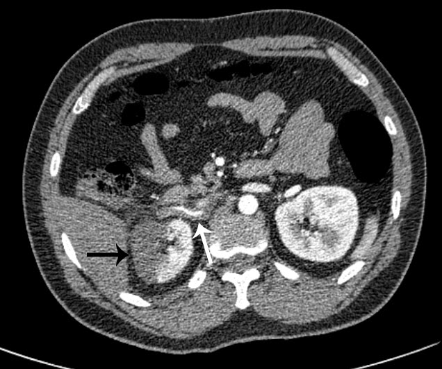
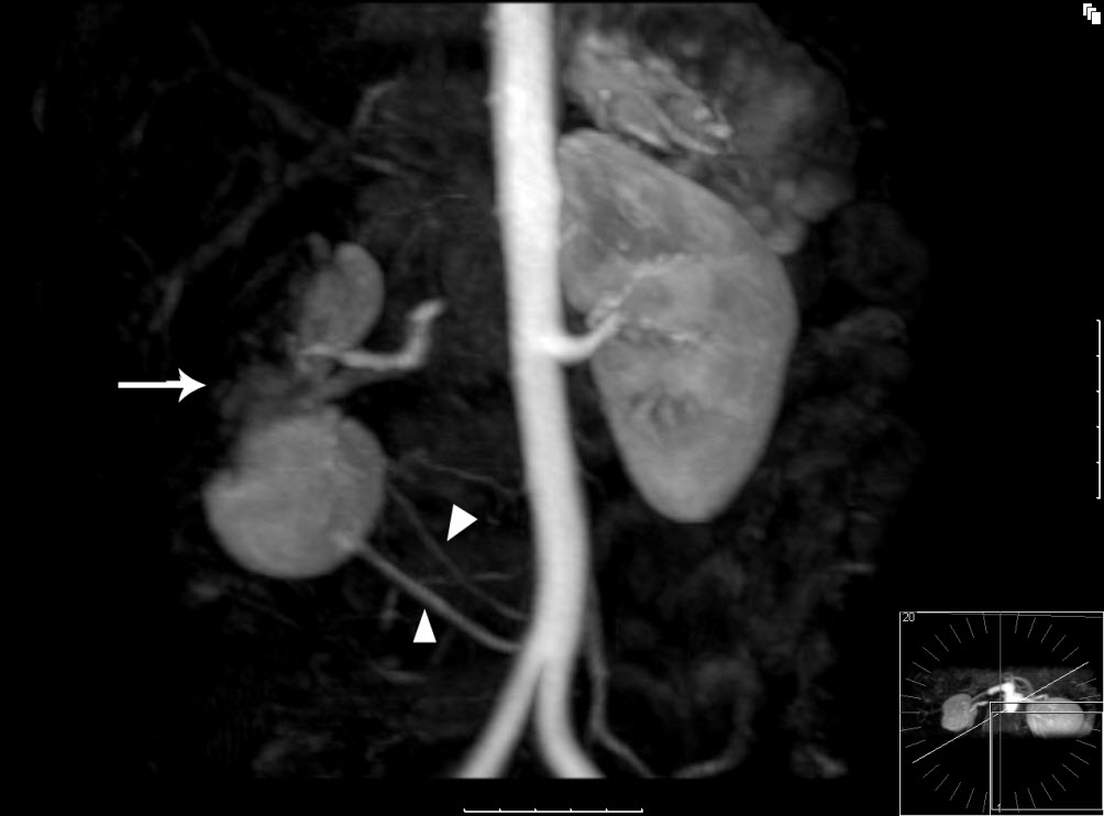
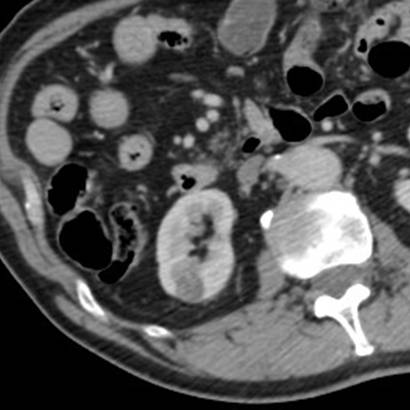
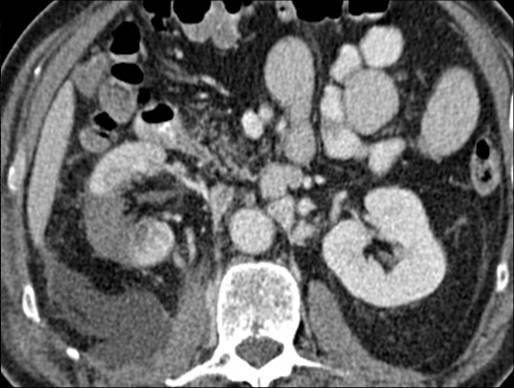
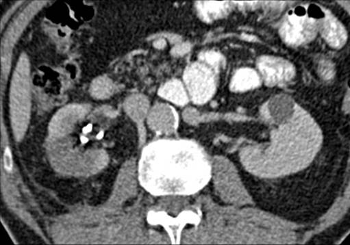
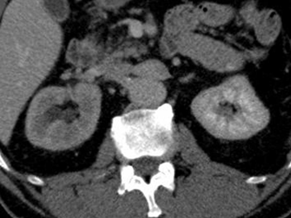
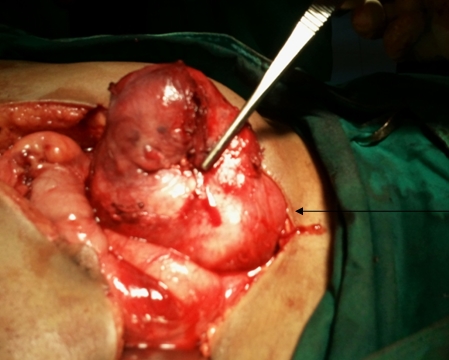
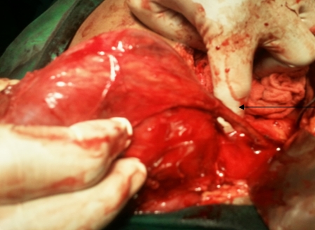
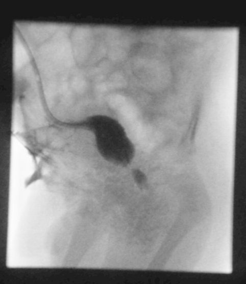
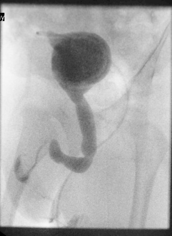
A 63-year-old female presented with a large left renal mass that was discovered during a work-up for abdominal pain of unknown duration. Computerized tomography of the abdomen revealed an exophytic, rounded mass arising from the upper pole of the left kidney with distortion of the renal collecting system. The patient subsequently underwent a left radical nephrectomy. The kidney specimen measured 16.8 x 10.0 x 6.8 cm and weighed 610 grams. On sectioning, the kidney showed a well-circumscribed tumor that measured 12.0 x 6.8 x 5.7 cm and occupied the upper pole, compressing the renal pelvis and calyceal system. The cut surface of the mass was homogeneously tan-pink in color with focal brown areas suggestive of old hemorrhage; no tumor necrosis or central scar was seen (Figure 1). No lymph nodes were noted.

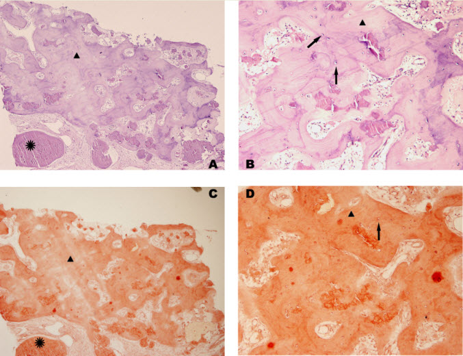
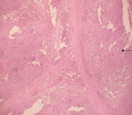
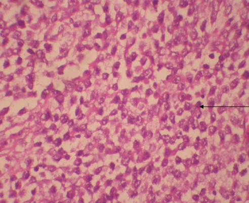
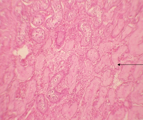
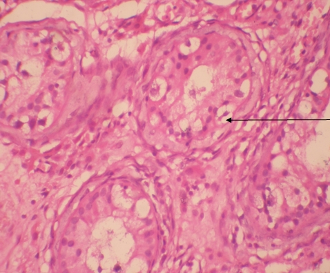
Histologic examination showed the mass to be composed of medium-sized polygonal cells, which displayed two architectural patterns. The majority of tumor cells were arranged in sheets or wide trabeculae (Figure 2). In adjacent areas, one could appreciate a tubulocystic growth pattern (Figure 3). The tumor cells had abundant granular eosinophilic cytoplasm with relatively well-defined cellular borders (Figure 4). There was mild to moderate nuclear pleomorphism, with an occasional thin-rim perinuclear halo. No sarcomatoid transformation was seen. The immunohistochemical stains showed diffuse immunoreactivity for pancytokeratin (AE1/3), cytokeratin 7, Ber-EP4, and CD117 (Figure 5). The stains with antibodies against RCC marker, CD10 and vimentin were negative. There was also cytoplasmic positive staining with Hale’s colloidal iron. Electron microscopy showed that abundant microvesicles, admixed with mitochondria, were distributed diffusely throughout the cytoplasm (Figure 6). On higher power, the microvesicles were bound by smooth single membranes, measuring 200 – 500 nm in diameter, and packing the spaces between mitochondria (Figure 7). A small number of lysosomes, lipid vacuoles and glycogen granules were noted in the cytoplasm, where the Golgi apparatus and the rough endoplasmic reticulum were poorly developed. Nuclei were rounded with irregular folded nuclear membrane and heterochromatin.

Discussion
When renal tumor cells show distinctive granular eosinophilic cytoplasm, the main diagnostic challenge is to separate an eosinophilic variant of ChRCC from the benign renal oncocytoma. Grossly, 33% of large oncocytomas may have a central stellate scar, which was not visible in the current tumor. Histologically, oncocytoma usually has tumor cells with inconspicuous cell borders arranged in nests or acini in a hypocellular hyalinized stroma, features that were not seen in this tumor either. Nuclear membrane irregularity and coarse chromatin are noted in this case, while oncocytomas usually have cells showing bland, uniform nuclei with smooth nuclear membranes. Hale’s colloidal iron stain showed diffuse cytoplasmic positivity in our case, whereas oncocytoma often displays focal positive staining confined to the luminal borders. Immunohistochemical stains performed on the current tumor showed that the cells were positive for cytokeratin 7, Ber-EP4, and CD117; and negative for RCC marker, CD10 and vimentin; a staining profile favoring a ChRCC. However, there is no established panel of immunohistochemical stains that can differentiate ChRCC from oncocytoma with certainty. This could, in part, be explained by the fact that these two tumors may share a common cell of origin in the distal nephron, namely intercalated cells of the collecting ducts.(5), (6) Electron microscopic analysis is considered the only definitive method to consistently distinguish these two entities.(7) The granular appearance of the cytoplasm on light microscopy corresponds to the ultrastructural finding of abundant microvesicles and scattered mitochondria in the cytoplasm of a majority of tumor cells, which is considered diagnostic for ChRCC.(7) In contrast; it is known that oncocytoma is characterized by the packing of the cytoplasm by mitochondria with paucity of microvesicles and other cell organelles. Unlike the classic ChRCC, the cytoplasm of this tumor was filled with crowded microvesicles diffusely interspersed between mitochondria, leading to less prominence or absence of perinuclear clearing (halo) by light microscopy. Previous studies have suggested that the microvesicles are derived from mitochondria because of the close relationship between microvesicles and mitochondria at the ultrastructural level;(8) although the possibility of endoplasmic reticulum being the origin has also been suggested.(1), (7) In addition, clear cell RCC can occasionally show a somewhat granular cytoplasm leading to potential confusion with ChRCC.(4), (7) However, clear cell RCC usually demonstrates alveolar or acinar growth patterns with delicate thin-walled blood vessels by light microscopy and abundant cytoplasmic lipid vacuoles and glycogen granules by electron microscopy. Immunohistochemically and histochemically, clear cell RCC is usually positive for antibodies against vimentin, RCC marker and CD10, and negative for CD117 and colloidal iron.

ChRCC was first described in 1985 as a tumor composed of cells with unique histomorphologic and ultrastructural features.(1) The tumor is presumably derived from intercalated cells of the collecting ducts.(5) Histologically, ChRCC is characterized by polygonal cells with distinct nuclear borders and evenly distributed, finely granular, translucent and pale cytoplasm. The classic architectural appearance is that of sheets or wide trabeculae of cells often separated by incomplete fibrovascular septations. Occasional, papillary, tubulocystic and nested patterns may be present.(1), (4) The nuclei often are hyperchromatic with nuclear membrane irregularity (raisinoid) and coarse chromatin. Binucleation or multinucleation are also common. Classically, perinuclear cytoplasmic clearing (halo) is present. Interestingly, it appears that nuclear grade as determined by the Fuhrman system may not reflect the clinical or biological behavior of chromophobe RCC as closely as it does for the other subtypes.(9)

The eosinophilic variant of ChRCC was first reported in 1997.(10) It usually shows small to medium-sized, eosinophilic granular/oncocytic cells arranged in sheets.(11) Compared with the classic ChRCC, our case showed smaller cell size, less prominent cell borders and thinner or absent perinuclear halos. However, similar to classic ChRCC, the eosinophilic variant is a tumor of low malignant potential with reported 5-year and 10-year survival rates of 78-100% and 80-90%, respectively.(1), (11) It is also believed that the cytogenetic abnormalities of this variant are similar to those of the classic ChRCC, including losses of chromosomes 1, 2, 6, 10, 13, 17 and 21.(12), (13)

ChRCC with microcystic and adenomatous patterns was first described in 1998.(14) Because of the tubulocystic architectures identified in the current case, a differential diagnosis that includes tubulocystic carcinoma of the kidney is needed. The tubulocystic carcinoma of the kidney is a recently recognized subtype of renal tumor that is not listed in the 2004 WHO classification.(3) Besides the packed tubules and cysts separated by bland fibrous stroma, the lining tumor cells of this newly described entity are cuboidal to columnar with abundant eosinophilic cytoplasm and usually large nuclei with prominent nucleoli.(15) These features are readily separable from the current case by routine histology. In addition, the genetic signatures of the tubulocystic carcinoma of the kidney are gains of chromosome 7 and 17.

In conclusion, we report for the first time a unique case of an eosinophilic variant of ChRCC that contained tubulocystic features. Because ChRCC is associated with a different prognosis from those conditions which mimic it, accurate identification of this tumor has important clinical implications. Even if the cytological features of this eosinophilic variant of ChRCC are extremely similar to that of renal oncocytoma, we believe that the combination of careful evaluation of the architectural patterns, cytologic characteristics and special stain profile of adequately sampled tumor will be very helpful for its accurate identification. If needed, ultrastructural study will yield a definitive diagnosis. Likewise, the unique morphologic and architectural patterns will help to distinguish the eosinophilic variant of ChRCC from clear cell RCC with granular cells and tubulocystic carcinoma of the kidney. Further elucidation of the characteristic cytogenetic abnormalities will provide a foundation for molecular classification of the neoplasm.

Acknowledgement
The authors thank Dr. Peter Kragel for his advices and critical comments on the manuscript and Debra Laich for her expert help in electron microscopy.

Declaration of Conflicting Interests
The author(s) declared no potential conflicts of interest with respect to the research, authorship, and/or publication of this article.

References
(1) Thoenes W, Storkel S, Rumpelt HJ. Human chromophobe cell renal carcinoma. Virchows Arch B Cell Pathol Incl Mol Pathol 1985;48(3):207-217.
(2) Przybycin CG, Cronin AM, Darvishian F, Gopalan A, Al-Ahmadie HA, Fine SW, et al. Chromophobe renal cell carcinoma: a clinicopathologic study of 203 tumors in 200 patients with primary resection at a single institution. Am J Surg Pathol 2011 Jul;35(7):962-970.
(3) Eble JN, Sauter G, Epstein JI, Sesterhenn IA. World Health Organization Classification of Tumours. Pathology and genetics of tumors of the urinary system and male genital organs. Lyon: IARC Press; 2004.
(4) Amin MB, Paner GP, Alvarado-Cabrero I, Young AN, Stricker HJ, Lyles RH, et al. Chromophobe renal cell carcinoma: histomorphologic characteristics and evaluation of conventional pathologic prognostic parameters in 145 cases. Am J Surg Pathol 2008 Dec;32(12):1822-1834.
(5) Truong LD, Shen SS. Immunohistochemical diagnosis of renal neoplasms. Arch Pathol Lab Med 2011 Jan;135(1):92-109.
(6) Ortmann M, Vierbuchen M, Fischer R. Sialylated glycoconjugates in chromophobe cell renal carcinoma compared with other renal cell tumors. Indication of its development from the collecting duct epithelium. Virchows Arch B Cell Pathol Incl Mol Pathol 1991;61(2):123-132.
(7) Tickoo SK, Lee MW, Eble JN, Amin M, Christopherson T, Zarbo RJ, et al. Ultrastructural observations on mitochondria and microvesicles in renal oncocytoma, chromophobe renal cell carcinoma, and eosinophilic variant of conventional (clear cell) renal cell carcinoma. Am J Surg Pathol 2000 Sep;24(9):1247-1256.
(8) Moreno SM, Benitez IA, Martinez Gonzalez MA. Ultrastructural studies in a series of 18 cases of chromophobe renal cell carcinoma. Ultrastruct Pathol 2005 Sep-Oct;29(5):377-387.
(9) Paner GP, Amin MB, Alvarado-Cabrero I, Young AN, Stricker HJ, Moch H, et al. A novel tumor grading scheme for chromophobe renal cell carcinoma: prognostic utility and comparison with Fuhrman nuclear grade. Am J Surg Pathol 2010 Sep;34(9):1233-1240.
(10) Erlandson RA, Shek TW, Reuter VE. Diagnostic significance of mitochondria in four types of renal epithelial neoplasms: an ultrastructural study of 60 tumors. Ultrastruct Pathol 1997 Sep-Oct;21(5):409-417.
(11) Manipadam MT, Korula A, Chandrasingh J, Devasia A. Chromophobe renal cell carcinoma: A report of two cases with unusual histological features. Indian J Urol 2008 Jan;24(1):123-125.
(12) Brunelli M, Eble JN, Zhang S, Martignoni G, Delahunt B, Cheng L. Eosinophilic and classic chromophobe renal cell carcinomas have similar frequent losses of multiple chromosomes from among chromosomes 1, 2, 6, 10, and 17, and this pattern of genetic abnormality is not present in renal oncocytoma. Mod Pathol 2005 Feb;18(2):161-169.
(13) Yusenko MV. Molecular pathology of chromophobe renal cell carcinoma: a review. Int J Urol 2010 Jul;17(7):592-600.
(14) Michal M, Hes O, Svec A, Ludvikova M. Pigmented microcystic chromophobe cell carcinoma: a unique variant of renal cell carcinoma. Ann Diagn Pathol 1998 Jun;2(3):149-153.
(15) Azoulay S, Vieillefond A, Paraf F, Pasquier D, Cussenot O, Callard P, et al. Tubulocystic carcinoma of the kidney: a new entity among renal tumors. Virchows Arch 2007 Nov;451(5):905-909.

Gross rz

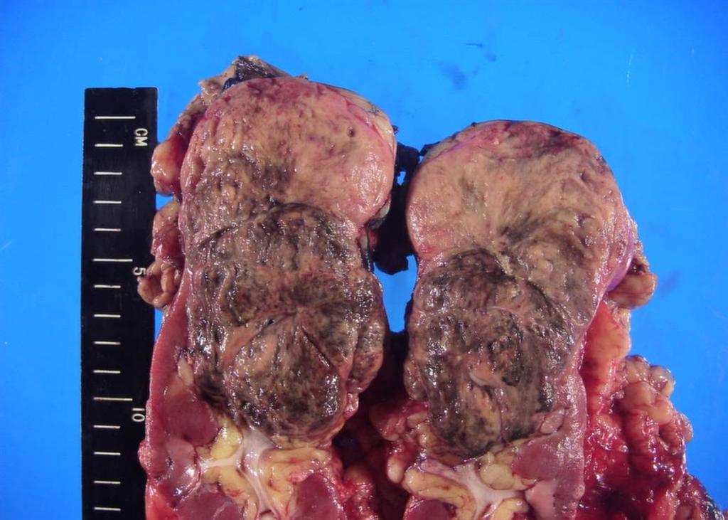
Figure 1. The upper pole mass was homogeneously tan-pink in color with dark brown areas suggestive of old hemorrhage.

Fig2 rz

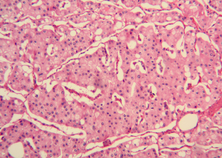
Figure 2. The majority of tumor cells were arranged in sheets or wide trabeculae. (hematoxylin and eosin, 100x)

Fig3 rz

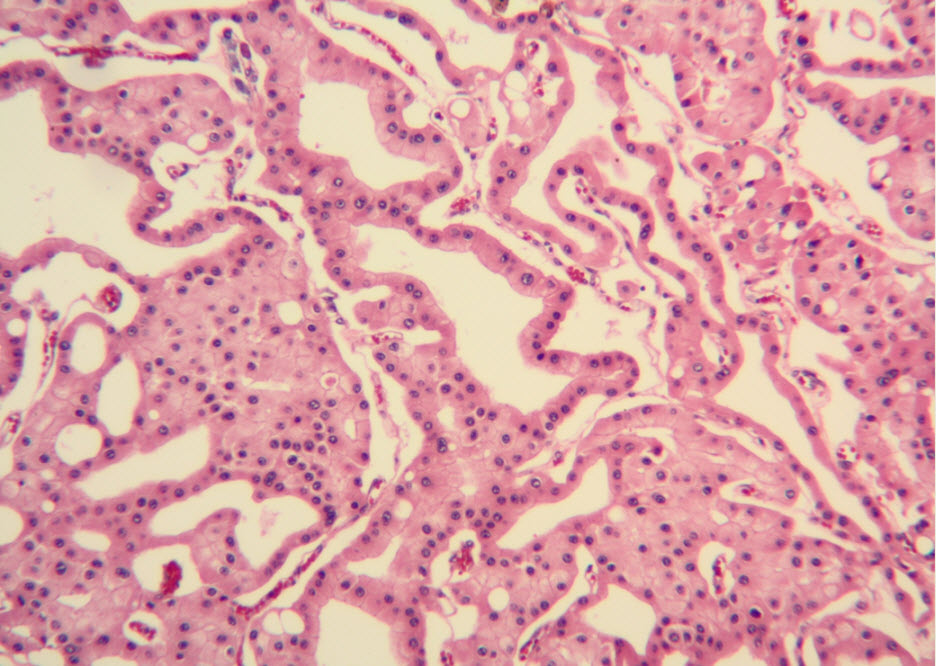
Figure 3. Adjacent to wide trabeculae, tumor cells show tubulocystic growth pattern. (hematoxylin and eosin, 100x)

Fig4 rz

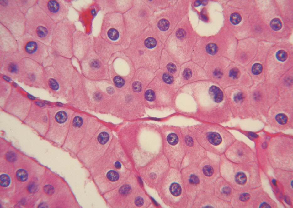
Figure 4. The polygonal tumor cells have abundant granular eosinophilic cytoplasm with relatively welldefined cellular borders. There was mild to moderate nuclear pleomorphism, with occasional thin-rim perinuclear halo. (hematoxylin and eosin, 400x )

Fig5 rz

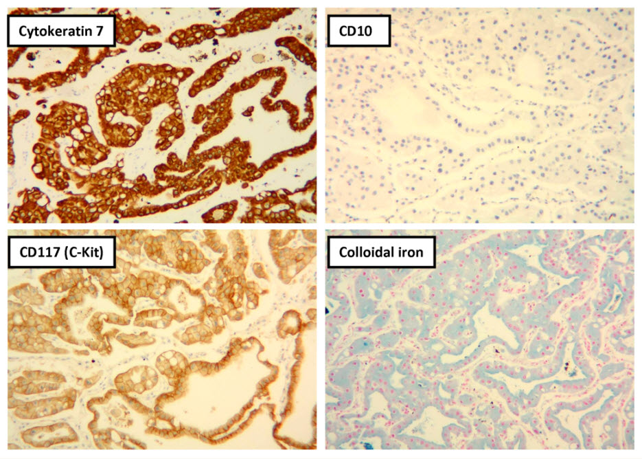
Figure 5. Selected photomicrographs of the renal mass with immunohistochemical and Hale’s colloidal iron stains are shown. (x100)

Fig6 rz

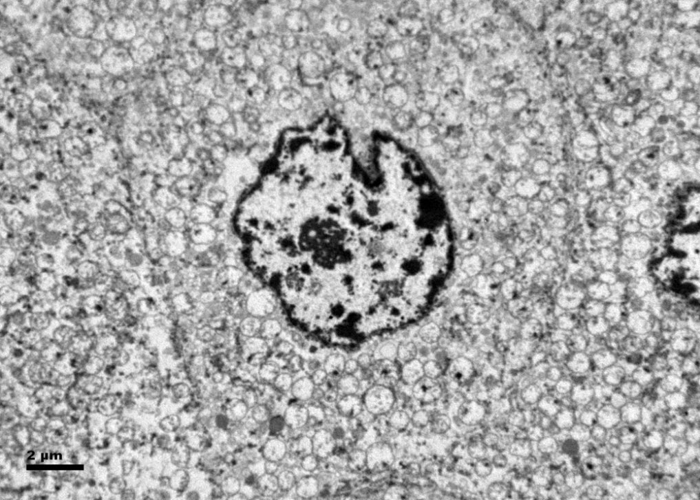
Figure 6. Ultrastructurally, the tumor cells show abundant microvesicles, admixed with mitochondria, distributed diffusely throughout the cytoplasm. Nuclei were rounded with irregular nuclear membranes and coarse chromatin.

Fig7 rz

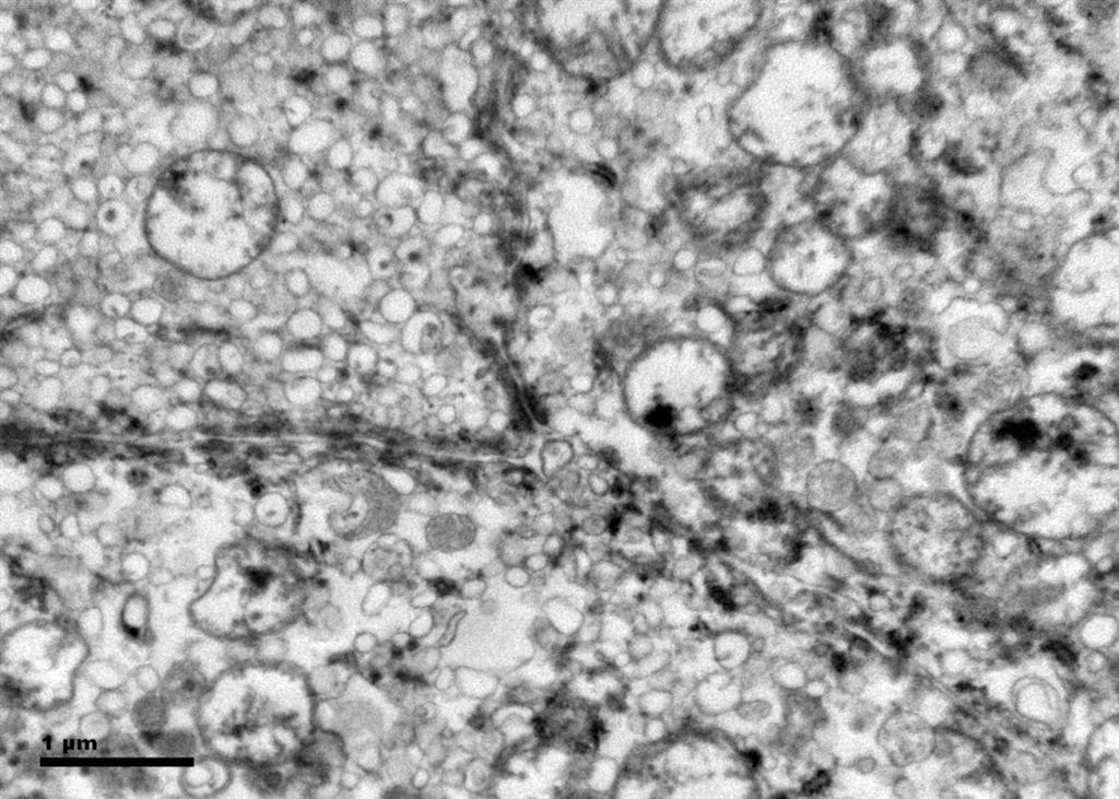
Figure 7. The crowded microvesicles show smooth single membranes, measure 200 – 500 nm in diameter, and pack the spaces between mitochondria. A small number of lysosomes, lipid vacuoles and glycogen granules are also present.

Date added to bjui.org: 06/11/2012

DOI: 10.1002/BJUIw-2012-048-web


Missed Double Urethral Obstruction

A 14 days old infant who presented to a regional hospital with inability to pass urine and severe bilateral hydronephrosis. He underwent a suprapubic cystostomy which was complicated by bladder rupture. He presented to us pyrexial with abdominal distention and deteriorating renal functions. Child underwent a laparotomy. Extra and intra-peritoneal bladder rents were indentified, repaired with mini-vesicostomy was performed . An MCU done through the vesicostomy suggested posterior urethral valves which was fulgurated. The child did not void per urethrally when the vesicostomy was blocked. A repeat micturating cysto-urethrography done later filled the anterior urethra fully and demonstrated an anterior urethral valve as well. The anterior urethral valves co existing with posterior urethral valves was missed at the initial study because of poor filling of the anterior urethra. Treatment of the Anterior urethral valves resulted in normal voiding after closure of the vesicostomy.
The radiologic technique of studying the urethra in MCU must ensure that anterior urethra is completely outlined, in order to avoid missing anterior urethral obstruction co-existing with Posterior urethral valves.

Authors: Narasimhan, Kannan Laksmi; Chua, Joyce; Rai, Rambha; Fortier, Marielle
Corresponding Author: Narasimhan, Kannan Laksmi

Chua Joyce,#
Rambha Rai,#
Marielle Fortier,*
Narasimhan KL,#

Department of Pediatric Surgery #, and Diagnostic Imaging * KK Women’s and Children’s Hospital, Singapore (SIN)

Address for communication
K.L.Narasimhan
Dept of Pediatric Surgery.
KK women’s and children’s hospital.
100, Bukit Timah Road,
Singapore 229899
Email: narasimhankl@ gmail.com


Case Report
A term infant (1.9 Kg) who had a normal 28 week antenatal ultrasound scan presented at birth with an inability to pass urine, severe bilateral hydronephrosis and an enlarged bladder. The child could not be catheterized per urethra by the treating surgeon and a suprapubic cystostomy was performed. The catheter drained 200cc – 240 cc of urine per day for 14 days, but stopped draining subsequently. This was associated with abdominal distension, and leakage of urine from the abdominal wound. Contrast studies suggested that the silicone catheter had migrated into the rectum and the child was re-explored. Intra-operatively the catheter was found to be in the bladder with copious urine tracking into the skin and subcutaneous tissue. The bladder was closed with a supra–pubic catheter left in situ. Post-operatively the child developed progressive abdominal distension with scrotal edema.
The child was shifted to our center at parental request. On arrival he was pyrexial with a distended and shiny abdomen. Imaging with ultrasound, cystogram and CT scans confirmed intra and extra-peritoneal leak of urine from the bladder (figure1).The child was acidotic with a raised urea 25.9 (1.4-7.7mmol/L ) , creatinine 187 ( 27-80mmol/L) and bicarbonate 13.2 ( 18-27mmol/L). He was re-explored via the previous incision on Day 17 after birth. There was urine leaking into the main wound from the bladder suture line (extra -peritoneal leak). The bladder wall was thickened and friable. A 5 mm defect on the right postero-lateral wall of the dome of the bladder communicating with peritoneal cavity was identified (intra-peritoneal leak). This was repaired. The peritoneal cavity drained purulent fluid, from which a Klebsiella species was cultured. The dome of bladder was bought out via a sub-umbilical incision as a minivesicostomy by a technique described (1) and intubated with an 8 Fr catheter. The child was discharged home with urine draining from the minivesicostomy. Occasionally he passed urine in drops per-urethra. An MCU (micturating cysto-urethrogram) on day 14 showed a dilated posterior urethra (Figure 2) but failed to outline the anterior urethra even under pressure. A DMSA scan showed bilaterally functioning non scarred kidneys. A follow up ultrasound showed no hydronephrosis. The renal function returned to normal post-operatively. The child was readmitted and underwent cystoscopy at 4 months of age. The anterior urethra seemed normal and posterior urethral valves (Type 1 -Young’s) were identified and fulgurated at the 5, 7 and 12o’clock position using a 9 mm resectoscope. Post fulguration, when the vesicostomy was occluded, he had difficulty in passing urine and had a breakthrough urinary tract infection. Hence, his vesicostomy was not closed. A repeat MCU done at 9 months of age revealed dilatation of the anterior urethra up to the peno-scrotal junction with a cut off consistent with anterior urethral valves (Figure 3). The child was re cystoscoped and the anterior urethral valves were identified using hook electrodes and cut using a 9 mm resectoscope. The minivesicostomy was closed. Subsequently the child is voiding with a good stream, is thriving and has stable renal function. The volume of the bladder is small and the child is on oxybutynin. The bladder function has improved over the subsequent months.

Discussion
Anterior urethral valves have rarely been reported in large series. Hence unaware clinicians can easily overlook this cause of obstructive uropathy (2). They are congenital in nature and can be located anywhere in the anterior urethra (bulbar urethra 40%, penoscrotal junction 30%, penile urethra 30%). Some have no associated diverticulum. Both the anterior and posterior urethral valves share similar presentations and have similar effects on the kidney and bladder functions. The diagnosis of anterior urethral valves can be missed on retrograde urethrography and also during MCU if the whole of the anterior urethra is not included in the field of exposure (2). In the index patient it was missed on initial MCU as well as at cystoscopy.
The association of AUV and PUV is rare (2, 3, 5). The case reported by Kumar et al is another case of neonatal presentation of double urethral obstruction (5). At the first VCUG, the AUV was not picked up .The picture of the MCU in the paper does not outline the anterior urethra adequately. The baby failed to void after fulguration of posterior urethral valves and a fluctuant swelling was seen at the penoscrotal junction when he attempted to void. A repeat VCUG was done that showed a large urethral diverticulum. This time the authors have filled the anterior urethra with contrast adequately. The baby had a vesicostomy and a repair of urethral diverticulum with removal of the non functioning left kidney. The other 2 cases cited by us have been diagnosed without missing a double obstruction (3,4).
PUV is seven times commoner than AUV in the reported literature (2). The precise etiology of both the conditions remains elusive and various postulates exist. Due to the rarity of this association, one may not identify both the problems which may present as a diagnostic challenge.
Missing the anterior urethral valves in the index case could have been a disaster especially if we had embarked on closure of vesicostomy after fulguration at the first sitting. Diagnosis of anterior urethral valves would have been picked up on retrograde urethrography and MCU if the whole of the anterior urethra was filled and included in the field of exposure. This is an important learning point in uroradiology.

References
1. Monika Nanda, Monika Bawa, Narasimhan K.L.: Minivesicostomy in the management of PUV after valve ablation. Journal of Pediatric Urology (2010) Dec 14 Epub
2. Narasimhan K.L., Chowhary S K, Balpinder kaur: Anterior urethral valves .Indian Pediatrics, 2005:42-708-10.
3. Rao KLN, Eradi B, Menon P. Anterior and Posterior urethral valves: A rare association J Ped Surg 2003, 38: E24.
4. Bhagat SK, Gopalakrishnan, G, Kekre NS, Kumar S. Anterior and posterior urethral valves with sub-coronal hypospadias: a rare association. J Ped Surg (2008) 43, E 23- E25
5. Kumar A, Bajpai M, Gupta AK: Double urethral obstruction in a Neonate – a case report. Eur J Pediatr Surg 2005:15:449-451.


pic 1revised

Figure 1: Initial cytogram in the newborn period showing both intra and extra- peritoneal leak of contrast

pic2revised


Figure 2: Showing contrast injected through the vesicostomy showing a dilated Posterior urethra but the anterior urethra did not get filled despite pressure.

pic3 revised

Figure 3: Contrast injected through the vesicostomy showing a dilated anterior urethra upto the penoscrotal junction

Date added to bjui.org: 02/11/2012

DOI: 10.1002/BJUIw-2012-026-web


© 2024 BJU International. All Rights Reserved.