Archive for category: Case Studies

Metastatic Colon Adenocarcinoma to the Tunica Vaginalis Testis Presenting As Hydrocele

To the best of our knowledge, ours is the first case of tunica vaginalis metastasis originating from a primary adenocarcinoma of the ascending colon. 

Authors: Ji Zheng1, Gensheng Lu1, Chengping Xu2, Bo Song1, Zhansong Zhou1

1. Department of Urology, Southwest hospital, Third Military Medical University, Chongqing, China
2. Department of Pathology, Southwest hospital, Third Military Medical University Chongqing, China

Corresponding Author: Zhansong Zhou,  Department of Urology, Southwest hospital, Third Military Medical University, Chongqing, China.  E-mail: [email protected]


Case report
 
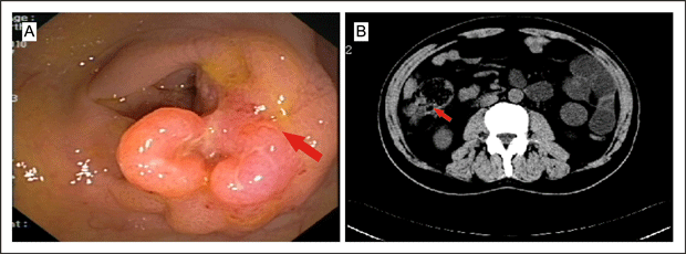
A 65-year-old man was referred to our urology clinic complaining of left scrotal enlargement for one month. Seven months before admission, colonoscopy (Figure 1A) and computed tomography (CT) of his abdomen (Figure 1B) showed a mass in his ascending colon and enlarged mesenteric lymph nodes.


Figure 1A and B. CT scan of the abdomen 
Fig1colonoscopy-and-computed-tomography-of-abdomen-


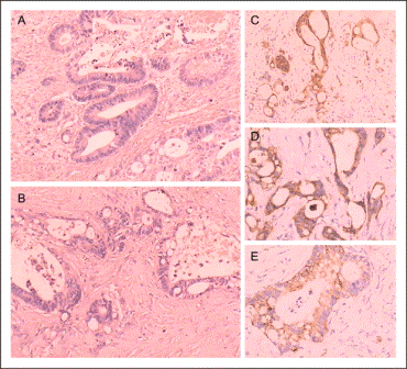
Biopsy was consistent with a moderately differentiated adenocarcinoma. He was diagnosed preoperatively with ascending colonic adenocarcinoma, and mesenteric lymph nodes metastases were suspected. Due to his experiencing severe intestinal spasm and bloody stools, the patient requested surgical treatment, though systematic chemotherapy had been advised. He underwent a radical right hemicolectomy, and this pathology revealed moderately differentiated adenocarcinoma with lymph node metastases (Figure 3A).


Figure 3A. Pathology revealed moderately differentiated adenocarcinoma with lymph node metastases
Fig3adenocarcinoma-with-lymph-node-metastases


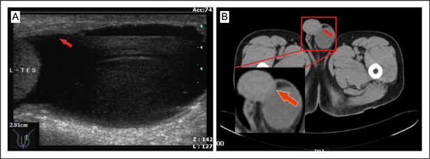
Two weeks after surgery, in view of his having lymphatic metastases, our patient agreed to and completed the first cycle of a combined chemotherapy regimen with oxaliplatin (150 mg intravenous [IV] day 2), tegafur (600 mg IV days 1 to 3), and calcium folinate (400 mg IV days 1 to 3). Another four cycles of combined chemotherapy were accomplished in the following four months. One month before referral to urology, an ultrasound examination had shown a hydrocele of the left tunica vaginalis testis, thickening of the left perididymis, with a normal appearance of the testis and epididymis (Figure 2A). Ten days previously, a painless intrascrotal nodule had been palpated accidentally and scrotal examination revealed a hard flat mass, 1.5×0.6×0.3 cm in diameter, adhering to the left tunica vaginalis testis. CT scan demonstrated a mass in this area (Figure 2B).


Figure 2A and B. CT scan 
Fig2hydrocele-of-the-left-tunica-vaginalis-testis


Serum levels of the tumor-associated proteins CA242 and CA19-9 were significantly elevated to 268 kU/L (normal range 0-20 kU/L) and 372 kU/mL (normal range 0-35 kU/mL), respectively, while those of cancer antigens, carcinoembryonic antigen, neuron-specific enolase, alpha-fetoprotein, prostate specific antigen, CA125, CA15-3 and beta human chorionic gonadotropin were within their normal ranges.
The patient underwent left orchiectomy through an inguinal approach and the mass was found to be densely fixed to the left tunica vaginalis testis but separated from the testis, epididymis and spermatic cord. Frozen sections suggested the mass to be an adenocarcinoma. Interestingly, a left oblique inguinal hernia with greater omentum inside was found and the greater omentum was in direct contact with the lesion of the tunica vaginalis testis. A hernia repair was performed accordingly.
Subsequent biopsy revealed a moderately-differentiated adenocarcinoma infiltrating the tunica vaginalis testis without involvement of the testis or epididymis (Figure 3B). The primary colonic tumor and this tumor shared identical neoplastic glands with intra-cytoplasmic mucin and moderate nuclear pleomorphism (Figure 3A, 3B). Immunohistochemistry was performed using the immunoperoxidase method. The tumor cells showed positivity for Villin (Figure 3C), CK(Figure 3D) and CK20(Figure 3E), but negative for D2-40, TTF1, WT1, Ki67, CK7, SMA, and PSA, which suggested an occult gastrointestinal or colorectal origin of the carcinoma, with a lesser probability of carcinoma of the lung, liver, thyroid, breast, prostate, or hematological system.[1,2]


Discussion
 
Solid tumor metastasis to the testis and paratesticular region is rare and principally encountered as an incidental autopsy finding.[3] Metastatic carcinoma accounted for respectively five of 112 and nine of 66 paratesticular tumors reviewed.[4,5] The common primary sites of metastasis of the paratesticular tissue are the prostate, kidney, gastrointestinal tract, lung, and breast.[6]
Tumors of the tunica vaginalis testis, both primary and secondary, and whether benign or malignant, are extremely rare.[7,8]  Fewer than 10 cases, including our case, have been reported to date in the English and non-English literature,[8–13] and the primary sites for metastatic tumor of tunica vaginalis testis include stomach,[8–10,12] rectum,[11] cecum ,[12] and colon (as in our case). Most cases present with paratesticular mass or hydrocele, and they may or may not be associated with pain.
Metastasis to the paratesticular tissues can be the first sign of tumor recurrence and more rarely present as the first manifestation of an occult primary neoplasm.[14] Our review revealed hydrocele of the tunica vaginalis testis presented as the first clinical manifestation of primary [9] and secondary [8,10–12] and our case  tunica vaginalis testis tumors, which suggested that cancerous lesions should be considered in the management of hydrocele, especially in the follow-up of cancer patients. CT or PET-CT is suggested as a rapid and reliable modality for facilitating a preoperative diagnosis.
Clinically, most patients with metastatic paratesticular tumors suffer from a painless intrascrotal mass and the diagnosis is frequently delayed. Hydrocele, inguinal hernia, epididymal cyst, spermatocele and spermatic cord tumors are the most common misdiagnoses of the tumor.[15] The consideration of these diseases in the differential diagnosis of a painless paratesticular mass is critical, as it may allow more precise diagnosis and more targeted treatment, especially in high-risk patients.
Infiltrative lesions of the epididymis [9] and spermatic cord [10,11] can be found in some metastatic tunica vaginalis testis tumor cases, which implies that other primary sites of tunica vaginalis testis metastases may be similiar to primary sites of paratesticular metastatic tumors, and include prostate, gastrointestinal tract, kidney, lung and breast.[15, 16]
Proposed routes of metastasis to the tunica vaginalis testis may include direct extension from adjacent organs, intraductal spread via the vas deferens, retrograde venous and lymphatic extension, arterial embolism [9], and transperitoneal seeding through a patent tunica vaginalis [17] or congenital hydrocele [18]. In our case, the possibility of lymphatic or hematogenous spread was suspected, particularly in view of the primary tumor with lymph node metastasis. However, the presence of a left oblique inguinal hernia in direct contact with the lesion of the tunica vaginalis testis made  local tissue invasion another possible mode of spread .[19]
In our review, the median age of patients with metastatic tumor of the tunica vaginalis testis is approximately 62 years, with a reported age range from 50 to 67 years. Tumors metastasising to the paratesticular tissue usually are identified in the setting of disseminated disease and occur only uncommonly as a solitary site of metastasis.[2]  The prognosis generally is poor, with the average survival period from the time of diagnosis of the metastasis being 9.1 months.[2] Chemotherapy is the only possible option for the treatment of a secondary tumor at the distant metastatic stage. Our patient completed four cycles of a combined chemotherapy regimen with oxaliplatin (120 mg intravenous [IV] day 1), fluorouracil (500 mg IV days 1 to 2), and calcium folinate (200 mg IV days 1 to 2). He is alive and has been under careful surveillance for about five months up to the time of this report.
In our case and in others reviewed, once paratesticular metastasis had occurred, disease control or cure was negligible.[20] Whether or not a more aggressive approach would lead to a different outcome remains unknown. In some patients, early diagnosis by cytological examination of hydrocele fluid [21] or ultrasound-guided fine-needle aspiration cytology [22] may be helpful for diagnosis and prognosis, as well as treatment of secondary tumors of the tunica vaginalis testis.
To the best of our knowledge, ours is the first case of tunica vaginalis metastasis originating from a primary adenocarcinoma of the ascending colon. Though rare, metastatic lesions of the tunica vaginalis testis should be included in the differential diagnosis of a patient with ambiguous paratesticular masses or in the setting of a hydrocoele. High awareness of this entity may lead to earlier diagnosis and more timely and appropriate treatment, despite the unfavorable prognosis for treatment of secondary tumor of tunica vaginalis testis at present.


References
1.Park SY, Kim HS, Hong EK, et al: Expression of cytokeratins 7 and 20 in primary carcinomas of the stomach and colorectum and their value in the differential diagnosis of metastatic carcinomas to the ovary. Hum Pathol 33:1078-1085, 2002
2.De Lott LB, Morrison C, Suster S, et al:CDX2 is a useful marker of intestinal-type differentiation: a tissue microarray-based study of 629 tumors from various sites. Arch Pathol Lab Med 129:1100-1105, 2005
3.Meacham RB, Mata JA, Espada R,et al:Testicular metastasis as the first manifestation of colon carcinoma. J Urol 116:621-622, 1988
4.Lioe TF, Biggart JD: Tumours of the spermatic cord and paratesticular tissue. A clinicopathological study. Br J Urol 71:600-606, 1993
5.Hartwick RW, Srigley JR, Burns B, et al: A clinicopathologic review of 112 paratesticular tumours[Abstract]. Laboratory Invest 56:83, 1987
6.Algaba F, Santaularia JM, Villavicencio H: Metastatic tumor of the epididymis and spermatic cord. Eur Urol 9:56-59, 1983
7.Plas E, Riedl CR, Pflüger H: Malignant mesothelioma of the tunica vaginalis testis: review of the literature and assessment of prognostic parameters. Cancer 83:2437-2446, 1998
8.Lee J, Kang SC, Ban JH, et al: Metastatic Tumor of Tunica Vaginalis Testis with Hydrocele in a Patient with Gastric Cancer. Korean J Urol 48:667-669, 2007
9.Yeo JK:Scrotal Hydrocele as the First Clinical Manifestation of Occult Gastric Cancer.Korean. J Urol 50:1151-1153, 2009
10.Kageyama Y, Kawakami S, Li G, et al: Metastatic tumor of spermatic cord and tunica vaginalis testis from gastric cancer: a case report . Hinyokika Kiyo 43:429-431, 1997
11.Yasuhide T, Heizo Y, Katsusuke N: Metastatic Tumor of the Tunica Vaginalis Testis Arising from Rectal Cancer:A Case Report. Nishinihon Journal of Urology 61:245-247, 1999
12.Wai HP, Yau TK, Sze WM, et al:Metastatic tumour of the tunica vaginalis testis from carcinoma of the stomach. Int J Clin Pract 54:685-686, 2000
13.Ruiz JM, Sanchis CM, López PC, et al: Metastasis to the tunica vaginalis testis from a primary mucinous tumor of the cecum. Arch Esp Urol 63:235-238, 2010
14.Bennett VS, Bailey DM: Cholangiocarcinoma presenting as a solitary epididymal metastasis: a case report and review of the literature. Diagn Pathol 2:33, 2007
15.Beccia DJ, Krane RJ, Olsson CA. Clinical management of non-testicular intrascrotal tumors. J Urol 116:476-479, 1976
16.Algaba F, Santaularia JM, Villavicencio H: Metastatic tumor of the epididymis and spermatic cord. Eur Urol 9:56-59, 1983
17.Dutt N, Bates AW, Baithun SI: Secondary neoplasms of the male genital tract with different patterns of involvement in adults and children. Histopathology  37:323-331, 2000
18.Hanash KA, Carney JA, and Kelalis PP: Metastatic tumors to the testicles:routes of metastasis. J Urol 102: 465-468, 1969
19.Galanis I, Chatzimavroudis G, Katsougiannopoulos A, et al: Spermatic cord metastasis presenting as strangulated inguinal hernia – first manifestation of a multifocal colon adenocarcinoma: a case report. Cases J 2:61, 2009
20.Charles W, Joseph G, Hunis B, et al: Metastatic colon cancer to the testicle presenting as testicular hydrocele. J Clin Oncol 23:5256-5257, 2005
21.Gupta SC, Gupta AK, Misra V, et al: Pre-operative diagnosis of malignant mesothelioma of tunica vaginalis testis by hydrocele fluid cytology. Eur J Surg Oncol 24:153-154, 1998
22.Bruno C, Minniti S, Procacci C:Diagnosis of malignant mesothelioma of the tunica vaginalis testis by ultrasound-guided fine-needle aspiration. J Clin Ultrasound 30:181-183, 2002

 
Date added to bjui.org: 19/02/2012 


DOI: 10.1002/BJUIw-2011-067-web


Splenogonadal Fusion: A Rare Congenital Anomaly in Children

This report presents a case of splenogonadal fusion in a 13-year-old boy and reviews the relevant literature.

Authors: Osman Zeki Karakus1, Mustafa Ozcetin2, Fikret Erdemir3

1. Maternity and Children’s Hospital, Department of Pediatric Surgery, Tokat, Turkey.
2. Zonguldak Karaelmas University , School of Medicine, Department of Pediatrics, Zonguldak, Turkey.
3. Gaziosmanpasa University, School of Medicine, Department of Urology, Tokat, Turkey

 
Corresponding Author: Mustafa OZCETIN, Zonguldak Karaelmas University , School of Medicine,  Department of Paediatrics, Zonguldak, Turkey.  E-mail: [email protected]

Abstract
 
Splenogonadal fusion is a rare, benign, congenital anomaly characterized by fusion of the spleen and a gonad; it is frequently associated with other organ abnormalities. Approximately 175 cases of splenogonadal fusion have been reported. The diagnosis is rarely suspected preoperatively. Splenogonadal fusion is most commonly an incidental finding during inguinal exploration for an undescended testis or hernia. We report a 13-year-old boy who was admitted to our clinic with symptoms of an undescended testis and diagnosed with splenogonadal fusion laparoscopically, and discuss the relevant literature.


Introduction
 
The prevalence of congenital anomalies is 23.9~32.4 per 1000 births (1). According to the European Surveillance of Congenital Anomalies (EUROCAT), congenital heart defects are the most common non-chromosomal subgroup, at 6.5 per 1000 births, followed by limb defects (3.8 per 1000), urinary system anomalies (3.1 per 1000), and nervous system defects (2.3 per 1000) (2). Splenogonadal fusion is a rare congenital anomaly. Approximately 175 cases of splenogonadal fusion have been reported since Bostroem first described this condition in 1883. Most of the cases have been reported with cryptorchidism. Splenogonadal fusion is infrequently diagnosed preoperatively because of its rarity. It was reported that orchiectomy is done unnecessarily in these cases (3). Therefore, a correct diagnosis of splenogonadal fusion using a laparoscopic approach can save the affected gonad.


Case Report
 
A 13-year-old boy was admitted to our clinic with symptoms of an undescended left testis and intermittent swelling in the left scrotal region. On physical examination, while the right testis was normal in size and location in the scrotum, the left testis was palpated in the mid-inguinal region. In addition, a left inguinal hernia was detected with the Valsalva maneuver. No other physical abnormalities were detected in a systemic evaluation. Routine hematologic and biochemical analyses were normal. With the diagnosis of an undescended testis, a left orchiopexy was planned via a standard inguinal incision. Left inguinal exploration revealed that the left testis was in the inguinal canal. The associated hernia sac was dissected and repaired. At this point, a fleshy, smooth, reddish-brown vascular structure was seen on the superior part of the testis lying in the abdominal cavity (Figure 1).
 

Figure 1. The vascular shaped structure and testis are seen


This structure was closely related to the spermatic cord and vessels. At this step, a laparoscopy was performed. The structure was traced to the lower pole of the spleen and excised completely laparoscopically (Figure 2).


Figure 2. The vascular shaped structure which connected to the spleen 


The vascular structure was approximately 15 cm long and 1 cm wide. The procedure was concluded with a left orchiopexy. Histopathologically, the specimen was normal splenic tissue (Figure 3).


Figure 3: Pathological appearance of the dissected tissue (H&E, 10x)


The patient was discharged 24 hours postoperatively, with no complications.


Discussion


Some congenital anomalies of the spleen are common, such as splenic lobulation and accessory spleens, while other conditions are rare, such as wandering spleen, polysplenia, and splenogonadal fusion (4). Splenogonadal fusion is a rare congenital anomaly characterized by fusion of the spleen and a gonad. Although the age of reported cases ranges from stillborn to 81 years, approximately half of the cases presented in patients younger than 10 years and 82% in those younger than 30 years (5). In 1956, Putschar and Manion reviewed 30 cases and categorized this anomaly into continuous and discontinuous types (6). The discontinuous type lacks any anatomic connection between the gonad and spleen. By contrast, as in our case, the continuous type involves a direct anatomical connection, between the spleen and gonad, by a cord that may be totally splenic, beaded with multiple splenic nodules, or composed of fibrous tissue. Both types are equally frequent. However, the continuous type carries a 5-fold higher risk of associated anomalies than the discontinuous type, such as peromelia (absence or malformation of the limbs), micrognathia, cardiac defects, cleft palate, anal anomalies, craniosynostosis, spina bifida, Möbius syndrome, microgastria, thoracopagus, and osteogenesis imperfecta (5,6). One third of the cases reviewed by Gouw et al. were associated with one or more congenital defects, excluding congenital inguinal hernia or cryptorchidism (7). In our case, no organ anomaly except cryptorchidism was detected.
Although the aetiology of this condition is not clear, several mechanisms have been proposed to explain it. All of the theories are based on the close proximity of the left gonadal ridge and splenic anlage between the 5th and 8th weeks of gestation. This explains why splenogonadal fusion is almost always seen on the left. During this period, several groups of cells derived from the coelomic epithelium and mesenchyme of the mesogastrium aggregate to form the splenic anlage in the dorsal mesogastrium. This occurs at approximately the same time as the gonadal ridge is formed between the dorsal mesogastrium and mesonephros on either side. During weeks 6 and 7, rotation of the stomach to the left and growth of the dorsal mesogastrium translocate the spleen to the left side of the abdominal cavity, bringing it into close proximity with the gonadal ridge (6). In addition, it was proposed that cryptorchidism and continuous splenogonadal fusion are caused by abnormal development of the cranial suspensory ligaments of the testis and failure of their involution (8).
Splenogonadal fusion is most commonly seen incidentally during inguinal exploration for cryptorchidism, a hernia, or a hydrocele. Cryptorchidism is the genital anomaly seen most commonly in splenogonadal fusion. In a review of 111 cases with splenogonadal fusion, cryptorchidism was reported in 31% (9). The most common complaint is testicular swelling. Another presentation is acute painful scrotal swelling secondary to swelling of the ectopic splenic tissue as a consequence of various processes, such as malaria, leukaemia, mononucleosis, and traumatic rupture of the ectopic spleen (5, 10). Splenogonadal fusion is seldom diagnosed or suspected preoperatively. Almost one in six cases of splenogonadal fusion has been diagnosed at autopsy (3). When suspected clinically, technetium isotope scanning is used to detect accessory splenic tissue (11). Computed tomography (CT) and ultrasonography have also been used for the diagnosis (12). In most cases, the splenic tissue can be dissected off the gonadal structures easily; if there are doubts concerning the nature of the swelling, an intraoperative frozen section may confirm the diagnosis. Diagnostic and operative laparoscopic approaches are often indicated in children for cryptorchidism, appendectomy, herniorrhaphy, and an examination of the abdominal organs. In the continuous type of splenogonadal fusion there is a direct anatomical connection between the spleen and gonad through a cord of splenic tissue. In this context, a few authors have reported that diagnostic laparoscopy can be performed via an open umbilical approach. With this approach, vascular connections, the vas deferens, and other anomalies can be seen clearly. Since laparoscopy is helpful in diagnosing this rare condition, and for planned surgical treatment, as in our case, a few cases of splenogonadal fusion have been diagnosed and managed with laparoscopy (13, 14). A laparoscopic approach prevents the loss of time due to secondary operations and frozen section examination or radiological imaging methods.
Another problem with this anomaly is unnecessary orchiectomy. Indeed, patients can undergo an unnecessary orchiectomy because a primary testicular neoplasm is suspected. In one study, 37% of the patients with splenogonadal fusion underwent an unnecessary orchiectomy for a suspected primary testicular neoplasm (3).
In summary, although extremely rare, splenogonadal fusion should be considered during the evaluation of an undescended testis.


References


1-     Dastgiri S, Stone DH, Le-Ha C, Gilmour WH. Prevalence and secular trend of congenital anomalies in Glasgow, UK. Arch Dis Child 2002;86:257-63.
2-     Dolk H, Loane M, Garne E. The prevalence of congenital anomalies in europe. Adv Exp Med Biol 2010;686:349-64.
3-     Karaman MI, Gonzales ET. Splenogonadal fusion: report of 2 cases and review of the literature. J Urol 1996;155:309-11
4-     Varga I, Galfiova P, Adamkov M, Danisovic L, Polak S, Kubikova E, Galbavy S. Congenital anomalies of the spleen from an embryological point of view. Med Sci Monit 2009;15:269-76.
5-     Khairat AB, İsmail AM. Splenogonadal fusion: Case presentation and literature review. J Pediatr Surg 2005;40:1357-60.
6-     Putschar WGJ, Manion WC. Splenic-gonadal fusion. Amer J Pathol 1956;32:15-33.
7-     Gouw AS, Elema JD, Bink-Boelkens MT. The spectrum of splenogonadal fusion. Case report and review of 84 reported cases. Eur J Pediatr 1985;144:316-33.
8-     Kaya C, Koca O, Karaman MI, Radmayr C. Splenogonadal fusion in a 13-year-old boy with contralateral displaced intraabdominal testis. Urology 2010;75:173-5.
9-     Cortes D, Throup JM, Visfeldt J. The pathogenesis of cryptorchidisim and Splenogonadal fusion: A new hypothesis. Br J Urol 1996;77:285-90.
10- Andrews RW, Copeland DD, Fried FA. Splenogonadal fusion. J Urol 1985;133:1052-3.
11- Guarin U, Dumitrieva Z, Ashley SJ. Splenogonadal fusion—rare congenital anomaly demonstrated by 99mTc-sulphur colloid imaging: case report. J Nucl Med 1975;16:922-4.
12- Li YH. Preoperative detection of splenogonadal fusion by CT. Surg Radiol Anat 2009;31:733-5.
13- Papparella A. Laparoscopy in the Diagnosis and Management of Splenogonadal Fusion: Case Report. Eur J Pediatr Surg 2011;21:203-4.
14- Braga LH, Braga MM, Dias MA. Laparoscopic diagnosis and treatment of splenogonadal fusion associated with intra-abdominal cryptorchidism in a child. Pediatr Surg Int 1999 ;15:465-6.


Date added to bjui.org: 16/02/2012


DOI: 10.1002/BJUIw-2011-097-web


Renal infarction a cause of acute flank pain

We report a case of a 34-year old woman who presented with acute pain in the left flank and lower abdomen, with a definitive diagnosis idiopathic renal infarction.


Authors: R.P. Meijer1, L.M.C.L. Fossion2, K.R. van IJzendoorn2, H.P. Beerlage3

1. Department of Urology, Antoni van Leeuwenhoek Ziekenhuis, Amsterdam, the Netherlands
2. Department of Urology, MMC Veldhoven, Veldhoven, the Netherlands
3. Department of Urology, Jeroen Bosch Ziekenhuis, ‘s-Hertogenbosch, the Netherlands

Corresponding Author: Kevin van IJzendoorn, Department of Urology, MMC Veldhoven, De Run 4600, 5500 MB Veldhoven, The Netherlands.  E-mail: [email protected] or [email protected]


Abstract


Renal infarction can mimic more common entities causing acute flank or abdominal pain, thereby delaying the commencement of proper treatment. Such a delay may lead to irreversible loss of renal function. We report a case of a 34-year old woman who presented with acute pain in the left flank and lower abdomen. Blood biochemistry revealed an elevated white blood cell count (23.6/microL) and high lactate dehydrogenase (1699U/L). Urinalysis showed microscopic hematuria and mild leukocyturia. A contrast-enhanced CT scan revealed multiple segmentary wedge shaped hypo-echoic lesions in the parenchyma of the left kidney, indicating ischemia. The definitive diagnosis was an idiopathic renal infarction. The patient was started on anti-coagulants and recovered gradually. Many urologists and physicians are unfamiliar with the diagnosis of renal infarction. This often leads to a delay in the process of decision-making. One should not be reluctant to perform an early contrast-enhanced CT-scan in such a case.


Introduction


Renal infarction is a diagnosis which is often missed in clinical practice. It can mimic more common entities causing acute flank pain, thereby delaying the commencement of proper treatment. In the literature only a few articles, mostly case reports, can be found on this subject. Many urologists and physicians are unfamiliar with the diagnosis of renal infarction. This often leads to a delay in the process of decision-making, and may lead to irreversible loss of renal function.


Case report


We report the case of a 34-year old woman, with no previous medical history, who presented with acute pain in the left flank and lower abdomen. She had no urinary complaints She appeared moderately unwell, but without a fever. Clinical examination indicated pain in the left loin and lower abdomen. Laboratory results revealed an elevated white blood cell count (23.600/microL) and a high lactate dehydrogenase (1699 U/L). Urinalysis showed microscopic hematuria and mild leukocyturia. Abdominal X-ray (KUB) did not show any calculi. Initial ultrasound of the abdomen and the kidneys did not show abnormality. An empirical intravenous antibiotic regimen was started, on the likelihood that the diagnosis was acute pyelonephritis of the left kidney. However, the patient failed to respond to this treatment, was therefore reinvestigated. Ultrasound of the kidneys was repeated the following day and showed an oedematous left kidney with  diminished venous blood flow on Doppler. Contrast-enhanced computer tomography (CT) revealed multiple segmentary wedge shaped hypo-echoic lesions in the parenchyma of the left kidney (Fig 1).


Figure 1. Contrast-enhanced CT-scan, with multiple wedge shaped lesions in the parenchyma of the left kidney, indicating ischemia.


Angiography was performed, and showed multiple thrombi in several segmental arteries, primarily interpolar and in the lower pole of the left kidney; however no abnormalities of the vascular endothelium were seen (Fig 2).


Figure 2. Renal angiography. A filling defect can be seen due to thrombi in segmental  arteries, primarily interpolar and in the lower pole of the left kidney.


In order to identify any possible underlying pathology, which may have caused the renal ischemia, the patient underwent further investigation. Coagulation disorders were excluded. Electrocardiography (ECG) did not show any dysrhythmia. Echocardiography was performed, and showed  good ventricular function and no signs of valvular pathology. The patient was started on anti-coagulants. In the following days she recovered progressively and the pain resolved gradually. Creatinine clearance stayed within the normal range (100 mL/min). During initial follow-up she did not develop hypertension.


Discussion


Renal infarction may cause a variety of symptoms: acute abdominal or flank pain (65%)[1], nausea and vomiting (20%), fever, hypertension and hematuria[1,2], but it may also be asymptomatic. Patients with a history of thrombo-embolic disease (e.g. atrial fibrillation, coagulation disorders) are more at risk for renal ischemia[2-4]. Biochemistry may reveal an elevated white blood cell count (>11.000/microL) and mostly a raised serum lactate dehydrogenase (LDH) level (>450 U/L) (95%)[1,2,4,5]. Urinalysis may show hematuria (55-70%), leukocyturia (50%) or proteinuria (70%)[1,2]. The diagnosis may be confirmed by the following imaging techniques: abdominal ultrasound with Doppler,  contrast-enhanced CT, angiography and DTPA renography. The CT scan has proven to be a valuable diagnostic tool in renal infarction because of its high sensitivity and its non-invasive characteristics[1,2,6]. Depending on the duration of ischemia,  renal angiography should be performed. This invasive procedure, with the possibility to apply thrombolytic agents (e.g. streptokinase) locally, will not help to preserve renal function if it is started after the period of ischemia tolerance of the kidney has elapsed[7]. The tolerance of the kidney for warm ischemia ranges from 30 to 60 minutes[8,9]. In case of a longer delay or a total occlusion of the renal artery, analgesia should be followed by conservative treatment, consisting of anti-coagulation (e.g. heparin)[1,2]. To find possible causes of  renal infarction, ECG and echocardiography should be performed in all patients. Furthermore coagulation disorders should be ruled out. Several causes of renal infarction are listed in Table 1.


Table 1. Causes of Renal Infarction 


After considering the possible causes, it was concluded that in this patient, the definitive diagnosis was an idiopathic renal infarction. Possible complications following renal infarction include: transient hypertension, which may resolve spontaneously, or permanent hypertension, which may require treatment. Hypertension ensues from renal hypoperfusion and the following activation of the renin-angiotensin system. Permanent hypertension may be treated with an angiotensin converting enzyme (ACE)-inhibitor[10]. In case of therapy-resistant hypertension, surgical treatment of the occlusion or even nephrectomy may be necessary. Many urologists and physicians are unfamiliar with the diagnosis of renal infarction. This often leads to a delay in the process of decision-making. Therefore we stress the need to consider the possibility of a renal infarction in all patients who present with acute flank or abdominal pain, high serum lactate dehydrogenase and an elevated white blood cell count. One should not be reluctant to perform an early contrast-enhanced CT-scan in such a case.


References


1. Hazanov N, Somin M, Attali M, et al. Acute renal embolism: forty-four cases of renal infarction in patients with atrial fibrillation. Medicine 2004; 83(5): 292-99.
2. Domanovits H, Paulis M, Nikardjam M, et al. Acute renal infarction: clinical characteristics of 17 patients. Medicine 1999; 78(6): 386-94.
3. Braun DR, Sawczuk IS, Axelrod SA. Idiopathic renal infarction. Urology 1995; 45(1): 142-45.
4. Lessman RK, Johnson SF, Coburn JW, Kaufman JJ. Renal artery embolism: Clinical features and long-term follow-up of 17 cases. Ann Intern Med 1978; 89(4): 477-82.
5. Winzelberg GG, Hull JD, Agar JW, Rose BD, Pletka PG. Elevation of serum lactate dehydrogenase levels in renal infarction. JAMA 1979; 242(3): 268-69.
6. Glazer GM, Francis IR, Brady TM, Teng SS. Computed tomography of renal infarction: Clinical and experimental observations. AJR Am J Roentgenol 1983; 140(4): 721-27.
7. Blum U, Billmann P, Krause T, et al. Effect of local lowdose thrombolysis on clinical outcome in acute embolic renal artery occlusion. Radiology 1993; 189: 549-54.
8. Kane CJ, Mitchell JA, Meng MV, Anast J, Carroll PR, Stoller ML. Laparoscopic partial nephrectomy with temporary arterial occlusion: description of technique and renal functional outcomes. Urology 2004; 63(2): 241-6.
9. Shekarriz B, Shah G, Upadhyay J. Impact of temporary hilar clamping during laparoscopic partial nephrectomy on postoperative renal function: a prospective study. J Urol 2004; 172(1):54-7.
10. Tullis MJ, Caps MT, Zierler RE, et al. Blood pressure, antihypertensive medication, and atherosclerotic renal artery stenosis. Am J Kidney Dis 1999; 33(4): 675-81.

 
Date added to bjui.org: 13/02/2012 


DOI: 10.1002/BJUIw-2011-120-web


Complete remission of rapidly-progressing, unilateral, primary adrenal diffuse large B-cell lymphoma with surgery and rituximab-chop chemotherapy

We report a patient with a massive adrenal tumor treated with early surgical intervention due to concern for that an adrenocortical carcinoma was present.


Authors: Michael J. Lyons1, Anthony J. Kubat1, Stephen N. Huang1, Richard J. Kahnoski1,2,

Brian R. Lane1,2,3  

1. Spectrum Health Hospital System, Grand Rapids, MI
2. Michigan State University College of Human Medicine, Grand Rapids, MI
3. Van Andel Research Institute, Grand Rapids, MI

 
Corresponding Author: Brian R. Lane, M.D., Ph.D. Urology Division, Spectrum Health Medical Group, 4069 Lake Drive, Suite 313, Grand Rapids, MI 49546.  Tel: 616-267-7333; E-mail: [email protected]


Abstract
 
We report a patient with a massive adrenal tumor treated with early surgical intervention due to concern for that an adrenocortical carcinoma was present. Although initial abdominal ultrasound for abdominal pain detected only a large simple renal cyst, CT scan obtained one1 week later identified the 11 cm left renal cyst and a 13 cm left adrenal tumor containing solid and cystic components. About 1Approximately one week later, pathologic analysis of the adrenalectomy specimen revealed a 19 cm lymphoma expressing CD20, CD45 and vimentin. For stage 1A diffuse large B cell lymphoma, she the patient received 4 cycles of rituximab, cyclophosphamide, doxorubicin, vincristine and prednisone (R-CHOP) and remains disease-free two2 years after diagnosis. Despite the rapid initial growth of this diffuse large B-cell lymphoma involving one adrenal gland, an excellent outcome has been achieved.


Case Report
 
The patient is a 54 year old female that who presented with a 3-day history of left lower abdominal pain radiating intermittently to the left flank.  Past medical history includeds hypothyroidism and JAK-2-negative essential thrombocythemia. Physical examination was unremarkable, except for mild left lower quadrant and left costovertebral angle tenderness with palpation.  Urinalysis revealed microscopic hematuria and urine culture was negative.  KUB revealed no urinary calcifications.  Urinary cytology and cystoscopy were negative. Over the ensuing week, the urinary symptoms and flank pain did not resolve and the patient developed a low-grade temperature. Renal ultrasound was performed to evaluateas an investigaiton for pyelonephritis, a repeat urine culture was obtained, and she was started on empirical antibiotics. Abdominal ultrasound showed the left kidney to contain two simple cysts, including an 11.0 cm exophytic mid-pole cyst and 2.4 cm lower pole cyst, without evidence of hydronephrosis or calcification. The right kidney and remainder of the abdomen was unremarkable.
The patient presented to the emergency department with worseneningd left-sided abdominal pain one1 week later.  Her white blood cell count was 9,000 and her platelet count was 659,000; other laboratory studies results were within normal limits.  Computed tomography with IV contrast revealed a large complex solid and cystic mass in the left retroperitoneal space measuring 13 x 10 x 7 cm. Coronal CT imaging showed that the adrenal tumor displaced the left kidney, which contained a similarly-sized simple renal cyst (Figure 1).


Figure 1. Coronal CT imaging showing the adrenal tumor 


No retroperitoneal lymph nodes or visceral metastases were identified and subsequent chest x-ray was normal. At consultation with a urologic oncologist, she reported no history suspicious for a functional adrenal tumor, denying uncontrolled blood pressure, new hair growth or other features of Cushing’s syndrome. Re-examination of the ultrasound images failed to demonstrate concern forany evidence of a suprarenal lesion.  Functional work-up was pursued with serum potassium, urinary cortisol, and plasma and urinary metanephrines. After this negative functional evaluation was completed, the patient underwent open adrenalectomy and renal cyst decortication.
Surgery at three weeks after initial presentation was uneventful and she was discharged after an uncomplicated four4 day hospital stay. Frozen section analysis of a 19 cm adrenal mass containing a 13.5 cm multilobular, tan-pink, firm mass and an 11 cm unilocular cystic component, revealed concern forpossible lymphoma, rather than adrenocortical carcinoma. Immunohistochemical staining for CD-3, CD-20, CD-45, CD-10, CD-5, CD-30, Ki-67, pancytokeratin (cam 5.2/AE1/AE3), vimentin, ALK-1 and inhibin, for aas work-up of for lymphoma and differentiation from adrenocortical carcinoma, was obtained. Pathologic analysis showed large CD-20 and CD-45 positive B cells with irregular nuclei, prominent nucleoli, and scant cytoplasm. The normal architecture of the adrenal gland was essentially obliterated by sheets of large cells, without a discernable growth pattern (Figure 2).


Figure 2. The normal architecture of the adrenal gland was essentially obliterated by sheets of large cells, without a discernable growth pattern.


Large areas of necrosis were interspaced in the sheets of CD-20 positive cells (Figure 3).


Figure 3. Large areas of necrosis were interspaced in the sheets of CD-20 positive cells.


As is typical of lymphoma, the cells tend to disaggregate, and single cells are observed. A number of cells have a plasmacytoid appearance and rare multinucleated cells are also present.  The chromatin is open to vesicular, and many nuclei are lobulated. Fibrous bands, as seen in an adrenocortical carcinoma, are absent, as is the typical trabecular growth pattern with formation of sinusoids.  Immunohistochemical staining revealed the cells are negative for inhibin and pancytokeratin (cytokeratin AE1/AE3/cam 5.2); pancytokeratin is variably expressed in adrenocortical carcinoma, and inhibin can be positive.1  Vimentin was positive, although this is a somewhat non-specific finding. Staining results for CD-10, CD-30, and ALK-1 were negative. CD-3 and CD-5 were positive in background T cells. Ki-67 revealed a proliferation index of approximately 90%. Fluorescent in situ hybridization for an 8;14 translocation of Burkitt’s lymphoma was negative. A diagnosis of diffuse large B cell lymphoma (DLBCL), not otherwise specified (WHO 2010), was rendered, and reported to the operating surgeon.
Referral to medical oncology was made and staging was completed with whole body PET-CT and bone marrow biopsy, both of which were negative. For stage 1A DLBCL, she received 4 cycles of rituximab, cyclophosphamide, doxorubicin, vincristine and prednisone (R-CHOP) and tolerated this regimen without dose reduction or treatment interruption.  Postoperative CT imaging showed no evidence of disease 2 years after diagnosis (Figure 4) and the patient continues to be disease-free at most recent follow-up.


Figure 4. Postoperative CT imaging showed no evidence of disease 2 years after diagnosis. 


Discussion
 
Primary adrenal lymphoma is a rare entity with fewer than 120 cases reported in the worldwide literature.2-7 Prognosis is generally poor with most patients succumbing to disease within one year of diagnosis.2 The present case has been associated with an unexpectedly good prognosis and several elements deserve attention and comment. First, presentation with a huge, symptomatic, and rapidly-progressing adrenal mass should always suggest the diagnosis of adrenocortical carcinoma, despite the final pathologic outcome in the present case.8-10 When an adrenal tumor is suspected, abdominal ultrasound may not be the best imaging modality. Radiographic characterization of adrenal tumors can be performed with dedicated adrenal-protocol CT or MR imaging, which allow the differentiation of adenomas and myelolipomas from more concerning lesions.8-10  Second, imaging should be done in conjunction with laboratory studies to evaluate for a functional adrenal tumor, such as pheochromocytoma or adrenocortical carcinoma.8-10  Identification of a catecholamine-producing tumor prior to intervention can be a life-saving maneuver.10  In the present case, the rapid growth of a non-functional tumor was indicative of a lymphoma, but about 50% of adrenocortical carcinomas are non-functional as well.10  Third, consideration for adrenal biopsy was made, but this was not performed for fear of tumor seeding or sampling error given the large cystic component of the tumor. 10-12  Ultimately, given the diagnosis of DLBCL, surgery may have been omitted if adrenal biopsy revealed this diagnosis, but the excellent outcome here suggests that adrenalectomy may have been of therapeutic benefit. Fourth, the coexistence of essential thrombocythemia and DLBCL is unusual, and literature review confirms scant correlation between these entities. Since the lymphoma in this case report arises from the B-lymphocyte lineage and essential thrombocythemia arises from the distinctly separate megakaryocyte lineage, the likelihood of a unifying hematopoietic disorder is low, and chance could explain a patient acquiring both diagnoses independently.
Lymphoma of the adrenal gland accounts for 25% of non-Hodgkin’s lymphoma and typically presents as secondary involvement with bilateral masses.  Primary lymphoma of the adrenal gland represents less than 1% of non-Hodgkin’s lymphomas and occurs unilaterally in between 30 and 50% of cases.2,10  Secondary involvement of the adrenal gland by lymphoma is generally associated with widespread disseminated disease. Patients with adrenal lymphoma typically present with a variety of complaints such as fatigue, weight loss, fever, or abdominal pain (as in the present case).  Patients may also present with endocrine abnormalities related to adrenal insufficiency.  In a review of 55 cases of primary adrenal lymphoma, the median age was 68 years old with male:female ratio of 2.2:1.2  Poor prognostic indicators in patients with adrenal lymphoma include age, high LDH, bilateral disease, secondary involvement (vs. primary), and adrenal insufficiency.2  Most primary adrenal lymphomas are DLBCL’s, as in the present case.  Mozos and colleagues reported that lymphomas with non-germinal B-cell genotype and containing BCL-6 rearrangement are more commonly aggressive and with significant tumor bulk.7  Despite the large cystic component present radiographically and pathologically, no carcinoma elements were present in this pure lymphoma (Figure B), and despite the large size of this DLBCL, the outcome thus far has been remarkable.
Chemotherapy remains the central antineoplastic approach for lymphoma of all types. Various regimens have been used for various lymphomas involving the adrenal gland, including R-CHOP, CHOP, CVP (cyclophosphamide, vincristine, prednisone), and MACOP-B (addition of methotrexate and bleomycin.) and treatment should be tailored to the histologic subtype.3  R-CHOP appears to be a highly active regimen for DLBCL and has been associated with a statistically superior survival benefit when compared to CHOP alone.5  Chemotherapeutic intervention with R-CHOP has resulted in complete remission in a few reported cases, including one patient who did not receive surgery or radiotherapy.3,13,14  In the 3 reported cases of complete remission of primary adrenal DLBCL, all patients had bilateral lymphoma and received  between 3 and 6 cycles of R-CHOP.2,13,14 The importance of surgery and/or radiation for local control has not been established at present.2  The literature would suggest a more limited role for radiation therapy, although complete remission was achieved in one patient treated with radiation for a local recurrence after completing chemotherapy.4 Our patient has achieved complete remission without adrenal insufficiency two years after surgery and 4 cycles of R-CHOP, suggesting that this is an effective approach.


Conclusion
 
We present the first reported complete remission of unilateral primary adrenal DLBCL.  Based on the current literature and results of this case, R-CHOP chemotherapy with or without adrenalectomy is the most promising therapeutic strategy for DLBCL involving the adrenal gland.


References
1. Fetsch PA, Powers CN, Zakowski MF, Abati A. Anti-alpha-inhibin: marker of choice for the consistent distinction between adrenocortical carcinoma and renal cell carcinoma in fine-needle aspiration. Cancer 87(3):168-72, 1999
2. Ho CH, Chueh SC, Pu YS, Chen SC, Yu HJ, Huang KH.  Primary Adrenal Lymphoma-A Rare Entity with Grave Prognosis.  JTUA 4:168-172, 2009
3. Kim KM, Yoon DH, Lee SG, Lim SN, Sug LJ, Huh J, Suh C.  A Case of Primary Diffuse Large B-Cell Lymphoma Achieving Complete Remission with Rituximab-CHOP Chemotherapy.  J Korean Med Sci 24:525-528, 2009
4. Yoon JH, Lee YY, Park CG, Koe BH, Kim IS.  A Case of Primary Adrenal Gland Lymphoma.  The Korean Journal of Internal Medicine:  18:122-124, 2003
5. Coiffer B.  Rituximab in Combination with CHOP Improves Survival in Elderly Patients with Aggressive Non-Hodgkin’s Lymphoma.  Semin Oncol 29 (2 Suppl 6): 18-22, 2002
6. Spyroglou A, Schneider HJ, Mussack T, Reincke M, von Werder K, Beuschlein F.  Primary Adrenal Lymphoma:  3 Case Reports with Different Outcomes.  Exp Clin Endocrinol Diabetes  119: 208-13, 2011
7. Mozos A, Ye H, Chuang WY, Chu JS, Huang WT, Chen HK, Hsu YH, Bacon CM, Du MQ, Campo E, Chuang S.  Most Primary Adrenal Lymphomas are Diffuse Large B-Cell Lymphomas with Non-Germinal Center B-Cell Phenotype, BCL6 Gene Rearrangement and Poor Prognosis.  Modern Pathology 22; 1210-1217, 2009
8. Arnold DT, Blumhoff-Reed J, Burt K.  Evaluation and Management of the Incidental Adrenal Mass.  Proc (Baylor Med Cent) 16; 7-12, 2003
9. Mannelli M, Colagrande S, Valeri A, Parenti G.Incidental and metastatic adrenal masses. Semin Oncol. 37(6):649-61, 2010
10. Lack, E. Tumors of the Adrenal Glands and Extraadrenal Paraganglia. 2007 ARP Press, Silver Spring, Maryland.
11. Paulsen SD, Nghiem HV, Korobkin M, Caoili EM, Higgins EJ.  Changing Role of Imaging-Guided Percutaneous Adrenal Masses:  Evaluation of 50 Adrenal Biopsies.  AJR 182; 1033-1037, 2004
12. Mazzaglia PJ, Monchik JM.  Limited Value of Adrenal Biopsy in the Evaluation of Adrenal Neoplasm.  Arch of Surg 144 (5);465-470, 2009
13. Shirao S, Kuroda H, Kida M, et al. [Effective combined modality therapy for a patient with primary adrenal lymphoma]. Rinsho Ketsueki 47:204-9, 2006.
14. Yang YL, Ting-ting Y, Wang Z, et al. Complete remission of primary bilateral adrenal lymphoma achieved by Rituximab-CHOP chemotherapy. Cetnral Eur J Med 6(6):778-782, 2011.

 
Date added to bjui.org: 06/02/2012


DOI: 10.1002/BJUIw-2011-119-web


Autologous cytotoxic T lymphocyte therapy was effective for M-VAC-refractory invasive bladder cancer

In the present study, we performed CTL therapy in a patient with metastatic bladder cancer, who had relapsed after M-VAC therapy.


Authors: Kogenta Nakamura MD, PhD1; Kazuhiro Yoshikawa PhD2; Yoshiaki Yamada MD, PhD1; Makoto Sumitomo MD, PhD1

1 Department of Urology, Aichi Medical University School of Medicine
2 Cell Therapy Center, Aichi Medical University Hospital

 
Corresponding Author: Kogenta Nakamura, MD, PhD, Department of Urology, Aichi Medical University School of Medicine. Nagakute-cho, Aichi 480-1195, Japan. Tel: +81-561-62-3311   E-mail address: [email protected]


Abstract
A report on the efficacy of CTL therapy for urogenital cancer already exists in the literature.1 In the present study, we performed CTL therapy in a patient with metastatic bladder cancer, who had relapsed after M-VAC therapy.


Introduction
Standard chemotherapy for invasive bladder cancer with metastases includes M-VAC (methotrexate, vinblastine, doxorubicin, and cisplatin) and GC (gemcitabine and cisplatin).2 Many chemotherapeutic regimens have been used as second-line treatment, but no regimen surpasses M-VAC and GC.  There are high expectations for taxanes and other similar drugs, and the results of a large-scale, randomized trial are awaited.
Autologous cytotoxic T lymphocyte (CTL) therapy using autologous tumor tissue is a technique in which autologous lymphocytes collected from the peripheral blood and an autologous tumor irradiated with γ-rays are cultured in vitro in the presence of interleukin-2 (IL-2), and anti-CD3 for induction and proliferation of killer cells that are expected to be specific to the tumor, and then the killer cells are aseptically returned to the body.  For this therapy, it is essential that MHC-class I antigen is expressed on tumor cells.


Case report
We performed total cystectomy with periaortic and pelvic lymphadenectomy, and bilateral cutaneous ureterostomy in a 61 year old Japanese male patient with a diagnosis of T4N2M0 bladder cancer made 6 years previously.  We chose  to perform urinary diversion to cutaneous ureterostomy instead of ileal conduit, to avoid intestinal complications and also because we wished to start systemic chemotherapy as soon as possible. Pathological examination revealed urothelial carcinoma, grade G3, pT4. The right external iliac lymph nodes, bilateral obturator lymph nodes and aortocaval lymph nodes were positive.  Since the patient subsequently developed enlarged mediastinal, supraclavicular and para-aortic lymph nodes (Figure 1a), three courses of M-VAC therapy were performed in combination with mild hyperthermia, which is known to lead to fewer adverse reactions.3  The efficacy of the chemotherapy was evaluated using the Response Evaluation Criteria In Solid Tumor classification.  CT scan, after completion of three courses of M-VAC therapy, revealed that the mediastinal lymph nodes and the right supraclavicular lymph nodes had disappeared and the para-aortic lymph nodes were reduced in size.  Since CT scan subsequently showed enlarged para-aortic lymph nodes, two further courses of M-VAC therapy were performed.  However, the para-aortic lymph nodes remained unchanged (Figure 1b).
After approval was obtained from the institutional review board of our hospital (# 152), four courses of CTL therapy in combination with IFN-γ and IL-2 administration were initiated (at intervals of two weeks) after surgery.  Two days before administration, 200 mg/body of Endoxan was administered by intravenous infusion.  An average of 22.6  108 cells were administered.  During each course, blood was collected on the day after administration to examine whether the CD8 level had increased or not.  After completion of four courses of treatment, a CT scan was performed and revealed a partial response (PR) of the para-aortic lymph nodes (Figure 1 c).


Figure 1 (a) Computed tomography showing enlargement of para-aortic lymph nodes before M-VAC therapy.


(B) Computed tomography showing para-aortic lymph nodes remaining unchanged after M-VAC therapy.


(C) Computed tomography showing reduced para-aortic lymph node size after CTL therapy.  The lymph node size deceased from 15 to 8 mm.


At present, 71 months after completion of CTL therapy, the patient has had recurrence and has maintained his PR status.


Conclusion
Autologous CTL therapy did not cause any adverse reactions in our patient.  It is suggested that this therapy can be performed safely and should also be considered for cases of progressive bladder cancer with metastasis.


References
 
1 Kawai K, Saijo K, Oikawa T et al : Clinical course and immune response of a renal cell carcinoma patient to adoptive transfer of autologous cytotoxic T lymphocytes. Clin Exp Immunol 2003; 134: 264-269.
2 von der Maase H, Sengelov L, Roberts JT et al : Long-term survival results of a randomized trial comparing gemcitabine plus cisplatin, with methotrexate, vinblastine, doxorubicin, plus cisplatin in patients with bladder cancer. J Clin Oncol 2005; 23: 4602-4608.
3 Yamada Y, Itoh Y, Aoki S et al. Preliminary results of M-VAC chemotherapy combined with mild hyperthermia, a new therapeutic strategy for advanced or metastatic transitional cell carcinoma of the urothelium. Cancer Chemother Pharmacol 2009; 64: 1079-1083.


Date added to bjui.org: 02/02/2012

DOI: 10.1002/BJUIw-2011-059-web
 

Nutcracker syndrome: a rare anatomic variant

We present a case of a young man referred to our hospital with severe haematuria, pre-renal lipoma and lumbar pain. 


Authors: Navarro, Joaquin; Azua-Romeo, Javier; Tovar, Maria Teresa; Lpoez Lopez, Jose Antonio; Ernest Lluch Hospital, Urology, Calatayud, Zaragoza, Spain 
Corresponding Author: Javier Azua-Romeo, Ernest Lluch Hospital, Urology, Calatayud, Zaragoza, Spain. Email: [email protected]


Abstract
Objective
Nutcracker syndrome is the consequence of compression between the superior mesenteric artery and the aorta over the left renal vein. Its manifestations are very varied and nonspecific, therefore, this syndrome may be misdiagnosed. There are several different treatment options, from conservative management to surgery.


Method
Here, we present a case of a young man referred to our hospital with severe haematuria, pre-renal lipoma and lumbar pain. The literature from 1950 onwards has been reviewed and the discussion regarding this unusual syndrome has been summarized.


Results
After undergoing surgical treatment (left renal vein transposition), our patient is asymptomatic, with complete resolution of his symptoms.


Conclusions
Due to the rarity of this syndrome and it being usually asymptomatic, it may be underdiagnosed. Following accurate diagnosis and, treatment, the symptoms usually resolve.


Introduction
Nutcracker Syndrome is the consequence of compression between the superior mesenteric artery and the aorta over the left renal vein (mesoaortic entrapment). It results in dilation of the vein and the appearance of varicosities in the renal pelvis as well as in the ureteric and gonadal veins1. In general terms, variants of the retroperitoneal vasculature are rare, even though, due to current imaging technologies, it is possible to diagnose these abnormalities, which otherwise could remain unnoticed.
The first reference to the condition dates from 1950 in a paper by by El Sadr and Mine. Nevertheless, it was De Schepper who named it the Nutcracker Syndrome (renal vein entrapment syndrome)1, comparing the aorta and the superior mesenteric artery with the arms of a nutcracker that compress the left renal vein, inducing, due to the difficulty of venous return of the left kidney, left renal venous hypertension, venous stasis and congestion of the kidney.
Here, we present a new case of Nutcracker Syndrome in a young male patient who came to our Hospital with severe lumbar pain and macroscopic haematuria requiring blood transfusion, and who presented with a huge  lipoma  adjacent to his left kidney which aggravated his symptoms.


Case Report
Our case is a 17 year-old male patient without significant medical history who was referred with dull left lumbar pain occasionally radiating to the left flank, which had lasted several months. The pain was eased by analgesia. The episodes of pain had gradually become more frequent and intense and recently had been associated with gross haematuria. During each episode the patient remained afebrile and hemodynamically stable, although on one occasion it was necessary to transfuse two units of packed red cells due to his being anaemic.
On examination, the patient has a mild left varicocele. Blood tests revealed a low hematocrit and haemoglobin, and proteinuria was found on urinalysis. Ultrasonography and urography did not demonstrate any significant findings, so a CT scan of the abdomen and pelvis was requested to further investigate his hematuria. Images revealed that the left renal vein was of large calibre. A large lipoma displacing the left kidney was also visible, accentuating the angle of the renal vein as it passed behind the superior mesenteric artery (Figure 1).
 

Figure 1 A: Abdominal CT image showing a large calibre left renal vein compressed between the abdominal aorta and superior mesenteric artery, and a large lipoma adjacent to the left kidney
B: Postoperative image showing the left renal vein now of normal size.

 
Given these findings and the severity of the patient’s symptoms, the lipoma was removed. This was performed via a subcostal approach  in order to mobilise the left kidney. Since the removal of the lipoma allowed mobilization of the kidney, to relieve the compression of the aorto-mesenteric clamp on the left renal vein, this was transposed approximately two centimeters caudally to the left lateral side of the inferior vena cava.
The surgery and the immediate postoperative course were uneventful, allowing the patient to be discharged on the third postoperative day (Figure2).
 

Figure 2. Intraoperative image: The left renal vein and superior mesenteric artery.


Follow up CT scan has shown that  the technique is effective, demonstrating the left renal vein to have a normal diameter. The patient remains completely asymptomatic.


Discussion
In 1950, Mina and El-Sadr described a secondary varicocele due to compression by the superior mesenteric artery of the left renal vein, thereby hampering venous return dependent on the left renal vein, resulting in varicose pelvic, ureteral and gonadal veins. Nutcracker syndrome has been classified as: Anterior, if compression is due to the superior mesenteric artery (often associated with renal ptosis or an abnormal origin of the superior mesenteric artery) and Posterior3 (or Nutcracker pseudosyndrome) if the compression occurs between the aorta and vertebral bodies, usually caused by the persistence of the posterior branch of fetal periaortic vascular ring1.
Epidemiologically, this entity occurs more frequently in women and is diagnosed within the 3rd and 4th decade of life, our case however was in a 17 year-old male. It is difficult to define the exact incidence and prevalence of Nutcracker Syndrome since the majority of patients are asymptomatic, and are diagnosed incidentally on imaging ordered for other reasons. Despite this,  our patient suffered severe lumbar pain and macroscopic haematuria which required a blood transfusion, and presented with a huge lipoma which aggravated the symptoms.
Overall, when abdominal CT and ultrasound were reviewed, 72%  showed compression of the renal vein between aorto-mesenteric clamp, although most patients would be, as already stated, asymptomatic10.
The typical clinical manifestations include back pain occasionally radiating to the gluteal area and hematuria (macro or microscopic), in few cases, patients develop anemia which may require blood transfusion. Other signs and symptoms may include proteinuria,6 hypertension, orthostatic hypotension, fatigue and weakness. Physical examination may demonstrate the presence of varicocele in males and pelvic varicose veins in females with symptoms of chronic pelvic pain or dysmenorrhea5. Also arteriovenous fistulas associated with Nutcracker Syndrome have been described.
Often the diagnosis is reached by exclusion. As with any other condition, assessment begins with the history and exploration.  The findings of laboratory results are nonspecific. With regard to imaging studies, it is usual start with an abdominal ultrasound due to its utility, safety and low cost. On ultrasound, the renal pedicle can be seen with a dilated vein and slightly enlarged kidney due to renal congestion. The next step would be a diagnostic CT or MRI. Both could be demonstrate the presence of a greater angulation in the superior mesenteric artery leaving the abdominal aorta, trapping the left renal vein 4, 9.
With regard to more invasive investigations, retrograde venography and video angiography to determine the renocaval pressure gradient will give a precise diagnosis of aorto-mesenteric clamp. Venography will demonstrate the area where the compression is, the existence of collateral circulation in periureteral vessels, reflux into the renal vein branches (adrenal vein and gonadal vein) and the stagnation of contrast in the renal vein. The pressure gradient between the renal vein and cava must be greater than 1 mm Hg to lead to a diagnosis of the Nutcracker syndrome, since the normal value is stated as between 0 and 1, nevertheless in advanced cases of Nutcracker Syndrome, due the development of collateral circulation, the gradient might be normal7.
After confirming the diagnosis of Nutcracker Syndrome different treatment alternatives can be chosen taking into account the symptoms of each individual patient. In asymptomatic or mildly symptomatic cases, expectant treatment may be an option, even moreso in younger patients where spontaneous resolution might be expected2.
Amongst conventional open surgery, autotransplantation, transposition of the renal vein, renal vein bypass or renal vein and fixation and medialization through nephropexy could be considered7.
Using open or laparoscopic surgery, and with good outcome in the short term, an extravascular prosthesis could be placed (ring-reinforced PTFE). It will enhance the renal vein and prevent the collapse of the portion between the renal vein and aorto-mesenteric clamp. This technique was described for the first time in 1988 by Barnes. Utilising interventional radiology techniques, endovascular stents can be placed. The medical literature describes good outcomes with endovascular stents, in terms of the disappearance of symptoms, although this technique is not exempt from complications (proximal migration, embolism and thrombosis of the prosthesis). This technique requires the mandatory prescription of antiplatelet therapy with the risks this entails7, 8.


Conclusions 
Nutcracker Syndrome is a rare entity, and is due to the compression exerted on the left renal vein from the clamp that forms from the superior mesenteric artery and the aorta. The usual clinical presentation is lumbar pain and haematuria. This is most frequent in females among in their fourth decade of life. However, it is more usual for patients to be asymptomatic, and this group do not require treatment. In most cases, common imaging techniques will confirm the diagnosis and it is rarely necessary to undertake invasive techniques such as venography. Usually treatment is minimally invasive, although open techniques are cited in the literature. In asymptomatic patients, especially if they are young, expectant treatment is the best option.


References 
1. Ahmed K, Sampath R, Khan MS. Current trends in the diagnosis and management of renal nutcracker syndrome: a review. Eur J Vasc Endovasc Surg. 2006; 31(4):410-416.
2. El Harrech Y, Jira H, Chafiki J, Ghadouane M, Ameur A, Abbar M. Actitud expectante en el Síndrome del Cascanueces. Act Urol Esp 2009;33(1):93-96
3. Muller Arteaga C, Martín Martín S, Cortiñas González J R, González Fajardo J A ,Fernández del Busto E. Síndrome del cascanueces posterior: Vena renal retroaórtica asociada a fístula arteriovenosa y carcinoma renal. A propósito de un caso y revisión de la literatura. Act Urol Esp 2009;33(1):101-104.
4. Bass JE, Redwine MD, Kramer LA, Huynh PT, Harris JH Jr. Spectrum of Congenital Anomalies of the Inferior Vena Cava: Cross-sectional Imaging Findings. Radiographics. 2000; 20(3): 639-652.
5. Gutiérrez E, Hernández E, Sánchez-Guerrero A,  Morales E, Gutiérrez-Solís E, Praga M. Mujer de 29 años con microhematuria persistente y episodios de hematuria macroscópica. NefroPlus 2008; 1(2):33-36.
6. Chang CT, Hung CC , Ng KK, Yen TH. Nutcracker syndrome and left unilateral haematuria. Nephrol Dial Transplant 2005; 20: 460-461.
7. Zhang H, Li M, Jin W, San P, Xu P, Pan S. The left renal entrapment syndrome: diagnosis and treatment. Ann Vasc Surg 2007; 21: 198-203.
8. Santos Arrontes D, Salgado Salinas R, Chiva Robles V, Gómez Vicente JM, Fernández González J, Costa Subías J. Síndrome del Cascanueces. A propósito de un caso y revisión de la literatura. Actas Urol Esp. 27 (9): 726-731, 2003.
9. Martínez-Salamanca I, Herranz Amo F, Gordillo Gutierrez I, Díez Cordero JM, Subirá Ríos D. Síndrome Nutcracker o Cascanueces: Demostración mediante TAC helicoidal con reconstrucción 3D. Actas Urol Esp. 28 (7): 549-552, 2004.
10. Russo D, Minutolo R, Laccarino V, Andreucci M, Capuano A, Savino F. Gross Hematuria of Uncommon Origin: The Nutcracker Syndrome Am J of Kid Dis, Vol 32, No 3, 1998.


Date added to bjui.org: 27/01/2012 


DOI: 10.1002/BJUIw-2011-092-web


Laparoscopic repair of aortic injury

We present a video on laparoscopic repair of aortic injury sustained during laparoscopic pyeloplasty and discuss the technical aspects of laparoscopic vascular suturing.

Authors: Dr George P Abraham, Head of the Department; Dr Krishanu Das, Senior Specialist Urologist; Dr Krishnamohan R, Senior Registrar Urology; Dr Datson George P, Senior Registrar Urology; Dr Jisha J Abraham, Resident Urology; Dr Thomas Thachill, Senior Consultant Urology; Dr Oppukeril S Thampan, Senior Consultant Urology

Urology Department, Lakeshore Hospital, Kochi, Kerala, India.
NH 47 Bye pass Maradu Nettoor PO Kochi Kerala 682040.

 
Corresponding Author: Dr Krishanu Das, Urology DepartmentLakeshore Hospital, Kochi, Kerala. NH 47 Bye pass Maradu Nettoor PO Kochi Kerala 682040. E-mail: [email protected]


Abstract
 
Laparoscopic procedures may be complicated by injuries to the neighbouring organs or vasculature. Major vascular injuries sustained during laparoscopy demand immediate attention and herald conversion to incisional approach. We present a video on laparoscopic repair of aortic injury sustained during laparoscopic pyeloplasty and discuss the technical aspects of laparoscopic vascular suturing. The procedure achieved a good morbidity profile and on long term evaluation satisfactory vascular intergrity was attained. To the best of our knowledge it is the first report of laparoscopic repair of aortic injury.


Introduction
 
Major vascular injuries are occasionally encountered during laparoscopic surgery.1,2 Although in most instances they are inflicted during initial access, injury may occur any time during the laparoscopic procedure. Haemorrhage is usually intraperitoneal, but may occasionally be concealed in the retroperitoneal space. Major vascular injuries result in haemodynamic compromise and demand immediate attention. The usual management of such scenarios dictates conversion to open approach. We report a case of aortic injury sustained during laparoscopic dismembered pyeloplasty. The repair was also performed by laparoscopic approach.


Case Report
 
A 31 year old male presented with complaints of intermittent incapacitating left flank pain. Detailed workup revealed significant left pelviureteric junction obstruction. A transperitoneal laparoscopic dismembered pyeloplasty was contemplated. During the dissection of the left ureterogonadal pedicle an injury was sustained to a vascular structure medial to the ureter. Unaccustomed haemorrhage was encountered. Proximal and distal control was secured immediately using clips. The bleeding persisted and the structure was widely dissected. The injured structure was identified as the aorta. A 6 millimeter linear rent in the aorta was delineated. Decision was taken to suture the defect through a laparoscopic approach. Equipment necessary if incisional approach became necessary were kept ready. Laparoscopic suturing was undertaken with 6-0 polypropelene suture. The presence of clips helped in achieving vascular control proximally and distally. 3 sutures were employed in an interrupted fashion. Handling the suture with laparoscopic instruments was challenging but was successfully achieved. The clips were removed sequentially and the area inspected for any residual bleeding. A focus of persistent bleeding was identified and 2 additional interrupted sutures were applied. Satisfactory haemostasis was attained. The repair area was wrapped with a piece of surgicel. Dismembered pyeloplasty was completed via laparoscopic approach. The entire procedure spanned over 255 minutes and the blood loss recorded was 550 millilitres. The postoperative recovery was uneventful and the patient was ambulatory from the first postoperative day. He tolerated orals 18 hours post-procedure. Drain removal was undertaken 74 hours post-procedure. He was on close observation for 4 days and resumed routine activities after 1 week. The patient is well at 2 years follow-up with good pelvicaliceal drainage pattern and a normal CT angiogram.


Discussion
 
Vascular injuries complicate 0.05-2% of laparoscopic procedures in various reported series.4,5 Injury to the abdominal aorta culminate in catastrophic consequences and herald conversion to incisional access to secure vascular control and reconstruction. This defeats the goal of minimally invasive access and delays recovery of the sufferer. In this case the vascular injury repair was attempted by laparoscopic approach. Throughout the procedure the necessities for incisional approach were kept ready. Handling of polypropelene suture with laparoscopic instruments was technically demanding, as any overtensioning leads to suture breakage. Also contrary to the practice of reconstruction of vascular defects with continuous suturing, interrupted suturing was employed in this scenario.
Laparoscopic attainment of vascular control demands wide mobilization sufficiently proximal and distal to the vascular defect. This mandates familiarity with laparoscopic anatomy and proficiency in laparoscopic dissection and suturing. Satisfactory haemostasis was achieved without any requirement of additional haemostatic agents. Additionally long-term follow-up CT angiogram confirmed preservation of vascular integrity without vascular leak or pseudoaneurysm. The greatest advantage obtained was the excellent morbidity profile and preservation of body image.


Conclusion 
 
Laparoscopic repair of aortic injury has been hitherto unreported to our knowledge. Wide exposure of the defect, adequate vascular control proximal and distal to the defect and maintaining optimum tension during suturing are the key issues that needs to be addressed. The procedure requires technical expertise. The surgical team should be prepared for conversion to incisional approach if any difficulty is encountered.


References
1. Geers J, Holden C. Major vascular injury as a complication of laparoscopic surgery: a report of three cases and review of the literature. Am J Surg 1996; 62: 377–9.
2. Pring CM. Aortic Injury Using the Hasson Trocar: A Case Report and Review of the Literature. Ann R Coll Surg Engl. 2007; 89(2): 186.
3. Apelgren KN, Scheeres DN. Aortic injury. Surgical endoscopy 1994; 8: 689-91.
4. Lynn SC Jr, Katz AR, Ross PJ. Aortic perforation sustained at laparoscopy. J Reprod Med 1982; 27: 217–9.
5. Complications. In. Nezhat CR, Luciano AA, Siegler AM, et al., eds. Operative gynecologic laparoscopic: principles and techniques. New York: McGraw-Hill, 1995: 287–310.

 
Date added to bjui.org: 24/01/2012


DOI: 10.1002/BJUIw-2010-087-web


Possible Genetic Links in Development of Syringocoele

We report two sets of brothers with the diagnosis of syringocoele, raising the possibility of genetic implications in its development.

Authors: Dr Handoo Rhee, Urology Registrar, Mater Children’s Hospital, Raymond Terrace,South Brisbane,  QLD  4101

Dr David Winkle, Consultant Urologist, Mater Children’s Hospital, Raymond Terrace, South Brisbane, QLD  4101

Corresponding Author: Dr Handoo Rhee, Urology Registrar, Mater Children’s Hospital, Raymond Terrace,South Brisbane,  QLD  4101Corresponding author. +61 407766382E-mail address: [email protected]


Abstract
Syringocoele of Cowper’s gland is a rare congenital abnormality that can cause obstructive or irritative symptoms.  There have been many hypotheses in the past regarding the embryogenesis and aetiology of the condition.  However, due to its rarity, there have been few clues to proceed with further investigation.  We report two sets of brothers with the diagnosis of syringocoele, raising the possibility of genetic implications in its development.  A literature search has been performed to provide evidence for this theory’s plausibility.


Case reports
Syringocoele of Cowper’s gland is a rare congenital abnormality that can present as an obstructive or an irritative condition in the paediatric population.[1]  Cowper described bulbourethral glands in 1705 as “two small glands, placed on each side of the urethra, a little above the bulb of its cavernous body”. [2]The ducts cohere in the mid bulbar urethra, and their dilatation was first recognized as a syringocoele by Fenwick in 1896, and classified by Maizel in 1983.[3-4]  Typically diagnosed in male infants and children, it is a relatively uncommon condition.
We present two sets of brothers with voiding difficulties and later diagnosed as having syringocoele, which raises the possibility of hereditary components in its embryogenesis and aetiology.


Brothers 1
The older brother presented with post micturition dribbleing at age of 12.  The symptom was initially described at the age of 6.  At the time of cystourethroscopy, Type III Cowper’s duct syringocoele was identified and deroofed (Figure 1).


Figure 1. Cystourethroscopy of older brother 1. (a) Urethral sphincter. (b) Type III syringocoele. (c,d) Endoscopic deroofing of the lesion.


Eight years later, his younger brother aged 14 at the time was diagnosed with the same condition.  He was also increasingly suffering from post void dribbling.  Ascending urethrogram demonstrated a filling defect in the bulbar urethra on the inferior aspect, representing a syringocoele (Figure 2).
Figure 2. Retrograde urethrogram of untreated Cowper’s syringocoele


This was again marsupialized endoscopically with a good result.


Brothers 2
These brothers were both diagnosed with Type III syringocoele during cystoscopies to treat cystine stones.  Both had been diagnosed with cystinuria requiring approximately 200 procedures altogether including open pyelolithotomy, percutaneous nephrolithotomy and lasertripsy. Both patients eventually required treatment for their syringocoeles to alleviate developing symptoms.


Discussion
Cowper’s gland cyst or syringocoele is a rare condition that is well documented and classified.  Unfortunately, its embryogenesis and aetiology are not well understood.  We performed extensive literature search via PubMed, Ovid, Sciencedirect, Google Scholar and Elsevier databases to find 43 case reports since 1983 when Maizel described and classified the lesion.  The collected case reports described 149 patients in total.


History of Understanding Syringocoele
Many investigators have attempted to delineate the aetiology of syringocoele.  Lebowitz (1978), together with Fenwick’s description in 1896, stated that the cystic development in the bulbourethral duct was due to stenoses of the ductal orifice(s) from urethritis or other inflammatory disease. [5] However, due to the lack of convincing evidence, the focus turned to a congenital aetiology as more patients presented from a very early age without the history of infections or instrumentation.  Cook (1961) hypothesised that the cysts in the glands were more common when there is anomalous development of the ducts, such as when the paired Cowper’s ducts have joined to form a single distal duct. [6]  So far, there have been no reports to develop the hypothesis.


Associations of Syringocoele
Syringocoele has been described with other conditions, although without any consistency.  Some of the conditions associated with syringocoele include urethral diverticulum, anterior urethral valves, posterior urethral valves, ureterocoele, acontractile bladder and Cobb’s collar. [7-11]  Recently Lo described syringocoele in a 5 week old boy with posterior urethral valves, and bilateral small simple single system ureterocoeles. In this report, we described a pair of brothers both with cystinuria and syringocoele.  There are no other reports of such association.


Syringocoele in Animals
In an attempt to explain the aetiology of our serendipitous findings, the literature search was extended to include experimental animals.  Kiupel et al (2000) described an extremely high incidence of syringocoele in certain inbred mice colonies.  The article described the necropsy report of particular mice strains (SJL/J and RBF/DnJ), where cystic bulbourethral glands were found in 83.8% of mice, without other significant abnormalities. [12] A high incidence of bulbourethral gland cysts and ductal dilatations were also found in certain strains of bulls and sheep (up to 26%). [13-14] Some have implicated the oestrogenic effects of particular clover pastures in development of low fertility and high incidence of syringocoeles in wethers.  In Western Australia, the subterranean clover content of pastures is strictly monitored to maintain fertility and well being of animals. [15]


Transforming growth factor and the bulbourethral gland
The bulbourethral gland is often studied in isolation to demonstrate the effects of various agents on androgen dependent development.  Observing the ultrastructural features of the human bulbourethral glands demonstrate complex parenchyma made up of secretory tubules and alveoli lined by mucoid cells and excretory duct systems, supported by stroma and smooth muscle cells. [16] These are the result of complex interactions between dihydrotestosterone, mesonephric duct and urogenital sinus, which triggers epithelial-mesenchymal reactions.
Transforming growth factor (TGF) beta is an agent that appears to aid epithelial-mesenchymal interactions in many organs including the prostate and Cowper’s glands. TGF-Beta related mutations have been associated with disturbances in the regulations of immunity, cancer, heart disease, Marfan’s syndrome, and Loeys-Dietz syndrome. [17]
Dunker and Aumuller in 2002 [18] described development of cystic dilatation of Cowper’s gland in mutant mice with a deletion of transforming growth factor beta subtype 2 gene.  They postulated that the hyperplasia of Cowper’s gland epithelium and cystic dilatation is the result of disturbance in epithelial-stromal interactions secondary to reduced TGF-Beta subtype 2 level in heterozygous mutants and eventual decrease in apoptosis of these cells. However, given the in-utero mortality associated with homogenous TGF-Beta subtype 2 mutation, and its roles in a variety of cells, it is unlikely to be the entire cause for the development of isolated syringocoele without other medical conditions. [19-21]


Cystinuria
Cystinuria is an autosomal recessive disorder of cystine and dibasic amino acid transporters found in the kidneys and small intestine.  SLC3A1 and SLC7A9 are gene mutations that cause different types of cystinuria.  Cystinuria is not known to cause hormonal or stromal-epithelial interaction abnormalities.  During the literature search, no articles that links mutations in TGF and cystinuria could be found.


Future Directions
Syringocoele appears to develop in a variety of environments across different species.  The literature search has provided us with some clues to the future directions in understanding the aetiology of syringocoele.  They include genetic mutations that affect stromal-epithelial interactions with or without the effects of disturbances in hormone balance and amino acid transporting pathways.


Conclusion
Historically, there has been significant interest in understanding the aetiology of syringocoele.  Due to the rarity of the reports, there have been little clues to direct research.  Although these cases are not proof enough to firmly associate genetic implications on the development of syringocoele, the report does provide some clues for future research.


References
 
1. Campobasso P, Schieven E, Fernandes EC. Cowper’s syringocoele: an analysis of 15 consecutive cases. Arch Disease Child. 1996; 75: 71-73.
2. Cowper W: Two new glands near the prostate glands, with their excretory ducts, lately discover’d.  The Philosophical Transactions and Collections, London 3: 194, 1705.
3. Fenwick EH. Retention cysts of Cowper’s glands as a cause of chronic gleet, spasmodic stricture, organic stricture, extravasation of urine and of so-called “false passages” in the bulbous urethra.  Br Med J.  1896; 1:4.
4. Maizels M, Stephens FD, King LR, et al.  Cowper’s syringocoele: a classification of dilatations of Cowper’s gland duct based upon clinical characteristics of 8 boys. J Urol.  1983; 19: 129: 111-114.
5. Colodny AH, Lebowitz RL. Lesions of Cowper’s ducts and glands in infants and children. J Urol. 1978; 11:4:321-325.
6. Cook FE, Shaw JL. Cystic anomalies of the ducts of Cowper’s glands. J Urol. 1961; 85:659.
7. Salas RJ, Corominas CI. Diverticulum of the anterior urethra or syringocoele of the Cowper glands. Anal Esp Ped. 1989; 31:6:605-6.
8. Lo A, Upadhyay V, Teele R. Syringocoele of the bulbourethral duct with additional lower genitor-urinary anomalies. Pediatr Radiol. 2011; 41: 1201-1204.
9. Turker Koksal I, Erdogru T, Usta M, Ates M, Baykara M. Unexpected presentation of syringocele: Acontractile bladder. Urol Intern. 2003; 71:2:222-3.
10. Mutlu N, Culha M, Mutlu B, Acar O, Turkan S, Gokalp A. Cobb’s collar and syringocoele with stone. Intern J Clin Prac. 1998; 52:5:352-3.
11. McLellan DL, Gaston MV, Diamond DA, Lebowitz RL, Mandell J, Atala A et al. Anterior urethral valves and diverticula in children: a result of ruptured Cowpers duct cyst? Brit J Urol Intern. 2004; 94: 375-378.
12. Kiupel M, Brown K, Sundberg J. Bulbourethral gland abnormalities in inbred laboratory mice. J Exp Anim Sci. 2000; 40: 178-188.
13. Campero C, Ladds P, Thomas A.  Pathological findings in the bulbourethral glands of bulls.  1988. Austr. Vet. J. 65: 241-244.
14. McEntee K, 1990. Reproductive pathology of Domestic Mammals. Academic Press, Inc., San Diego.
15. Department of Agriculture.  Farmnote No 41/2005 [newsletter]. Keith Croker et al; Government of Western Australia; 2007.
16. Riva A, Usai E, Cossu M, et al. Ultrastructure of human bulbourethral glands and of their main excretory ducts. Arch Androl. 1990; 24: 177-184.
17. Carlson B. Human Embryology and Developmental Biology, 4th Edition. Philadelphia, Pennsylvania: Mosby/Elsevier, 2009.
18. Dunker N, Aumuller G. Transforming growth factor-beta 2 heterozygous mutant mice exhibit Cowper’s gland hyperplasia and cystic dilations of the gland ducts (Cowper’s syringocoeles). J Anat. 2003. 201: 173-183.
19. Sims Lucas S, Caroona G, Dowling J, et al. Augmented and accelerated nephrogenesis in TGF-beta2 heterozygous mutant mice. Pediatr Res. 2008; 63:6:607-12.
20. Dunker N, Krieglstein K. Targeted mutations of transforming growth factor-beta genes reveal important roles in mouse development and adult homeostasis. Eur J Biochem. 2000; 267: 6982-6988.
21. Sanford LP, Ormsby I, Gittenberger-de Groot A, et al. TGF Beta 2 knockout mice have multiple developmental defects that are non-overlapping with other TGF beta knockout phenotypes. Dev. 1997; 127; 2659-2670.

Date added to bjui.org: 23/01/2012


DOI: 10.1002/BJUIw-2011-117-web

Recurrent Arteriovenous Malformation of the Spermatic Cord

To our knowledge this is the first report of a recurrent AVM of the spermatic cord.


Authors: Harbin, Andrew MD; Anup Vora, MD; Bandi, Gaurav

Georgetown University Hospital, Department of Urology, 3800 Reservoir Road NW, Washington, DC 20007


Corresponding Author: Andrew Harbin MD, Georgetown University Hospital, Urology, Washington DC, USA. Email: [email protected]

Case Report
 
A 30 year old Hispanic male with no past medical history presented with a painful midline scrotal mass.  He reported a history of a similar mass which had been resected 5 years previously. Histologic analysis at that time showed a benign vascular lesion representing an arteriovenous malformation.  Physical examination at this latest presentation demonstrated a 3 cm tender, firm mass superiomedial to the right testis but attached to the spermatic cord.  Ultrasound showed a discrete 3.4×2.1×3.6cm cystic mass with thin, hypervascular internal septations (Figure 1).


Figure 1. Ultrasound image of the spermatic cord superior to the right testis displaying a cystic mass with multiple internal septations. 

 
The mass was adjacent to but separate from the right testis. Due to his persistent discomfort,  the patient elected to undergo surgical excision of the mass.


A midline scrotal incision was made and the testis was delivered.  Upon further dissection; a 3cm, dark, cystic mass with multiple septations was seen. The mass appeared to be attached to the spermatic cord (Figure 2).


Figure 2. Photograph demonstrating the AVM, arising from the spermatic cord cephalad to the right testis.


The mass was carefully resected from the cord, and did not obviously invade or arise from any particular cord structure.  Pathologic examination showed a benign vascular lesion consistent with an arteriovenous malformation.  At follow up several weeks later, the patient had complete resolution of his symptoms.


Discussion
AVM of the genital area is rare, and there is only a few documented cases of AVM of the spermatic cord (1-6).  To our knowledge this is the first report of a recurrent AVM of the spermatic cord.
AVM of the spermatic cord is typically found incidentally as a painless mass or on imaging during infertility workup.  There is one case in which the patient presented (as in our case) with a painful mass in the scrotum (6).  Scrotal AVM, on the other hand, frequently presents with bleeding, ulcerated lesions or a large, painful mass (7,8).  Differential diagnosis should include lipoma of the cord, adenomatoid tumor, leiomyoma, malignancy (sarcoma), or other vascular lesions, such as varicocele, hemangioma, lymphangioma or hamartoma (5,8).
Patients are usually young men in their second, third or fourth decade of life with little past medical history (1-6).  Several patients with genital AVM report a prior history of trauma to the area (5).  Physical examination may reveal palpable, dilated tortuous veins, skin discoloration, and possibly a bruit or thrill on auscultation (8).  Workup of the suspected AVM may include ultrasound evaluation or arterial angiography.  Doppler images will show increased vascularity in the cystic septations (5,6,8,9).  Arterial angiography is preferred for cases of larger masses or when the diagnosis is uncertain (6,10).  This modality offers the advantage of possible embolization at the time of diagnosis.  Magnetic resonance imaging has also been employed for imaging of these lesions (9).
Management options include observation (9), angiographic embolization (10), and surgical excision (5).  Surgical excision is the definitive therapy if less invasive therapies fail, and may involve orchiectomy or cord resection (6).  While AVM of the spermatic cord is a rare lesion, it should be considered in the workup of a scrotal mass.  This is the first documented case of a recurrent AVM of the spermatic cord.  While prior trauma (5) or scrotal surgery (1) are well established risk factors, history of scrotal AVM should also be considered when evaluating a patient with a mass of the spermatic cord.


References
1. Auman JR. Spermatic cord arteriovenous fistula: an unusual complication of vasectomy. J Urol. 1985 Oct; 134 (4) :768.
2. Bumpers PM Jr, Hulbert WC Jr, Jimenez JF. Arteriovenous malformation of the spermatic cord. J Urol. 1989 Jan; 141 (1) :103-4.
3. Guz BV, Ziegelbaum M, Pontes JE. Arteriovenous malformation of spermatic cord. Urology. 1989 May; 33 (5) :427-8.
4. Oktay B, Ozyurt M, Erol O, Simşek U. Arteriovenous malformation of the spermatic cord. Br J Urol. 1991 Feb; 67 (2) :216. PubMed PMID:2004242.
5. Kang TW, Choi YD, Jeong YY, Kwon DD, Park K, Ryu SB, Park YI. Intrascrotal extratesticular arteriovenous malformation. Urology. 2004 Sep; 64 (3) :590. PubMed PMID:15351608.
6. Sountoulides P, Bantis A, Asouhidou I, Aggelonidou H. Arteriovenous malformation of the spermatic cord as the cause of acute scrotal pain: a case report. J Med Case Reports. 2007 Oct 16; 1:110. PubMed PMID:17939869; PubMed Central PMCID: PMC2194703.
7. Hatten BW, Bryant E. Bleeding scrotal arteriovenous malformation. J Emerg Med. 2009 Aug 12; PubMed PMID:19682823.
8. Bandi G, Bianco FJ, Dhabuwala CB. Recurrent scrotal arteriovenous malformation. J Urol. 2004 Apr; 171 (4) :1628. PubMed PMID:15017239.
9. Skiadas V, Antoniou A, Primetis H, Moulopoulos L, Vlahos L. Intratesticular arteriovenous malformation Clinical course, ultrasound and MRI findings of an extremely rare lesion on a 7 year follow-up basis. Int Urol Nephrol. 2006; 38 (1) :119-22. PubMed PMID:16502065.
10. Monoski MA, Gonzalez RR, Thomas AJ, Goldstein M. Arteriovenous malformation of scrotum using virtual azoospermia. Urology. 2006 Jul; 68 (1) :203.e5-6. PubMed PMID:16808963.


Date added to bjui.org: 18/01/2012 


DOI: 10.1002/BJUIw-2011-101-web

Serous borderline tumor of the paratestis in a child

We report a rare case of serous borderline tumor of the paratestis in a child.


Authors: HARUO KATO1, YOSHITAKA SEKINE1, MASASHI NOMURA1, MASAHIRO NISHII1, HIROSHI MATSUI1, MOTOAKI HATORI1, KAZUTO ITO1, JUNKO HIRATO2 AND KAZUHIRO SUZUKI1

Departments of Urology1 and Pathology2 Gunma University Graduate School of Medicine, Maebashi, Gunma, Japan

Corresponding Author: Haruo Kato,  Departments of Urology, Gunma University Graduate School of Medicine, Maebashi, Gunma, Japan.   Email: [email protected]


Abstract
 
Reported herein is a rare case of serous borderline tumor (SBT) of the paratestis in an 11-year-old boy. The patient presented with left scrotal hydrocele and paratesticular induration. Tumor markers were negative, and an inflammatory disease was suspected from magnetic resonance imaging. Three months later, left scrotal pain developed, and a left scrotal exploration was undertaken to examine the acute scrotum. Intraoperative findings showed the possibility of a malignant tumor and a left inguinal orchiectomy was performed. Microscopic examination revealed papillary epithelial proliferations lined by multiple layers of columnar epithelium with mild nuclear pleomorphism. On immunohistochemical analysis, the tumor cells were negative for calretinin. SBT of the paratestis identical to ovarian epithelial tumor is very rare, especially in a child. Although no SBT of the paratestis has recurred or metastasized, the long-term prognosis has not been extensively studied. In pediatric cases such as the present case, long-term follow-up is considered necessary.


Introduction
 
While serous tumors of borderline malignancy (serous borderline tumor, SBT) are common in the ovary, the corresponding tumors in the paratestis including tunica vaginalis and tunica albuginea are very rare. SBT of the paratestis was first reported by Young and Scully in 19861, and subsequently approximately only 15 cases of SBT of the paratestis have been reported. Most have been found in adults, and few cases in children. We here report the youngest case of SBT of the paratestis in an 11-year-old boy.


Case report
 
An 11-year-old boy was seen at our hospital because of painless induration in the left hemi-scrotum. He had a diagnosis of left hydrocele 2 years earlier. Physical examination revealed a left scrotal in which a hard mass other than the testis was palpable. The right testis was normal. Scrotal ultrasound revealed hyperechoic lesions at upper and lower poles of the left testis. Serum human chorionic gonadotropin (hCG) and alpha fetoprotein levels were normal. Magnetic resonance imaging (MRI) revealed 2 enhancing nodular lesions 15 mm and 9 mm in diameter at the upper and lower poles of the left testis, respectively (Fig.1).


Fig 1. Magnetic resonance imaging (MRI)


We suspected inflammatory disease originating from a tumor identified by MRI and followed up the patient at the outpatient clinic.
Three months later, the patient presented with acute left scrotal pain after an exercise activity that lasted for 5 hours. Left scrotal tenderness and swelling were noted. Hematological and urine test results were normal. Scrotal ultrasound findings were the same as before. A left scrotal exploration was performed for the acute scrotum. The intraoperative findings showed various lesions that originated from the tunica vaginalis of the left testis, including nodular, papillary, and calcified lesions. The scrotum contained bloody fluid. We could not discount the possibility of a malignant tumor and performed a left inguinal orchiectomy (Fig.2).


Fig 2. Specimen 


The cytology of the bloody fluid contained in the scrotum was class Ⅲ. Microscopic examination revealed papillary epithelial proliferations lined by multiple layers of columnar epithelium without stromal invasion. The tumor cells had mild nuclear pleomorphism and lightly eosinophilic cytoplasm. Focal deposits of calcium were seen between the tumor cells (Fig.3).


Fig 3. Focal deposits of calcium were seen between the tumor cells


On immunohistochemical analysis, the tumor cells were negative for calretinin (Fig.4).


Fig 4. Immunohistochemical analysis


The definitive histological examination showed a “serous papillary tumor of borderline malignancy.”
 The scrotal pain was identified as being caused by bleeding from the tumors. The pain disappeared after surgery, and the patient was discharged on the 4th postoperative day. Computed tomography (CT) was negative for lymph nodes and metastasis, and follow-up was conducted at the outpatient clinic without additional treatment.


Discussion
 
Epithelial tumors identical to ovarian mullerian tumors are rare in the paratestis. The characteristics of these tumors are not well defined. Most mullerian-type tumors of the paratestis are SBTs. Although an origin from remnants of mullerian ducts in the male appendix or mullerian metaplasia of the lining surface of the mesothelium of tunica vaginalis has been suggested, the pathogenesis of this tumor is controversial.1,2,3
In the ovary, SBTs are composed of cell types resembling those of the fallopian tubes, and have low malignant potential. Atypical epithelial proliferation of serous cells is greater than that of their benign counterparts but without destructive stromal invasion. 4 SBT of the paratestis has pathological features identical to ovarian mullerian tumors.
McClure et al. reported the largest series of 7 cases of SBT of the paratestis. The patients ranged from 14 to 77 years of age (mean 56 years, median 65 years) and the tumors ranged from 1 to 6 cm in size, arising from the tunica albuginea or the tunica vaginalis. 5 There have been few childhood cases reported with respect to the paratestis, and this is the youngest case of SBT of the paratestis to date.
All patients were treated with orchiectomy except for one patient who underwent wedge resection. Follow-up data showed no recurrence or metastasis of the tumors for a period from 4 months to 18 years, suggesting a good prognosis.5 However, in ovarian SBTs, late-onset recurrence, metastasis, or occasionally transformation to invasive carcinoma can occur.6 Therefore a prolonged follow-up is recommended. This is particularly necessary for SBT of the paratestis, especially in a childhood cases similar to the present case.
Most patients with SBT of the paratestis have a painless mass in the paratestis. The most evident initial symptom is hydrocele with or without an associated mass. In the present case, although the patient had a painless mass in the left hemiscrotum, inflammatory disease was suspected rather than the tumor indicated by MRI. It might be necessary to explain the possibility of the tumor sufficiently and take early operation into consideration in case a paratesticular nodule with hydrocele is detected.
It is important to distinguish SBT of the paratestis from malignant mesothelioma. Histologically, a typical SBT is a noninvasive proliferative neoplasm characterized by multiple fibrous papillae with extensive and complex hierarchical branching. Detachment and exfoliation of cells from the papillae are characteristic features. Some of the exfoliated cells are eosinophilic and have a rounded shape. The epithelial cells generally show only mild to moderate nuclear atypia.7 On the other hand, well-differentiated mesothelioma contains fibrous papillae lined by a single layer of cuboidal mesothelial cells. Immunohistochemical analysis is helpful for differentiating between SBT and mesothelioma. A large number of immunohistochemical markers have become available for diagnosis of malignant mesothelioma. Among these markers, the calcium binding protein calretinin is one of the most used in the diagnosis of malignant mesothelioma because of its high sensitivity and specificity, and is frequently expressed in all histologic types of malignant mesothelioma, in contrast with other highly sensitive mesothelioma markers. The best combination appears to be calretinin and cytokeratin 5/6 (or WT1) for the positive markers and CEA and MOC-31 (or B72.3, Ber-EP4, or BG-8) for the negative markers because of their specificity and sensitivity for mesothelioma.8
 In conclusion, this is a report of a rare case of SBT of the paratestis in an 11-year-old boy, representing the youngest case to date. SBT of the paratestis is rare and not well studied. Although no SBT of the paratestis has recurred or metastasized after resection to date, the length of the follow up period is controversial, especially in a childhood case. More experience with this type of tumor is necessary, and we hope to continue  longterm follow up of the present case.


References
 
1 Young RH, Scully RE. Testicular and paratesticular tumors and tumor-like lesions of ovarian common epithelial and Mullerian types. Am. J. Clin. Pathol. 1986; 86: 146–52.
2 Remmele W, Kaiserling E, Zerban U et al. Serous papillary cystic tumor of borderline malignancy with focal carcinoma arising in testis: case report with immunohistochemical and ultrastructural observations. Hum. Pathol. 1992; 23:  75–9.
3 DelaHunt B, Nacey JN. Ovarian-type papillary serous cystadenocarcinoma of the testis. Br. J. Urol. 1996; 77: 156–7.
4 Lee KR, Tavassoli FA, Prat J et al. Surface epithelial-stromal tumours. In: Tavassoli FA, Devilee P, eds. Pathology and Genetics of Tumours of the Breast and Female Genital Organs. WHO Classification of Tumours. Lyon: IARC Press, 2003; 117–24.
5 McClure RF, Keeney GL, Sebo TJ et al. Serous borderline tumor of the paratestis: a report of seven cases. Am. J. Surg. Pathol. 2001; 25: 373–8.
6 Plat J, De Nictolis M. Serous borderline tumors of the ovary: a long-term follow-up study of 137 cases, including 18 with a micropapillary pattern and 20 with microinvasion. Am J Surg Pathol. 2002; 26: 1111–28
7 Hart WR. Borderline epithelial tumors of the ovary. Mod Pathol. 2005; 18: S33–50
8 Ordonez NG. The immunohistochemical diagnosis of mesothelioma: a comparative study of epithelioid mesothelioma and lung adenocarcinoma. Am J Surg Pathol. 2003; 27: 1031–51


Date added to bjui.org: 12/01/2012 


DOI: 10.1002/BJUIw-2011-089-web

© 2024 BJU International. All Rights Reserved.