Archive for category: Infographics

Article of the Month: SMP vs retrograde intrarenal surgery for the treatment of 1–2 cm lower‐pole renal calculi: an international multicentre RCT

Every month, the Editor-in-Chief selects an Article of the Month from the current issue of BJUI. The abstract is reproduced below and you can click on the button to read the full article, which is freely available to all readers for at least 30 days from the time of this post.

In addition to the article itself, there is an accompanying editorial written by a prominent member of the urological community. This blog is intended to provoke comment and discussion and we invite you to use the comment tools at the bottom of each post to join the conversation. There is also a video, provided by the authors, showing SMP.

If you only have time to read one article this week, it should be this one.

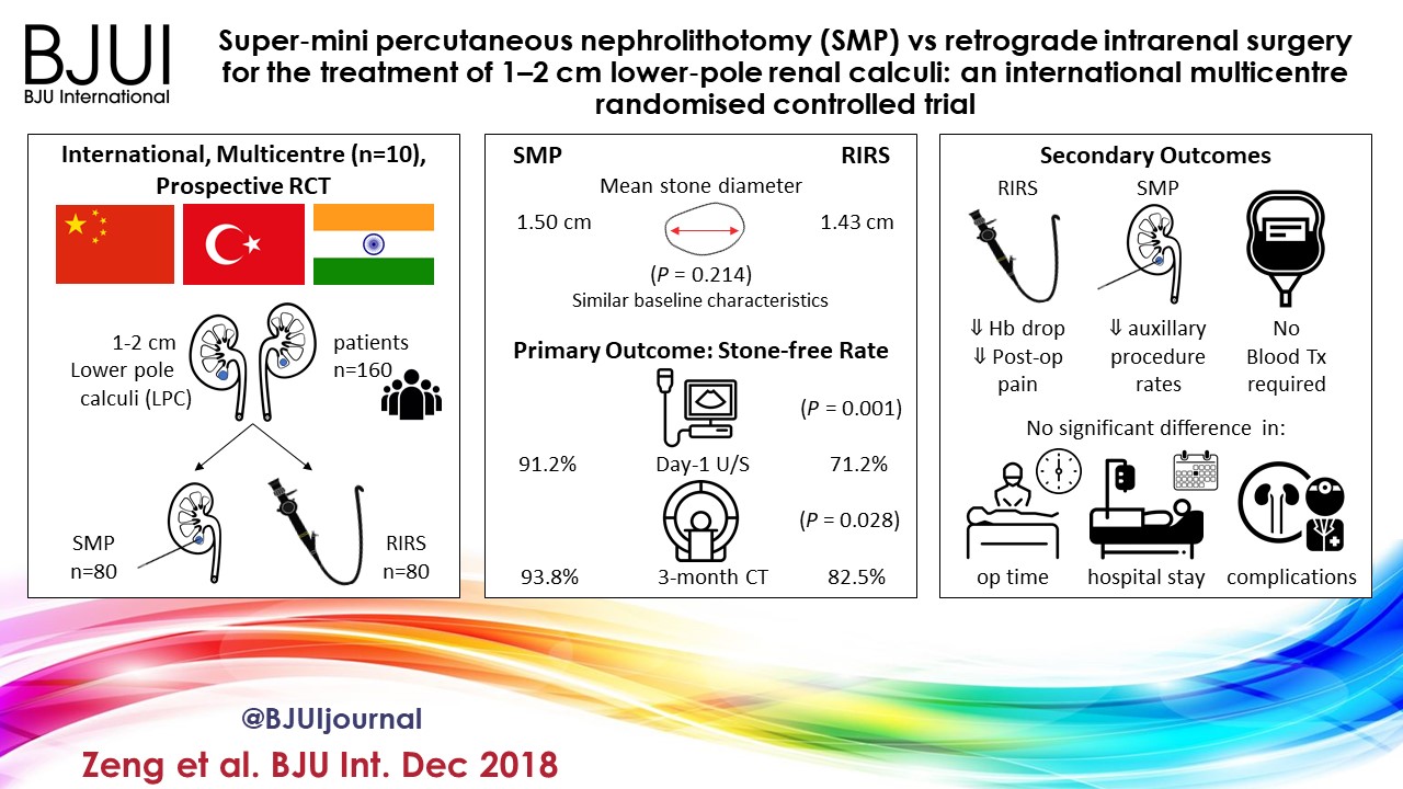
Super‐mini percutaneous nephrolithotomy (SMP) vs retrograde intrarenal surgery for the treatment of 1–2 cm lower‐pole renal calculi: an international multicentre randomised controlled trial

Guohua Zeng* , Tao Zhang* , Madhu Agrawal, Xiang He, Wei Zhang§, Kefeng Xiao, Hulin Li**, Xuedong Li††, Changbao Xu‡‡, Sixing Yang§§, Jean J. de la Rosette¶¶***, Junhong Fan*, Wei Zhu* and Kemal Sarica†††


Department of Urology *Minimally Invasive Surgery Center, The First Afliated Hospital of Guangzhou Medical University, **Guangdong Key Laboratory of Urology, ZhuJiang Hospital of Southern Medical University, Guangzhou, Zhejiang Provincial Peoples Hospital, Zhejiang, §The First Afliated Hospital With Nanjing Medical University, Nanjing, Shenzhen Peoples Hospital, Shenzhen, ††The Second Afliated Hospital of Harbin Medical University, Harbin, ‡‡The Second Afliated Hospital of Zhengzhou University, Zhengzhou, §§Renmin Hospital of Wuhan University, Wuhan, China, Centre for Minimally Invasive Endourology, Global Rainbow Healthcare, Agra, India, ¶¶Istanbul Medipol University, Istanbul, Turkey, ***AMC University Hospital, Amsterdam, The Netherlands, and †††Dr. Lut Kirdar Kartal Research and Training Hospital, Istanbul, Turkey

Abstract

Objectives

To compare the safety and effectiveness of super‐mini‐percutaneous nephrolithotomy (SMP) and retrograde intrarenal surgery (RIRS) for the treatment of 1–2 cm lower‐pole renal calculi (LPC).

Patients and Methods

An international multicentre, prospective, randomised, unblinded controlled study was conducted at 10 academic medical centres in China, India, and Turkey, between August 2015 and June 2017. In all, 160 consecutive patients with 1–2 cm LPC were randomised to receive SMP or RIRS. The primary endpoint was stone‐free rate (SFR). Stone‐free status was defined as no residual fragments of ≥0.3 cm on plain abdominal radiograph of the kidneys, ureters and bladder, and ultrasonography at 1‐day and on computed tomography at 3‐months after operation. Secondary endpoints included blood loss, operating time, postoperative pain scores, auxiliary procedures, complications, and hospital stay. Postoperative follow‐up was scheduled at 3 months. Analysis was by intention‐to‐treat. The trial was registered at https://clinicaltrials.gov/ (NCT02519634).

Results

The two groups had similar baseline characteristics. The mean (sd) stone diameters were comparable between the groups, at 1.50 (0.29) cm for the SMP group vs 1.43 (0.34) cm for the RIRS group (P = 0.214). SMP achieved a significantly better 1‐day and 3‐month SFR than RIRS (1‐day SFR 91.2% vs 71.2%, P = 0.001; 3‐months SFR 93.8% vs 82.5%, P = 0.028). The auxiliary procedure rate was lower in the SMP group. RIRS was found to be superior with lower haemoglobin drop and less postoperative pain. Blood transfusion was not required in either group. There was no significant difference in operating time, hospital stay, and complication rates, between the groups.

Conclusions

SMP was more effective than RIRS for treating 1–2 cm LPC in terms of a better SFR and lesser auxiliary procedure rate. The complications and hospital stay were comparable. RIRS has the advantage of less postoperative pain.


Article of the week: Does the introduction of prostate multi-parametric MRI into the AS protocol for localized PCa improve patient re-classification?

Every week, the Editor-in-Chief selects an Article of the Week from the current issue of BJUI. The abstract is reproduced below and you can click on the button to read the full article, which is freely available to all readers for at least 30 days from the time of this post.

In addition to the article itself, there is an accompanying editorial written by a prominent member of the urological community. This blog is intended to provoke comment and discussion and we invite you to use the comment tools at the bottom of each post to join the conversation.

If you only have time to read one article this week, it should be this one.

Does the introduction of prostate multiparametric magnetic resonance imaging into the active surveillance protocol for localized prostate cancer improve patient re-classification?

Richard J. Bryant*† , Bob Yang* , Yiannis Philippou*, Karla Lam*, Maureen Obiakor*, Jennifer Ayers*, Virginia Chiocchia†‡, Fergus Gleeson§, Ruth MacPherson§, Clare Verrill†¶, Prasanna Sooriakumaran†**, Freddie C. Hamdy*† and Simon F. Brewster*

*Department of Urology, Oxford University Hospitals NHS Foundation Trust, Oxford, UK, †Nuffield Department of Surgical Sciences, University of Oxford, Oxford, UK, ‡National Perinatal Epidemiology Unit, Nuffield Department of Population Health, University of Oxford, Oxford, UK, §Department of Radiology, Oxford University Hospitals NHS Foundation Trust, Oxford, UK, Oxford NIHR Biomedical Research Centre, University of Oxford, Oxford, UK, and **Department of Uro-Oncology, University College London Hospital NHS Foundation Trust, London, UK

Read the full article

Abstract

Objectives

To determine whether replacement of protocol‐driven repeat prostate biopsy (PB) with multiparametric magnetic resonance imaging (mpMRI) ± repeat targeted prostate biopsy (TB) when evaluating men on active surveillance (AS) for low‐volume, low‐ to intermediate‐risk prostate cancer (PCa) altered the likelihood of or time to treatment, or reduced the number of repeat biopsies required to trigger treatment.

Patients and Methods

A total of 445 patients underwent AS in the period 2010–2016 at our institution, with a median (interquartile range [IQR]) follow‐up of 2.4 (1.2–3.7) years. Up to 2014, patients followed a ‘pre‐2014’ AS protocol, which incorporated PB, and subsequently, according to the 2014 National Institute for Health and Care Excellence (NICE) guidelines, patients followed a ‘2014–present’ AS protocol that included mpMRI. We identified four groups of patients within the cohort: ‘no mpMRI and no PB’; ‘PB alone’; ‘mpMRI ± TB’; and ‘PB and mpMRI ± TB’. Kaplan–Meier plots and log‐rank tests were used to compare groups.

Results

Of 445 patients, 132 (30%) discontinued AS and underwent treatment intervention, with a median (IQR) time to treatment of 1.55 (0.71–2.4) years. The commonest trigger for treatment was PCa upgrading after mpMRI and TB (43/132 patients, 29%). No significant difference was observed in the time at which patients receiving a PB alone or receiving mpMRI ± TB discontinued AS to undergo treatment (median 1.9 vs 1.33 years; P = 0.747). Considering only those patients who underwent repeat biopsy, a greater proportion of patients receiving TB after mpMRI discontinued AS compared with those receiving PB alone (29/66 [44%] vs 32/87 [37%]; P = 0.003). On average, a single set of repeat biopsies was needed to trigger treatment regardless of whether this was a PB or TB.

Conclusion

Replacing a systematic PB with mpMRI ±TB as part of an AS protocol increased the likelihood of re‐classifying patients on AS and identifying men with clinically significant disease requiring treatment. mpMRI ±TB as part of AS thereby represents a significant advance in the oncological safety of the AS protocol.

Read more Articles of the week


Article of the month: The US opioid epidemic

Every month, the Editor-in-Chief selects an Article of the Month from the current issue of BJUI. The abstract is reproduced below and you can click on the button to read the full article, which is freely available to all readers for at least 30 days from the time of this post.

In addition to the article itself, there is an accompanying editorial written by a prominent member of the urological community. This blog is intended to provoke comment and discussion and we invite you to use the comment tools at the bottom of each post to join the conversation.

If you only have time to read one article this week, it should be this one.

The United States opioid epidemic: a review of the surgeon’s contribution to it and health policy initiatives

Katherine Theisen, Bruce Jacobs, Liam Macleod and Benjamin Davies
Department of Urology, University of Pittsburgh Medical Center, Pittsburgh, PA, USA
 
Read the full article

Abstract

Visual abstract created by Abdullatif Aydın and Rebecca Fisher

Opioid abuse and addiction is causing widespread devastation in communities across the USA and resulting in significant strain on our healthcare system. There is increasing evidence that prescribers are at least partly responsible for the opioid crisis because of overprescribing, a practice that developed from changes in policy and reimbursement structures. Surgeons, specifically, have been subject to scrutiny as ‘adequate treatment’ of post‐surgical pain is poorly defined and data suggest that many patients receive much larger opioid prescriptions than needed. The consequences of overprescribing include addiction and misuse, dispersion of opioids into the community, and possible potentiation of illicit drug/heroin use. Several solutions to this crisis are currently being enacted with variable success, including Prescription Drug Monitoring Programmes, policy‐level interventions aimed to de‐incentivize overprescribing, limiting opioid exposures through Enhanced Recovery After Surgery protocols, and the novel idea of creating surgery‐ and/or procedure‐specific prescribing guidelines. This problem is likely to require not one, but several potential solutions to reverse its trajectory. It is critical, however, that we as physicians and prescribers find a way to stop the needless overprescribing while still treating postoperative pain appropriately.


Read more Articles of the week

Infographic: The UK‐ROPE Study

Infographic: Efficacy and safety of prostate artery embolization for benign prostatic hyperplasia: an observational study and propensity‐matched comparison with transurethral resection of the prostate (the UK‐ROPE study)

Read the full article
See more infographics

Alpha‐blockers for uncomplicated ureteral stones: a clinical practice guideline

M Vermandere, T Kuijpers, J S Burgers, I Kunnamo, J van Lieshout, E Wallace,  J Vlayen, E Schoenfeld, R A Siemieniuk, L Trevena, X Zhu, F Verermen, B Neuschwander, P h Dahm, K A O Tikkinen, K Aubrey‐Bassler, R W M Vernooij, B Aertgeerts, G E Bekkering

Abstract

Background

The role of medical expulsive therapy for uncomplicated ureteral stones remains controversial in light of new contradictory trial evidence. A Cochrane review was recently published to summarize the current best evidence on this topic.

Aim

To develop an evidence‐based recommendation concerning the use of alpha‐blockers for uncomplicated ureteral stones, based on an up‐to‐date Cochrane review.

Method

We applied the Rapid Recommendations approach to guideline development, which represents an innovative approach by an international collaborative network of clinicians, researchers, methodologists and patient representatives seeking to rapidly respond to new, potentially practice‐changing evidence with recommendations developed according to standards for trustworthy guidelines.

Results

The panel suggests the use of alpha blockers in addition to standard care over standard care alone in patients with uncomplicated ureteral stones (weak recommendation based on low quality evidence). The panel judged that the net benefit of alpha‐blockers was small and that there was considerable uncertainty about patients’ values and preferences. This means that the panel expects that most patients would choose treatment with alpha‐blockers but that a substantial proportion would not. This recommendation applies to both patients in whom the presence of a ureteral stones is confirmed by imaging as well as patients in whom the diagnosis is made based on clinical grounds only.

Conclusion

The Rapid Recommendations panel suggests the use of alpha‐blockers for patients with ureteral stones. Shared decision‐making is emphasized in making the final choice between the treatment options.

This article is protected by copyright. All rights reserved.

Access the full article

Infographic: Impact of dutasteride/tamsulosin combination therapy on sexual function in men with LUTS secondary to BPH

Infographic: Impact of dutasteride/tamsulosin combination therapy on sexual function in men with LUTS secondary to BPH

Read the full article
PDF

Download Slides of this infographic:

PowerPoint


See more infographics

Infographic: Long-term sexual health outcomes in men with classic bladder exstrophy

Infographic: Long-term sexual health outcomes in men with classic bladder exstrophy

Read the full article
See more infographics

Infographic: Partin Tables in the Contemporary Era

The Partin Tables in the Contemporary Era: Infographic to accompany the May 2017 Article of the Month

Read the full article

Download Slides of this infographic:

PDF
PowerPoint

See more infographics


Infographic: 68Ga-PSMA PET/CT for LN staging in PCa

Infographic to accompany the February 2017 Article of the Month

68ga-psma-pet-ct-infographic

Read the full article

See more infographics

Infographic: The origins of urinary stone disease: upstream mineral formations initiate downstream Randall’s plaque

bju13555-fig-0001

Figure 1 The medullo-papillary complex. A total of 8–12 paraboloid complexes are contained within each human kidney. Each complex can be separated into three zones (Zones 1–3) distinguished by distinct segments of the loop of Henle. There are short- and long-looped nephrons and vessels. Owing to the paraboloid geometry of the medullo-papillary complex, shorter looped nephrons and vessels are contained in the periphery, and the longest looped nephrons and vessels are located centrally. Non-fenestrated descending vasa recta are surrounded by layers of smooth muscle, in contrast to the ascending vasa recta comprised of fenestrated endothelium. Within Zone 3, a transition occurs where pericytes replace smooth muscle.

bju13555-fig-0002

Figure 2 Spatial relationships and size distributions of the tubules and vessels within the medullo-papillary complex. From Zone 1 to Zone 2, the ascending and descending vasa recta become organised into vascular bundles (dotted line) and interbundle regions. In Zone 3, the descending thin limbs join the vascular bundles (light dotted line), and these are separate from collecting duct clusters. Collecting ducts grow larger in diameter towards Zone 3 and coalesce to form the 6–12 ducts of Bellini. These anatomically specific compartments contribute to radial and axial concentration gradients along the course of the complex.

bju13555-fig-0003

Figure 3 Medullo-papillary function is characterised by pressure and chemical gradients. Pressure gradients are present from Zone 1 to Zone 3. Due to the paraboloid form of the complex, larger diameter vasa recta are located centrally within the vascular bundles and have higher pressure gradients and flow rates than in the peripherally located vasa recta. Poiseuille’s law relates flow rate as proportional to pressure and radius to the fourth power, and inversely proportional to fluid viscosity and tube length. Within each tube, velocity of fluid is highest at the centerline, but decreases near the wall due to resistance. Over time, within a concentrated fluid, solutes are expected to accumulate along the walls. From Zone 1 to Zone 3, an increasing osmolarity gradient, contributed by primarily sodium salts and urea, generates the urine concentrating ability through countercurrent exchange. Areas vulnerable to hypoxic injury include the tip of the Zone 3, and Zone 2 because of the metabolically active thick ascending limbs and their relative physical separation from the descending vasa recta.

bju13555-fig-0004

Figure 4 Biomineralisation of the medullo-papillary complex leading to Randall’s plaque. Over time, lower pressure gradients in the peripheral tubules relative to the centrally located tubules lead to intratubular mineralisation within Zones 1 and 2. The functional volume of the complex gradually decreases, and at a certain threshold, the change in pressure gradient drives a mechanoresponsive switch that leads to interstitial mineralisation in Zone 3. The accumulation of biominerals in the interstitial space eventually becomes endoscopically visible as Randall’s plaque, the foundation for a future urinary tract stone.


Abstract

Objectives

To describe a new hypothesis for the initial events leading to urinary stones. A biomechanical perspective on Randall’s plaque formation through form and function relationships is applied to functional units within the kidney, we have termed the ‘medullo-papillary complex’ – a dynamic relationship between intratubular and interstitial mineral aggregates.

Methods

A complete MEDLINE search was performed to examine the existing literature on the anatomical and physiological relationships in the renal medulla and papilla. Sectioned human renal medulla with papilla from radical nephrectomy specimens were imaged using a high resolution micro X-ray computed tomography. The location, distribution, and density of mineral aggregates within the medullo-papillary complex were identified.

Results

Mineral aggregates were seen proximally in all specimens within the outer medulla of the medullary complex and were intratubular. Distal interstitial mineralisation at the papillary tip corresponding to Randall’s plaque was not seen until a threshold of proximal mineralisation was observed. Mineral density measurements suggest varied chemical compositions between the proximal intratubular (330 mg/cm3) and distal interstitial (270 mg/cm3) deposits. A review of the literature revealed distinct anatomical compartments and gradients across the medullo-papillary complex that supports the empirical observations that proximal mineralisation triggers distal Randall’s plaque formation.

Conclusion

The early stone event is initiated by intratubular mineralisation of the renal medullary tissue leading to the interstitial mineralisation that is observed as Randall’s plaque. We base this novel hypothesis on a multiscale biomechanics perspective involving form and function relationships, and empirical observations. Additional studies are needed to validate this hypothesis.

Ryan S. Hsi*, Krishna Ramaswamy*, Sunita P. Ho† and Marshall L. Stoller*


*Department of Urology, and Division of Biomaterials and Bioengineering, Department of Preventive and Restorative Dental Sciences, School of Dentistry, University of California San Francisco, San Francisco, CA, USA


Read the full article
Letter to the Editor with response
See more infographics
© 2024 BJU International. All Rights Reserved.