Archive for category: Videos

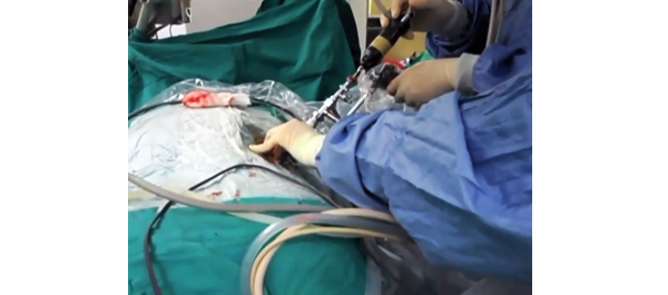
Video: Tract creation using plasma vaporization

Randomized and prospective trial comparing tract creation using plasma vaporization with balloon dilatation in percutaneous nephrolithotomy

Po Hui Chiang and Hsin Hao Su

Kaohsiung Chang Gung Memorial Hospital and Chang Gung University College of Medicine, Kaohsiung, Taiwan

OBJECTIVE

• To evaluate the efficacy and safety of plasma vaporization for tract creation in percutaneous nephrolithotomy (PCNL).

PATIENTS AND METHODS

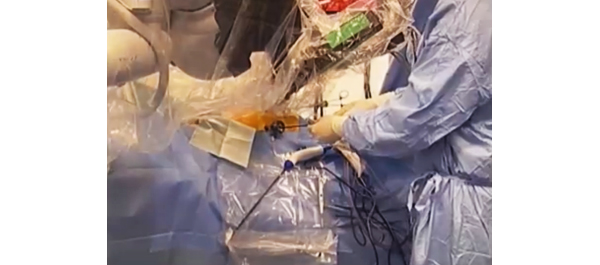
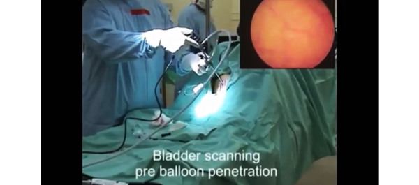
• In this randomized prospective trial we enrolled 65 patients and assigned each to one of two groups: 33 patients were randomly scheduled to undergo plasma vaporization and 32 were scheduled to undergo balloon dilatation for tract creation.

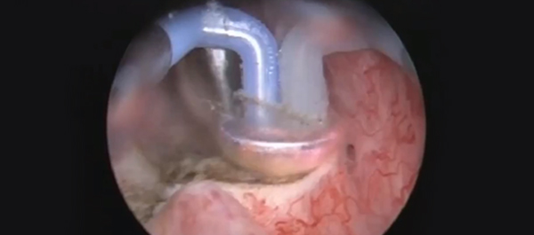
• A bipolar resectoscope mounted with a plasma vaporization button electrode or a traditional balloon dilator were used to create the nephrostomy tract.

RESULTS

• The mean blood loss, mean length of hospital stay and mean operating time, stone-free rates and postoperative complications in the two groups were compared using the t-test or chi-squared test (Fisher’s exact test).

• The plasma vaporization group had a significantly lower mean (SD) decrease in haematocrit level (3.5 [2.8]% vs 6.6 [3.3]%; P = 0.02) and a shorter mean (SD) hospital stay (2.6 [1.2] days vs 5.3 [3.4] days; P = 0.0).

• There were no significant differences in the operating time, stone-free rate or cases of postoperative fever between the two groups.

CONCLUSION

• The plasma vaporization technique is safe, leads to less blood loss than the other techniques, and is a simple solution for creating the nephrostomy tract for PCNL.


Step-by-Step: Robot-assisted AUS insertion


Robot-assisted laparoscopic artificial urinary sphincter insertion in men with neurogenic stress urinary incontinence

David R. Yates, Véronique Phé, Morgan Rouprêt, Christophe Vaessen, Jérôme Parra, Pierre Mozer and Emmanuel Chartier-Kastler

Academic department of Urology, Pitié-Salpétrière Hospital, Assistance Publique-Hôpitaux de Paris (AP-HP), Faculté de Médecine Pierre et Marie Curie, University Paris 6, Paris, France

The first two authors contributed equally to this article.

Read the full article
OBJECTIVES

• To describe for the first time the technique of robot-assisted artificial urinary sphincter (R-AUS) insertion in male patients with neurogenic incontinence.

MATERIALS AND METHODS

• From January 2011 to the present date, six patients with spinal cord injury have undergone R-AUS insertion at our academic institution and we have prospectively collected data on pre-, peri- and early postoperative outcomes.

• A transperitoneal five-port approach was used using a three-arm standard da Vinci® robot (Intuitive Surgical, Sunnyvale, CA, USA) in a 30° reverse Trendelenburg position.

• The artificial urinary sphincter (AUS) cuff was placed circumferentially around the bladder neck, the reservoir was left intra-abdominally in a lateral vesicular space and the pump was placed in a classic scrotal position.

RESULTS

• All six patients had successful robotic implantation of the AUS.

• The median patient age was 51.5 years, the median (range) operating time was 195 (175–250) min with no significant blood loss or intra-operative complications. The median (range) length of hospital stay was 4 (4–6) days.

• At a median (interquartile range) follow-up of 13 (6–21) months, all six patients had a functioning device with complete continence.

• To date, we have observed no incidence of early erosion, device infection or device malfunction.

CONCLUSIONS

• Allowing for the preliminary nature of our data, R-AUS insertion appears safe and technically feasible.

• Larger studies with long-term follow-up and comparison with open AUS insertion are necessary before definitive statements can be made for R-AUS in respect of complications and functional outcomes.

Video: Treating intrinsic sphincter deficiency with myofibre implantation


Periurethral skeletal myofibre implantation in patients with urinary incontinence and intrinsic sphincter deficiency: a phase I clinical trial

René Yiou1, Jean-Yves Hogrel8, Catherine-Marie Loche2, François-Jerome Authier3, Philippe Lecorvoisier7, Pauline Jouany4, Françoise Roudot-Thoraval5 and Jean-Pascal Lefaucheur6

1Hôpital Henri Mondor, Service d’Urologie and CRCDC, 2Hôpital Albert Chenevier, Service de Médecine Physique et de Réadaptation, 3Hôpital Henri Mondor, Département de Pathologie, 4Hôpital Henri Mondor, Unité de Recherche Clinique (URC), Pôle Recherche Clinique et Santé Publique, 5Hôpital Henri Mondor, Département de Santé Publique, Pôle Recherche Clinique et Santé Publique, UPEC, 6Hôpital Henri Mondor, Service de Physiologie – Explorations Fonctionnelles, Université Paris-Est, Faculté de Médecine, APHP, 7Hôpital Henri Mondor, INSERM, Créteil, and 8Université Paris 6, UMR S974, INSERM U974, CNRS UMR 7215, GH Pitié-Salpêtrière, Institut de Myologie, Paris, France

This open-label nonrandomized phase I clinical trial was registered on clinicalTrials.gov (#NCT00472069)

Read the full article
OBJECTIVES

• To assess the safety of periurethral myofibre implantation in patients with urinary incontinence due to intrinsic sphincter deficiency (ISD).

• To assess the resulting myogenic process and effects on urinary continence.

PATIENTS AND METHODS

• An open-label non-randomised phase I clinical trial was conducted in five men and five women with ISD (mean age, 62.5 years).

• A free muscle strip from the patient’s gracilis muscle was implanted around the urethra as a means to deliver locally myofibres and muscle precursor cells (MPCs).

• Patients were assessed for collection formation and incomplete bladder emptying.

• The maximum urethral closure pressure (MUCP) and concomitant periurethral electromyographic (EMG) activity were recorded before surgery and 1 and 3 months after surgery. Continence was assessed using the 24-h pad test and self-completed questionnaires, for 12 months.

RESULTS

• There were no serious side-effects.

• Continence improved significantly during the 12-month follow-up in four of the five women, including two who recovered normal continence. In the women, MUCP increased two-fold and de novo EMG periurethral activity was recorded. In the men, MUCP and EMG recordings showed similar improvements but the effect on continence was moderate.

• The few patients enrolled could affect these results.

CONCLUSIONS

• This is the first report of a one-step procedure for transferring autologous MPCs via myofibre implantation in patients with ISD.

• EMG and urodynamic assessments showed improvement of periurethral muscle activity.

• Further work is needed to confirm and improve the therapeutic efficiency of this procedure.


Video: In-Vivo Trials in Sheep


Novel anti-biofilm mechanism for wireless capsule endoscopy in the urinary tract: preliminary study in a sheep model

Amos Neheman*, Claude Schulman and Ofer Yossepowitch†§

*Urology Department, Meir Medical Center, Kfar-Saba, Sackler School of Medicine, Tel-Aviv University, Tel-Aviv, Israel, Department of Urology, University of Brussels, Brussels, Belgium, and §Institute of Urology, Rabin Medical Center, Beilinson, Petah Tikva, Israel

Read the full article
OBJECTIVE

• To develop and test the safety and feasibility of a novel anti-biofilm mechanism configured for wireless capsule endoscopy (WCE) in a sheep bladder model.

MATERIALS AND METHODS

• A WCE mechanism, designed for long-term bladder monitoring, was developed and introduced into a sheep bladder for 5 months.

• The transparency of the surface was assessed by evaluating a resolution target placed inside the capsule at serial intervals using cystoscopy under general anaesthesia.

• Animal behaviour, voiding patterns and urine cultures were monitored throughout the study.

• At study termination, the capsule was extracted and assessed using scanning electron microscopy.

RESULTS

• The resolution target was visualized clearly at all investigation points.

• No notable adverse effects were noted during the entire follow-up period and no urinary tract infection occurred.

• Scanning electron microscopy confirmed the efficacy of the technology to prevent biofilm formation and surface encrustation.

CONCLUSIONS

• We report a novel technology that effectively prevents biofilm formation on the outer surface of foreign objects in the urinary tract.

• Further studies are under way to test the applicability of this technology in bladder WCE to enable high-quality wireless image transmission.


Video: Androgen deprivation therapy in men with high fracture risk


Fracture after androgen deprivation therapy among men with a high baseline risk of skeletal complications

Yu-Hsuan Shao*, Dirk F. Moore*, Weichung Shih*, Yong Lin*, Thomas L. Jang* and Grace L. Lu-Yao*

*The Cancer Institute of New Jersey, Department of Medicine, The Robert Wood Johnson Medical School, New Brunswick, and Department of Biostatistics, UMDNJ School of Public Health, Piscataway, NJ, USA

Read the full article
OBJECTIVE

• To quantify the impact of androgen deprivation therapy (ADT) in men with a high baseline risk of skeletal complications and evaluate the risk of mortality after a fracture.

PATIENTS AND METHODS

• We studied 75 994 men, aged 66 years, with localized prostate cancer from the Surveillance, Epidemiology and End Results–Medicare linked data.

• Cox proportional hazard models were employed to evaluate the risk.

RESULTS

• Men with a high baseline risk of skeletal complications have a higher probability of receiving ADT than those with a low risk (52.1% vs 38.2%, P < 0.001).

• During the 12-year follow-up, more than 58% of men with a high risk and 38% of men with a low risk developed at least one fracture after ADT.

• The dose effect of ADT is stronger among men who received ADT only compared to those who received ADT with other treatments.

• In the high-risk group, the fracture rate increased by 19.9 per 1000 person-years (from 52.9 to 73.0 person-years) for men who did not receive ADT compared to those who received 18 or more doses of gonadotropin-releasing hormone agonist among men who received ADT only, and by 14.2 per 1000 person-years (from 45.2 to 59.4 person-years) among men who received ADT and other treatments.

•Men experiencing a fracture had a 1.38-fold higher overall mortality risk than those who did not (95% CI, 1.34–1.43).

CONCLUSIONS

• Men with a high baseline risk of skeletal complications developed more fractures after ADT.

• The mortality risk is 40% higher after experiencing a fracture.

• Consideration of patient risk before prescribing ADT for long-term use may reduce both fracture risk and fracture-associated mortality.


Video: Bipolar plasma enucleation vs open prostatectomy

Bipolar plasma enucleation of the prostate vs open prostatectomy in large benign prostatic hyperplasia cases – a medium term, prospective, randomized comparison

Bogdan Geavlete, Florin Stanescu, Catalin Iacoboaie and Petrisor Geavlete

Department of Urology, ‘Saint John’ Emergency Clinical Hospital, Bucharest, Romania

Read the full article
OBJECTIVES

• To evaluate the viability of bipolar plasma enucleation of the prostate (BPEP) by comparison with open transvesical prostatectomy (OP) in cases of large prostates with regard to surgical efficacy and peri-operative morbidity.

• To compare the medium-term follow-up parameters specific for the two methods.

PATIENTS AND METHODS

• A total of 140 benign prostatic hyperplasia (BPH) patients with prostate volume >80 mL, maximum flow rate (Qmax) <10 mL/s and International Prostate Symptom Score (IPSS) >19 were randomized in the two study arms.

• All cases were assessed preoperatively and at 1, 3, 6 and 12 months after surgery by IPSS, Qmax, quality of life score (QoL) and post-voiding residual urinary volume (PVR).

• The prostate volume and prostate specific antigen (PSA) level were measured at 6 and 12 months.

RESULTS

• The BPEP and OP techniques emphasized similar mean operating durations (91.4 vs 87.5 min) and resected tissue weights (108.3 vs 115.4 g).

• The postoperative haematuria rate (2.9% vs 12.9%) as well as the mean haemoglobin drop (1.7 vs 3.1 g/dL), catheterization period (1.5 vs 5.8 days) and hospital stay (2.1 vs 6.9 days) were significantly improved for BPEP.

• Recatheterization for acute urinary retention was more frequent in the OP group (8.6% vs 1.4%), while the rates of early irritative symptoms were similar for BPEP and OP (11.4% vs 7.1%).

• During the follow-up period, no statistically significant difference was determined in terms of IPSS, Qmax, QoL, PVR, PSA level and postoperative prostate volume between the two series.

CONCLUSIONS

• BPEP represents a promising endoscopic approach in large BPH cases, characterized by good surgical efficiency and similar BPH tissue removal capabilities compared with standard transvesical prostatectomy.

• BPEP patients benefited from significantly reduced complications, shorter convalescence and satisfactory follow-up symptom scores and voiding parameters.


John Davis, BJUI Associate Editor: Urological Oncology


John Davis, BJUI Associate Editor for urological oncology, talks about why authors should submit to the BJUI.

The aim of the Journal is to publish high-quality papers with high-impact statements. Along with rapid reviews and publication, BJUI is supporting the impact of papers through social media, such as Twitter and Facebook. The journal will still be printed monthly, but additionally have an exciting web interface.

The NERI Nocturia Advisory Conference 2012: focus on outcomes of therapy


The New England Research Institutes, Inc. (NERI) Nocturia Advisory Conference 2012: focus on outcomes of therapy

Jeffrey P. Weiss1,2, Jerry G. Blaivas1,2, Marco H. Blanker9, Donald L. Bliwise3, Roger R. Dmochowski4, Marcus Drake11, Catherine E. DuBeau5, Adonis Hijaz7, Raymond C. Rosen6, Philip E.V. Van Kerrebroeck10 and Alan J. Wein8

1Department of Urology, SUNY Downstate College of Medicine, Brooklyn, 2Weill Medical College of Cornell University, New York, NY, 3Emory University School of Medicine, Atlanta, GA, 4Department of Urologic Surgery, Vanderbilt University, Nashville, TN, 5Division of Geriatric Medicine, UMass Memorial Medical Center and UMass Medical School, Worcester, MA, 6New England Research Institutes, Watertown, MA, 7Urology Institute, University Hospitals Case Medical Center, Cleveland, OH, 8Division of Urology, Perelman School of Medicine at the University of Pennsylvania, Philadelphia, PA, 9Department of General Practice, University of Groningen, University Medical Center Groningen, Groningen, 10Department of Urology, Maastricht University Medical Center, Maastricht, The Netherlands, and 11University of Bristol, Bristol, UK

Read the full article
INTRODUCTION

Nocturia, awaking to void urine, is a common and sometimes bothersome symptom that may impose detrimental impacts on sleep-quality, mood, and overall health [1]. The multi-factorial aetiology of nocturia, coupled with the recent demonstration that this symptom is highly variable over time and often resolves spontaneously [2] , makes nocturia a challenging clinical entity. Although nocturia may have little health impact for some, for others it can be a highly bothersome, debilitating condition. Multiple studies have shown an association between nocturia and disturbed sleep, reduced well-being, and increased morbidity [3, 4, 5].

A consensus statement published in 2011 provided guidance to clinicians who are confronted with the wide range of clinical presentations of nocturia [1]. That paper focused primarily on a description of nocturia, its prevalence, its impact on health-related quality of life (QOL) and overall health, and an overview of available treatment options. The present paper extends and elaborates on the previous paper by examining the most recent research on diagnostic and treatment outcomes. Numerous papers have been published in the 2 years since the previous conference was organised, and the field, as a whole, has a large and dynamic research agenda. This paper summarises the findings resulting from a 2012 conference of key thought leaders in the field of nocturia who focused on updating outcome studies published since the previous conference was held.


Video: Androgen-ablation therapy (AAT) associated autophagy


Does androgen-ablation therapy (AAT) associated autophagy have a pro-survival effect in LNCaP human prostate cancer cells?

Haley L. Bennett, Jacqueline Stockley, Janis T. Fleming, Ranadip Mandal, Jim O’Prey*, Kevin M. Ryan*, Craig N. Robson and Hing Y. Leung

Urology Research Laboratory and *Tumour Cell Death Research Laboratory, Beatson Institute for Cancer Research, Glasgow, and Solid Tumour Target Discovery Group, Northern Institute for Cancer Research, Newcastle University, Newcastle upon Tyne, UK

Read the full article
OBJECTIVE

• To study the cellular effects of the anti-androgen bicalutamide on autophagy and its potential impact on response to androgen-ablation therapy (AAT) alone or combined with docetaxel chemotherapy in human prostate cancer LNCaP cells.

MATERIALS AND METHODS

• LNCaP cells were treated with bicalutamide  docetaxel, and cellular effects were assayed: lipidated LC3 (a microtubule-associated protein) for autophagy and its trafficking to fuse with lysosome; flow cytometry using propidium iodide or caspase 3 for cell death; and sulforhodamine B assay for cell growth.

RESULTS

• Bicalutamide treatment enhanced autophagy in LNCaP cells with increased level of autophagosome coupled with an altered cellular morphology reminiscent of neuroendocrine differentiation.

• Consistent with the literature on the interaction between androgen receptor activation and taxane chemotherapy, bicalutamide diminished docetaxel mediated cytotoxicity.

• Significantly, pharmacological inhibition of autophagy with 3-methyladenine significantly enhanced the efficacy cell kill mediated by AAT  docetaxel.

CONCLUSION

• Autophagy associated with bicalutamide treatment in LNCaP cells may have a pro-survival effect and strategy to modulate autophagy may have a potential therapeutic
value.

© 2024 BJU International. All Rights Reserved.