Tag Archive for: aotw 26-02-2020

Posts

Article of the week: Using spatial tracking with magnetic resonance imaging/ultrasound‐guided biopsy to identify unilateral prostate cancer

Every week, the Editor-in-Chief selects an Article of the Week from the current issue of BJUI. The abstract is reproduced below and you can click on the button to read the full article, which is freely available to all readers for at least 30 days from the time of this post.

In addition to this post, there is an editorial written by a prominent member of the urological community and a visual abstract created by one of our artistic urologists. Please use the comment buttons below to join the conversation.

If you only have time to read one article this week, we recommend this one. 

Using spatial tracking with magnetic resonance imaging/ultrasound‐guided biopsy to identify unilateral prostate cancer

Steve R. Zhou*, Alan M. Priester†‡, Rajiv Jayadevan, David C. Johnson§, Jason J. Yang*, Jorge Ballon*, Shyam Natarajan†‡ and Leonard S. Marks

*David Geffen School of Medicine, University of California, Department of Urology, University of California, Department of Bioengineering, University of California, Los Angeles, CA, and §Department of Urology, University of
North Carolina, Chapel Hill, NC, USA

Abstract

Objectives

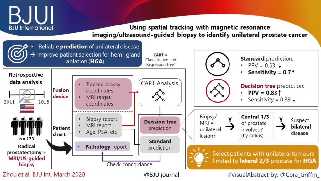
To create reliable predictive metrics of unilateral disease using spatial tracking from a fusion device, thereby improving patient selection for hemi‐gland ablation of prostate cancer.

Patients and Methods

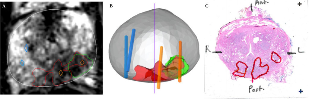
We identified patients who received magnetic resonance imaging (MRI)/ultrasound‐guided biopsy and radical prostatectomy at a single institution between 2011 and 2018. In addition to standard clinical features, we extracted quantitative features related to biopsy core and MRI target locations predictive of tumour unilaterality. Classification and Regression Tree (CART) analysis was used to create a decision tree (DT) for identifying cancer laterality. We evaluated concordance of model‐determined laterality with final surgical pathology.

Fig. 2. Correlation of MRI (A), spatial biopsy pathology (B), and WMP (C). Suspicious MRI lesion (green in A and B) is shown to underestimate true tumour volume (red in A and B, outlined in C). Positive ipsilateral cores (orange) confirm intermediate disease in the MRI lesion and near midline. Negative contralateral cores in blue erroneously imply unilaterality of disease. Only a subset of tracked cores is shown for clarity.

Results

A total of 173 patients were identified with biopsy coordinates and surgical pathology available. Based on CART analysis, in addition to biopsy‐ and MRI‐confirmed disease unilaterality, patients should be further screened for cancer detected within 7 mm of midline in a 40 mL prostate, which equates to the central third of any‐sized prostate by radius. The area under the curve for this DT was 0.82. Standard diagnostics and the DT correctly identified disease laterality in 73% and 80% of patients, respectively (P = 0.13). Of the patients identified as unilateral by standard diagnostics, 47% had undetected contralateral disease or were otherwise incorrectly identified. This error rate was reduced to 17% (P = 0.01) with the DT.

Conclusion

Using spatial tracking from fusion devices, a DT was more reliable for identifying laterality of prostate cancer compared to standard diagnostics. Patients with cancer detected within the central third of the prostate by radius are poor hemi‐gland ablation candidates due to the risk of midline extension of tumour.

Editorial: Can artificial intelligence optimize case selection for hemi‐gland ablation?

The victory of ‘AlphaGo’ over humans in Go, one of the most complex games with more than 10170 board configurations, has yielded tremendous attention worldwide [1]. The later version, ‘AlphaGo Zero’, has brought artificial intelligence (AI) to the next level by demonstrating an absolute superiority, winning 100‐0 against the champion‐defeating AlphaGo [2]. It is exciting, and perhaps shocking, to realize what AI can achieve.

In this issue of BJUI, the study by Zhou et al. [3] is the first to utilize AI to optimize case selection for hemi‐gland ablation. In this study, classification and regression tree (CART) analysis, which is a form of supervised machine‐learning algorithm, was used to identify laterality of prostate cancer. In the conventional approach, case selection was based on biopsy results and MRI findings. For the CART model, in addition to the common clinical variables (i.e. age, PSA, prostate volume, biopsy and MRI results), biopsy coordinate‐derived spatial features were also used as model inputs. The model output was the probability of unilateral clinically significant prostate cancer considered suitable for hemi‐gland ablation. Whole‐mount prostatectomy specimens were used as the standard of reference. The CART model correctly identified laterality in 80% of the cases, compared to 73% with the conventional approach. The positive predictive value of the CART model was 83%, compared to 53% with the conventional approach. The superiority of the CART model has been demonstrated, and the area under curve was 0.82.

Artifical intelligence has been widely adopted in the field of Urology [4]. For prostate cancer detection in particular, our group evaluated the diagnostic performances of four machine‐learning models based on clinical variables in a biopsy cohort of 1625 men [5]. The machine‐learning models achieved excellent performances in detecting clinically significant prostate cancer, with an accuracy of up to 95.3%. Algohary et al. [6] constructed three machine‐learning models to identify the presence of clinically significant prostate cancer based on MRI radiomic features in patients who underwent active surveillance. When compared with the Prostate Imaging–Reporting and Data System (PI‐RADS) scoring system, the machine‐learning models were able to improve overall accuracy by 30–80%.

Fehr et al. [7] developed an automated system to classify Gleason scores based on MRI images. The automated system could distinguish between Gleason scores of 6 and 7 or above cancers with an accuracy of up to 93%. The differentiation between Gleason score 3+4 and 4+3 disease also yielded an accuracy of up to 93%. Importantly, the performance of AI and machine‐learning models is highly dependent on the quality and accuracy of the data being input. In terms of prostate cancer detection, either mapping biopsy or whole‐mount prostatectomy specimens should be considered to represent the ‘ground truth’.

There are a number of challenges in implementing AI in clinical practice. First, decision‐making in healthcare requires logical deduction and explanation. The data processing in AI, however, is often described as a ‘black box’. Taking AlphaGo as an example, some ‘moves’ were considered incomprehensible even by world‐class players. Second, although results from AI are promising, there is in general a lack of regulations and standards to assess its safety, efficacy and validity. Liability issues can be problematic in case of medical mishaps. Third, doctors are human. Conflict of interest does exist, and how we can utilize AI in a complementary rather than a competitive manner is a challenging obstacle to overcome.

Nevertheless, AI has huge potential in improving healthcare. Collaborative effort is needed globally to develop and optimize AI systems, and to increase its acceptability and practicality upon implementation. Future studies answering clinically important questions using appropriate standards of reference will be of paramount importance in paving the way for the AI era in urology.

by Jeremy Yuen‐Chun Teoh, Edmund Chiong and Chi‐Fai Ng

References

  1. Silver DHuang AMaddison CJ et al. Mastering the game of Go with deep neural networks and tree search. Nature 2016529484– 9
  2. Silver DSchrittwieser JSimonyan K et al. Mastering the game of Go without human knowledge. Nature 2017550354– 9
  3. Zhou SRPriester AMJayadevan R et al. Using spatial tracking with magnetic resonance imaging/ultrasound‐guided biopsy to identify unilateral prostate cancer. BJU Int 2020125399– 406
  4. Chen JRemulla DNguyen JH et al. Current status of artificial intelligence applications in urology and their potential to influence clinical practiceBJU Int 2019124567– 77
  5. Wang GTeoh JYChoi KSDiagnosis of prostate cancer in a Chinese population by using machine learning methods. Conf Proc IEEE Eng Med Biol Soc 201820181– 4
  6. Algohary AViswanath SShiradkar R et al. Radiomic features on MRI enable risk categorization of prostate cancer patients on active surveillance: Preliminary findings. J Magn Reson Imaging 201848818– 28
  7. Fehr DVeeraraghavan HWibmer A et al. Automatic classification of prostate cancer Gleason scores from multiparametric magnetic resonance images. Proc Natl Acad Sci USA 2015112E6265– 73


Visual abstract: Using spatial tracking with MRI/ultrasound‐guided biopsy to identify unilateral PCa

See more infographics

© 2024 BJU International. All Rights Reserved.