Tag Archive for: Article of the Month

Posts

Editorial: Penile vibratory stimulation (PVS) a novel approach for penile rehabilitation post nerve sparing radical prostatectomy

The reported incidence of erectile dysfunction (ED) after nerve-sparing radical prostatectomy (NS-RP) varies in the literature from 30 to 80% [1]. This can be explained by the state of neuropraxia which affects the cavernosal nerves, even if the nerves are anatomically intact. During this period there is a lack of nocturnal tumescence which leads to tissue hypoxia and ischaemic damage to the cavernosal smooth muscles leading to smooth muscle necrosis and fibrosis, which in turn causes veno-occlusive dysfunction (VOD). A study by Mulhall et al. [2] showed that, at 12 months after NS-RP, 50% of patients will have VOD and ED. The role of penile rehabilitation, therefore, is to maintain adequate tissue oxygenation until the cavernosal nerves recover with the return of the spontaneous nocturnal tumescence; thus, penile rehabilitation should not be confused with ED treatment. If you see yourself as religious, addiction may make you feel guilty or get you to feel isolated among your friends at your religious organization. A spiritual Christian rehab center in Orlando may be the right choice for you. Not only do you get to meet like-minded people to share your experiences in your journey to sobriety, but the process may also help you to rediscover your faith in God. Legacy Healing Center Tampa offer programs that make spiritual guidance an important part of every type of addiction treatment. Orange County law enforcement has taken steps to make sure the drugs are not as easily available as they once were. This has helped manage Orlando’s drug problem and kept it from turning worse. As important as prevention is to saving lives, however, to the hundreds who are already addicted, rehab is what helps. If you are religious or spiritual, faith-based drug rehab can be the answer to the challenges that you face. It’s important to remember that faith-based rehab only works well for those who are deeply spiritual or religious. Trying faith-based rehab when you are ambivalent about religion can work against you. You may find that you aren’t able to accept what you’re asked to practice, and you may find yourself rebelling. It’s important to choose a treatment approach that you can go along with in good conscience.

Several lines of treatment, including phosphodiesterase 5 inhibitors, intracavernous injection of alprostadil and vacuum pump therapy, have been used in penile rehabilitation but an agreed rehabilitation programme in terms of agents used, timing and duration of therapy does not yet exist [1].

The present study by Fode et al. [3] reports a novel approach to penile rehabilitation using penile vibratory stimulation (PVS). The study looked into the effect of PVS on postoperative erection and continence. The Ferticare® vibrator (Fig. 1) was used at an amplitude of 2 mm and a vibration frequency of 100 Hz and applied to the frenulum once daily, with a sequence consisting of 10 s of stimulation followed by a 10-s rest and repeated 10 times.

The results showed a trend towards better erection in the PVS group (n = 30) compared with the control group (n = 38) as evidenced by the higher International Index of Erectile Function (IIEF) score, but the difference was not significant (P = 0.09). After 1 year, 16 patients (53%) in the PVS group had an IIEF score ≥18 compared with 12 (32%) patients in the control group (P = 0.07). The results did not show any effect of treatment on continence; at 12 months, 90% of the PVS group achieved continence compared with 94.7% of the control group (P = 0.46), although the PVS group had a significantly higher preoperative LUTS score which may explain the results.

The theory postulated is that application of PVS activates the parasympathetic erectile spinal centre (S2–S4), which in turn leads to activation of the cavernosal nerves, enhancing the healing process, and recovery from neuropraxia and restoration of spontaneous erections. Also this would lead to stimulation of the somatic S2–S4 spinal centre, which controls the pelvic floor muscles via the pudendal nerve, leading to the recovery of continence. Although this has been shown in patients with spinal cord injury as the authors mentioned; this may not be the case in post NS-RP with the nerves in a state of neurapraxia, whereas in patients with spinal cord injury the nerves are intact. It would have been of great value to conduct neurophysiological tests on these patients to demonstrate that, despite the cavernosal nerves being in a state of neurapraxia, nerve activity in response to PVS was actually present.

The rehabilitation protocol used in the present study started early but only continued for 6 weeks postoperatively. Studies have shown that the potential recovery time of erectile function after NS-RP is 6–36 months, with the majority recovering within 12–24 months [1,4]. The results might have shown statistical significance in favour of PVS, had treatment continued for a longer period. Starting PVS treatment in the early postoperative period may not be suitable in all patients; in this study six out of 36 patients (16.6%) were non-compliant with the protocol; four had prolonged catheterization and two experienced pain. Furthermore, neurophysiological testing is required to show that in the early postoperative period the cavernosal nerves are actually intact and therefore respond to PVS.

Although the results of the present study did not reach significance, they are encouraging, as there was a trend in favour of treatment with regard to erectile function. Further studies involving larger numbers of patients are warranted to investigate this new line of rehabilitation.

Amr Abdel Raheem* and David Ralph
*Andrology Department, Cairo University Hospital, Cairo, Egypt, and St. Peter’s Andrology Centre, Institute of Urology, London, UK

References

  1. Mulhall JP, Bivalacqua TJ, Becher EF. Standard operating procedure for the preservation of erectile function outcomes after radical prostatectomy. J Sex Med 2013; 10: 195–203
  2. Mulhall JP, Slovick R, Hotaling J et al. Erectile dysfunction after radical prostatectomy: hemodynamic profiles and their correlation with the recovery of erectile function. J Urol 2002; 167: 1371–5
  3. Fode M, Borre M, Ohl D, Lichtbach J, Sønksen J. Penile vibratory stimulation in the recovery of urinary continence and erectile function after nerve-sparing radical prostatectomy: a randomized, controlled trial. BJU Int 2014; 114: 111–7
  4. Rabbani F, Schiff J, Piecuch M et al. Time course of recovery of erectile function after radical retropubic prostatectomy: does anyone recover after 2 years? J Sex Med 2010; 7: 3984–90

Video: Penile vibratory stimulation after radical prostatectomy

Penile vibratory stimulation in the recovery of urinary continence and erectile function after nerve-sparing radical prostatectomy: a randomized, controlled trial

Mikkel Fode*, Michael Borre, Dana A. Ohl, Jonas Lichtbach§ and Jens Sønksen*

*Department of Urology, Herlev University Hospital, Herlev, Department of Urology, Aarhus University Hospital, Aarhus, Denmark, Department of Urology, University of Michigan, Ann Arbor, MI, USA, and §Department of Physiotherapy, Herlev University Hospital, Herlev, Denmark

OBJECTIVE

• To examine the effect of penile vibratory stimulation (PVS) in the preservation and restoration of erectile function and urinary continence in conjunction with nerve-sparing radical prostatectomy (RP).

PATIENTS AND METHODS

• The present study was conducted between July 2010 and March 2013 as a randomized prospective trial at two university hospitals. Eligible participants were continent men with an International Index of Erectile Function-5 (IIEF-5) score of at least 18, scheduled to undergo nerve-sparing RP.

• Patients were randomized to a PVS group or a control group. Patients in the PVS group were instructed in using a PVS device (FERTI CARE® vibrator).

• Stimulation was performed at the frenulum once daily by the patients in their own homes for at least 1 week before surgery. After catheter removal, daily PVS was re-initiated for a period of 6 weeks.

• Participants were evaluated at 3, 6 and 12 months after surgery with the IIEF-5 questionnaire and questions regarding urinary bother. Patients using up to one pad daily for security reasons only were considered continent. The study was registered at https://clinicaltrials.gov/ (NCT01067261).

RESULTS

• Data from 68 patients were available for analyses (30 patients randomized to PVS and 38 patients randomized to the control group).

• The IIEF-5 score was highest in the PVS group at all time points after surgery with a median score of 18 vs 7.5 in the control group at 12 months (P = 0.09), but the difference only reached borderline significance.

• At 12 months, 16/30 (53%) patients in the PVS group had reached an IIEF-5 score of at least 18, while this was the case for 12/38 (32%) patients in the control group (P = 0.07).

• There were no significant differences in the proportions of continent patients between groups at 3, 6 or 12 months. At 12 months 90% of the PVS patients were continent, while 94.7% of the control patients were continent (P = 0.46).

CONCLUSION

• The present study did not document a significant effect of PVS. However, the method proved to be acceptable for most patients and there was a trend towards better erectile function with PVS. More studies are needed to explore this possible effect further.


Article of the month: MRI and active surveillance for prostate cancer

Every week the Editor-in-Chief selects the Article of the Week from the current issue of BJUI. The abstract is reproduced below and you can click on the button to read the full article, which is freely available to all readers for at least 30 days from the time of this post.

In addition to the article itself, there is an accompanying editorial written by a prominent member of the urological community. This blog is intended to provoke comment and discussion and we invite you to use the comment tools at the bottom of each post to join the conversation.

Finally, the third post under the Article of the Week heading on the homepage will consist of additional material or media. This week we feature a video from Dr. Hyun M. Lee discussing his paper.

If you only have time to read one article this week, it should be this one.

Role of multiparametric 3.0-Tesla magnetic resonance imaging in patients with prostate cancer eligible for active surveillance

Bong H. Park, Hwang G. Jeon, Seol H. Choo, Byong C. Jeong, Seong I. Seo, Seong S. Jeon, Han Y. Choi and Hyun M. Lee

Department of Urology, Samsung Medical Center, Sungkyunkwan University School of Medicine, Seoul, Korea
Current address: Bong H. Park, Department of Urology, Incheon St. Mary’s Hospital, The Catholic University of Korea College of Medicine, Seoul, Korea

Read the full article
OBJECTIVE

• To evaluate predictors of more aggressive disease and the role of multiparametric 3.0-T magnetic resonance imaging (MRI) in selecting patients with prostate cancer for active surveillance (AS).

PATIENTS AND METHODS

• We retrospectively assessed 298 patients with prostate cancer who met the Prostate Cancer Research International: Active Surveillance (PRIAS) criteria, defined as T1c/T2, PSA level of ≤10 ng/mL, PSA density (PSAD) of <0.2 ng/mL2, Gleason score <7, and one or two positive biopsy cores.

• All patients underwent preoperative MRI, including T2-weighted, diffusion-weighted, and dynamic contrast-enhanced imaging, as well as radical prostatectomy (RP) between June 2005 and December 2011.

• Imaging results were correlated with pathological findings to evaluate the ability of MRI to select patients for AS.

RESULTS

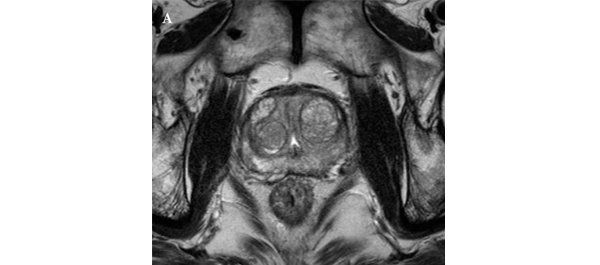
• In 35 (11.7%) patients, no discrete cancer was visible on MRI, while in the remaining 263 (88.3%) patients, a discrete cancer was visible.

• Pathological examination of RP specimens resulted in upstaging (>T2) in 21 (7%) patients, upgrading (Gleason score >6) in 136 (45.6%), and a diagnosis of unfavourable disease in 142 (47.7%) patients.

• The 263 patients (88.3%) with visible cancer on imaging were more likely to have their cancer status upgraded (49.8% vs 14.3%) and be diagnosed with unfavourable disease (52.1% vs 14.3%) than the 35 patients (11.7%) with no cancer visible upon imaging, and these differences were statistically significant (P < 0.001 for all).

• A visible cancer lesion on MRI, PSAD, and patient age were found to be predictors of unfavourable disease in multivariate analysis.

CONCLUSION

• MRI can predict adverse pathological features and be used to assess the eligibility of patients with prostate cancer for AS.


Editorial: Multiparametric MRI and active surveillance for prostate cancer: future directions

A growing body of data exists suggesting an important role of MRI in selecting men with prostate cancer for active surveillance (AS). In the present study, Park et al. [1] show that a suspicious lesion on MRI was independently predictive of adverse pathology after radical prostatectomy (RP). This finding supports existing data suggesting that suspicious lesions on MRI confer an increased risk of disease reclassification among men enrolled in AS [2]. Indeed, in our institutional AS experience we found that men with a suspicious lesion on MRI were more likely to have biopsy reclassification with extended follow-up [3].While these data are provocative, much work remains to be done before the adoption of MRI as a standard screening tool for entry into AS for men with very low-risk prostate cancer.

Introduction of functional sequence imaging into multiparametric MRI protocols has resulted in improved detection and characterisation of clinically localised prostate cancer. However, before widespread implementation into AS protocols can occur, increased rigor and standardisation in image interpretation is needed. As in the present study, 5-point Likert scales have become an increasingly popular method of quantifying a lesions likelihood of representing cancer [1]. Still other authors have quantified a lesions level of suspicion using both weighted and non-weighted scoring systems based on the number of positive MRI sequences [3,4]. While useful for statistical analysis, these reporting methods are fraught with concerns of inter-observer variability and generalizability. Additionally, a recent report by Lee et al. [5] found that a simple measurement of lesion diameter on diffusion-weighted MRI was predictive of insignificant disease after RP. Combining the plethora of functional and morphological data obtained by multiparametric MRI into a standardised, reproducible tool will greatly facilitate implementation of MRI into current AS screening protocols.

As a step in the right direction, Stamatakis et al. [4] recently generated a nomogram for predicting biopsy reclassification in men on AS after taking into consideration both functional and morphological characteristics of MRI lesions. Adding an additional layer of complexity, they also assessed the utility of calculated values, e.g. lesion density (lesion volume/prostate volume), in predicting biopsy reclassification. Briefly, their analysis showed that the number of lesions, lesion suspicion, and lesion density were predictive of biopsy reclassification. While nomogram validation and testing of its predictive value on pathological outcomes is needed, this represents a major advance in the standardised application of MRI to AS cohorts.

Despite great strides in the application of multiparametric MRI to AS cohorts, a significant concern about the false-negative rate exists. Considering the present report, of the 35 men with no visible lesion on MRI, 14.3% men had unfavourable pathology after RP [1]. This is similar to previous studies reporting disease reclassification rates of <18% [2,6]. These men with normal imaging and high-grade cancer highlight the importance of incorporating imaging and clinical data when selecting men for AS. Better defining the false-negative rate of multiparametric MRI, and effectively identifying men with a normal MRI and high-grade disease remain major challenges.

Considering all of the available data, it is becoming increasingly clear that MRI has the potential for improving the identification of patients for whom AS would be safe. It is currently the practice at our institution to refer eligible men for multiparametric MRI before enrolment in AS. Our future scholarly efforts should be directed at the standardisation of reporting MRI data and the development of user-friendly AS criteria that synthesise MRI results with clinicopathological data.

Jeffrey K. Mullins and H. Ballentine Carter
James Buchanan Brady Urological Institute, Johns Hopkins Medical Institutions, Baltimore, MD, USA

Read the full article

References

  1. Park BH, Jeon HG, Choo SH et al. Role of multiparametric 3.0-Tesla magnetic resonance imaging in patients with prostate cancer eligible for active surveillance. BJU Int 2014; 113: 864–70
  2. Margel D, Yap SA, Lawrentschuk N et al. Impact of multiparametric endorectal coil prostate magnetic resonance imaging on disease reclassification among active surveillance candidates: a prospective cohort study. J Urol 2012; 187: 1247–52
  3. Mullins JK, Bonekamp D, Landis P et al. Multiparametric magnetic resonance imaging findings in men with low-risk prostate cancer followed using active surveillance. BJU Int 2013; 111: 1037–45
  4. Stamatakis L, Siddiqui MM, Nix JW et al. Accuracy of multiparametric magnetic resonance imaging in confirming eligibility for active surveillance for men with prostate cancer. Cancer 2013; 119: 3359–66
  5. Lee DH, Koo KC, Lee SH et al. Tumor lesion diameter on diffusion weighted magnetic resonance imaging could help predict insignificant prostate cancer in patients eligible for active surveillance: preliminary analysis. J Urol 2013; 190: 1213–7
  6. Guzzo TJ, Resnick MJ, Canter DJ et al. Endorectal T2-weighted MRI does not differentiate between favorable and adverse pathologic features in men with prostate cancer who would qualify for active surveillance. Urol Oncol 2012; 30: 301–5


Video: MRI can assess the eligibility of patients with prostate cancer for AS

Role of multiparametric 3.0-Tesla magnetic resonance imaging in patients with prostate cancer eligible for active surveillance

Bong H. Park, Hwang G. Jeon, Seol H. Choo, Byong C. Jeong, Seong I. Seo, Seong S. Jeon, Han Y. Choi and Hyun M. Lee

Department of Urology, Samsung Medical Center, Sungkyunkwan University School of Medicine, Seoul, Korea
Current address: Bong H. Park, Department of Urology, Incheon St. Mary’s Hospital, The Catholic University of Korea College of Medicine, Seoul, Korea

Read the full article
OBJECTIVE

• To evaluate predictors of more aggressive disease and the role of multiparametric 3.0-T magnetic resonance imaging (MRI) in selecting patients with prostate cancer for active surveillance (AS).

PATIENTS AND METHODS

• We retrospectively assessed 298 patients with prostate cancer who met the Prostate Cancer Research International: Active Surveillance (PRIAS) criteria, defined as T1c/T2, PSA level of ≤10 ng/mL, PSA density (PSAD) of <0.2 ng/mL2, Gleason score <7, and one or two positive biopsy cores.

• All patients underwent preoperative MRI, including T2-weighted, diffusion-weighted, and dynamic contrast-enhanced imaging, as well as radical prostatectomy (RP) between June 2005 and December 2011.

• Imaging results were correlated with pathological findings to evaluate the ability of MRI to select patients for AS.

RESULTS

• In 35 (11.7%) patients, no discrete cancer was visible on MRI, while in the remaining 263 (88.3%) patients, a discrete cancer was visible.

• Pathological examination of RP specimens resulted in upstaging (>T2) in 21 (7%) patients, upgrading (Gleason score >6) in 136 (45.6%), and a diagnosis of unfavourable disease in 142 (47.7%) patients.

• The 263 patients (88.3%) with visible cancer on imaging were more likely to have their cancer status upgraded (49.8% vs 14.3%) and be diagnosed with unfavourable disease (52.1% vs 14.3%) than the 35 patients (11.7%) with no cancer visible upon imaging, and these differences were statistically significant (P < 0.001 for all).

• A visible cancer lesion on MRI, PSAD, and patient age were found to be predictors of unfavourable disease in multivariate analysis.

CONCLUSION

• MRI can predict adverse pathological features and be used to assess the eligibility of patients with prostate cancer for AS.


Flying high as a kite

Some of my happiest memories are from my childhood. Part of it was spent in Lucknow where my mother had her ancestral home. An important city in Northern India, Lucknow was the seat of the Nawabs who built many beautiful palaces. One of these has a labyrinth, which many have entered only to get lost within its many chambers. Another, the Chhota Imambara is pictured on the cover. Lucknow is also famous for its cuisine with street vendors selling tasty kebabs. Above all, I remember many hours perched on the roof top of our home in the old town, flying kites, with my family. The sky above became a riot of colours. Today there is even a touring company offering nostalgic kite flying holidays in this ancient city.

In May, our Article of the Month comes from the King George Medical University, Lucknow. In a prospective, longitudinal comparison over six years, of a large number of patients undergoing urinary diversion after radical cystectomy, the authors demonstrate better quality of life after orthotopic neobladder rather than ileal conduit formation [1]. The mean age of the patients was in the mid 50s, which is perhaps why a significant number underwent neobladder formation. This article and the accompanying editorial from Urs Studer [2] are must reads for anyone involved in the management of bladder cancer. In the UK many of our patients are generally older with multiple co-morbidities and end up having ileal conduits. For the younger patients it is perhaps time for a rethink?

We also feature an excellent multi-institutional collaboration reporting on PCNL outcomes in England from the Hospital Episode Statistics (HES) database over a five year period. Mortality is rare after this procedure but 9% of patients have a readmission within 30 days [3]. While the HES like most other databases has its inherent limitations, the authors should be congratulated for analysing complex outcomes on nearly 6000 patients; in particular John Withington who is writing his thesis on the subject.

And finally – an invitation. If you are attending the AUA, we are again having a BAUS–BJUI–USANZ session on the afternoon of the 18 May. The faculty is international and the program even more exciting than it was last year. This is a further testament to the strong friendship that exists between our organisations and the AUA. The Coffey–Krane prize for the best paper published in the BJUI by a trainee, will be presented at the end of this session followed by the BJUI reception.

Many of you have loved our new design, layout and quality although this has led to a precipitous drop in our acceptance rate in favour of only the very best papers. Thank you for your support, which has given us the strength and resolve to fly high. The sky is the limit.

Prof. Prokar Dasgupta
Editor-in-Chief, BJUI

King’s College London, Guy’s Hospital#

References

  1. Singh V, Yadav R, Sinha RJ, Gupta DK. Prospective comparison of quality-of-life outcomes between ileal conduit urinary diversion and orthotopic neobladder reconstruction after radical cystectomy: a statistical model. BJU Int 2014; 113: 726–732
  2. Studer UE. Life is good with orthotopic bladder substitutes! BJU Int 2014; 113: 686–687
  3. Armitage JN, Withington J, van der Meulen J, et al. Percutaneous nephrolithotomy in England: practice and outcomes described in the Hospital Episode Statistics database. BJU Int 2014; 113: 777–782


Article of the month: Better QOL with orthotopic neobladders

Every week the Editor-in-Chief selects the Article of the Week from the current issue of BJUI. The abstract is reproduced below and you can click on the button to read the full article, which is freely available to all readers for at least 30 days from the time of this post.

In addition to the article itself, there is an accompanying editorial written by a prominent member of the urological community. This blog is intended to provoke comment and discussion and we invite you to use the comment tools at the bottom of each post to join the conversation.

Finally, the third post under the Article of the Week heading on the homepage will consist of additional material or media. This week we feature a video from Dr. Singh of orthotopic neobladder reconstruction by sigmoid colon.

If you only have time to read one article this week, it should be this one

Prospective comparison of quality-of-life outcomes between ileal conduit urinary diversion and orthotopic neobladder reconstruction after radical cystectomy: a statistical model

Vishwajeet Singh, Rahul Yadav, Rahul Janak Sinha and Dheeraj Kumar Gupta
Department of Urology, King George Medical University, Lucknow, Uttar Pradesh, India

Read the full article
OBJECTIVE

• To conduct a prospective comparison of quality-of-life (QoL) outcomes in patients who underwent ileal conduit (IC) urinary diversion with those who underwent orthotopic neobladder (ONB) reconstruction after radical cystectomy for invasive bladder cancers.

PATIENTS AND METHODS

• Between January 2007 and December 2012, 227 patients underwent radical cystectomy and either IC urinary diversion or ONB (sigmoid or ileal) reconstruction.

• Contraindications for ON were impaired renal function (serum creatinine >2 mg/dL), chronic inflammatory bowel disease, previous bowel resection and tumour involvement at the bladder neck/prostatic urethra. Patients who did not have these contraindications chose to undergo either IC or ONB reconstruction, after impartial counselling.

• Baseline characteristics, including demographic profile, body mass index, comorbidities, histopathology of the cystoprostatectomy (with lymph nodes) specimen, pathological tumour stage, postoperative complications, adjuvant therapy and relapse, were recorded and compared.

• The European Organization for Research and Treatment of Cancer QoL questionnaire C30 version 3 was used to analyse QoL before surgery and 6, 12 and 18 months after surgery.

RESULTS

• Of the 227 patients, 28 patients in the IC group and 35 in the ONB group were excluded. The final analysis included 80 patients in the IC and 84 in the ONB group.

• None of the baseline characteristics were significantly different between the groups, except for age, but none of the baseline QoL variables were found to be correlated with age.

• In the preoperative phase, there were no significant differences in any of the QoL domains between the IC or the ONB groups. At 6, 12 and 18 months in the postoperative period, physical functioning (P < 0.001, P < 0.001 and P = 0.001, respectively), role functioning (P = 0.01, P = 0.01 and P = 0.003, respectively), social functioning (P = 0.01, P = 0.01 and P = 0.01, respectively) and global health status/QoL (P < 0.001, P < 0.001 and P = 0.002, respectively) were better in patients in the ONB group than in those in the IC group and the differences were significant.

• The financial burden related to bladder cancer treatment was significantly lower in the ONB group than in the IC group at 6, 12 and 18 months of follow-up (P = 0.05, P = 0.05 and P = 0.005, respectively)

CONCLUSIONS

• ONB is better than IC in terms of physical functioning, role functioning, social functioning, global health status/QoL and financial expenditure.

• ONB reconstruction provides better QoL outcomes than does IC urinary diversion.


Editorial: Life is good with orthotopic bladder substitutes!

In the present issue of the BJUI, Singh et al. [1] present the results of a non-randomized prospective study comprising 80 patients who underwent ileal conduit diversion and 84 who underwent orthotopic bladder substitution. Quality of life was assessed using the European Organisation for the Research and Treatment of Cancer quality-of-life questionnaire, the QLQ-30C, at 6, 12 and 18 months postoperatively. Physical and social functioning and global health status were significantly better in patients with orthotopic bladder substitution than in those who underwent ileal conduit diversion. Moreover, the postoperative financial burden was significantly lower for patients in the orthotopic bladder group than for those in the ileal conduit group, who required stoma appliances, a finding of particular importance not only in India, where the study was performed, but worldwide. The authors’ results are particularly impressive given their use of a questionnaire that included many items (‘Were you short of breath?’, ‘Did you need to rest?’, ‘Have you lacked appetite?’, ‘Have you been constipated?’, ‘Did you feel tense?’, ‘Did you worry’ or ‘Did you feel irritable?’, etc.) that can hardly discriminate between the quality of life of patients who underwent orthotopic bladder substitution and those who underwent ileal conduit diversion. To find significant differences between the two types of urinary diversion, despite such dilution factors, speaks strongly in favour of orthotopic bladder substitution.

The results of this prospective single-centre trial are of particular importance because, as the authors state, other investigators could not show such differences, presumably for a variety of reasons, such as too few patients or single follow-up assessments given at time points that varied from patient to patient. Quality-of-life assessment at similar follow-up time points, as performed by these authors, is important because, with adequate counselling, the postoperative function of orthotopic bladder substitutes improves over time.

Without a doubt, however, a poorly functioning orthotopic bladder substitute may lead to a poorer quality of life than a well-functioning ileal conduit diversion. Poor functional results and life-threatening complications can be largely avoided with ileal orthotopic bladder substitutes, provided the treating urologist has adequate knowledge of the procedure and the patient receives adequate postoperative education [2]. The major ways to ensure good results are:

  • appropriate patient selection (good renal function, regular follow-up possible);
  • the avoidance of damage to the sphincter apparatus and its innervation (individualized nerve-sparing cystectomy, minimum use of bipolar electrocautery near the pelvic plexus and membranous urethra);
  • the use of ileum instead of colon (better compliance) [3-5];
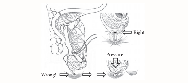
  • the avoidance of a funnel-shaped outlet that can result in kinking, outlet obstruction, residual infected urine and, in the worst case, lifelong need for clean intermittent catheterization (CIC) (Fig. 1).

By contrast to most other urological procedures, orthotopic bladder substitution requires proactive postoperative management [6] to ensure:

  • residual urine-free spontaneous voiding after catheter removal;
  • sterile urine to improve urinary continence and to reduce mucous production [7];
  • the prevention of salt loss syndrome and metabolic acidosis by increased salt intake and sodium bicarbonate substitution in the early postoperative period to ensure a base excess of +2;
  • a systematic increase in functional capacity by progressively expanding voiding intervals to obtain a reservoir capacity of ∼500 mL and, thus, a low end-fill pressure which ensures urinary continence day and night (the latter combined with the use of an alarm clock).

It is equally important to perform lifelong follow-up of patients and regularly at 6- to 12-month intervals so as to diagnose and treat early secondary complications, such as uretero-intestinal strictures or residual, infected urine. If the latter occurs, any form of outlet obstruction, such as ileal mucosa protruding in front of the bladder outlet, strictures or growth of inadvertently left prostatic tissue, must be looked for and treated. In our own experience, secondary outlet obstruction occurred in ∼20% of patients observed for 10 years. This rather high incidence is typical for intestinal bladder substitutes because when voiding, unlike the genuine bladder, there is no coordinated contraction of the reservoir wall which would result in an elevated voiding pressure which, in turn, would overcome an outlet resistance. Bladder substitutes empty mainly by gravitational force alone. If voiding is only possible by abdominal straining, then something must be wrong; therefore, instead of recommending CIC for patients who build up residual and consecutively infected urine, we strongly favour treating the outlet obstruction, usually on an outpatient basis. The avoidance of the need for CIC through surgical technique (no funnel-shaped outlet) and during regular follow-up by treating any potential cause of residual urine can substantially improve the patient’s quality of life. It also avoids the cost of catheters and the risk of infectious complications. Thanks to this active management and removal of any outlet obstruction, 96% of our patients followed for 10 years were still able to void spontaneously [8].

Urs E. Studer
Department of Urology, University Hospital Bern, Bern, Switzerland

Read the full article

References

  1. Singh V, Yadav R, Sinha RJ, Gupta DK. Prospective comparison of quality-of-life outcomes between ileal conduit urinary diversion and orthotopic neobladder reconstruction after radical cystectomy: a statistical model. BJU Int 2014; 113: 726–732
  2. Thurairaja R, Burkhard FC, Studer UE. The orthotopic neobladder. BJU Int 2008; 102: 1307–1313
  3. Berglund B, Kock NG, Myrvold HE. Volume capacity and pressure characteristics of the continent cecal reservoir. Surg Gynecol Obstet 1986; 163: 42–48
  4. Schrier BP, Laguna MP, van der Pal F, Isorna S, Witjes JA. Comparison of orthotopic sigmoid and ileal neobladders: continence and urodynamic parameters. Eur Urol 2005; 47: 679–685
  5. Varol C, Studer UE. Managing patients after an ileal orthotopic bladder substitution. BJU Int 2004; 93: 266–270
  6. Zehnder P, Dhar N, Thurairaja R, Ochsner K, Studer UE. Effect of urinary tract infection on reservoir function in patients with ileal bladder substitute. J Urol 2009; 181: 2545–2549
  7. Thurairaja R, Studer UE. How to avoid clean intermittent catheterization in men with ileal bladder substitution. J Urol 2008; 180: 2504–2509


Video: Orthotopic neobladder reconstruction by sigmoid colon

Prospective comparison of quality-of-life outcomes between ileal conduit urinary diversion and orthotopic neobladder reconstruction after radical cystectomy: a statistical model

Vishwajeet Singh, Rahul Yadav, Rahul Janak Sinha and Dheeraj Kumar Gupta
Department of Urology, King George Medical University, Lucknow, Uttar Pradesh, India


Read the full article
OBJECTIVE

• To conduct a prospective comparison of quality-of-life (QoL) outcomes in patients who underwent ileal conduit (IC) urinary diversion with those who underwent orthotopic neobladder (ONB) reconstruction after radical cystectomy for invasive bladder cancers.

PATIENTS AND METHODS

• Between January 2007 and December 2012, 227 patients underwent radical cystectomy and either IC urinary diversion or ONB (sigmoid or ileal) reconstruction.

• Contraindications for ON were impaired renal function (serum creatinine >2 mg/dL), chronic inflammatory bowel disease, previous bowel resection and tumour involvement at the bladder neck/prostatic urethra. Patients who did not have these contraindications chose to undergo either IC or ONB reconstruction, after impartial counselling.

• Baseline characteristics, including demographic profile, body mass index, comorbidities, histopathology of the cystoprostatectomy (with lymph nodes) specimen, pathological tumour stage, postoperative complications, adjuvant therapy and relapse, were recorded and compared.

• The European Organization for Research and Treatment of Cancer QoL questionnaire C30 version 3 was used to analyse QoL before surgery and 6, 12 and 18 months after surgery.

RESULTS

• Of the 227 patients, 28 patients in the IC group and 35 in the ONB group were excluded. The final analysis included 80 patients in the IC and 84 in the ONB group.

• None of the baseline characteristics were significantly different between the groups, except for age, but none of the baseline QoL variables were found to be correlated with age.

• In the preoperative phase, there were no significant differences in any of the QoL domains between the IC or the ONB groups. At 6, 12 and 18 months in the postoperative period, physical functioning (P < 0.001, P < 0.001 and P = 0.001, respectively), role functioning (P = 0.01, P = 0.01 and P = 0.003, respectively), social functioning (P = 0.01, P = 0.01 and P = 0.01, respectively) and global health status/QoL (P < 0.001, P < 0.001 and P = 0.002, respectively) were better in patients in the ONB group than in those in the IC group and the differences were significant.

• The financial burden related to bladder cancer treatment was significantly lower in the ONB group than in the IC group at 6, 12 and 18 months of follow-up (P = 0.05, P = 0.05 and P = 0.005, respectively)

CONCLUSIONS

• ONB is better than IC in terms of physical functioning, role functioning, social functioning, global health status/QoL and financial expenditure.

• ONB reconstruction provides better QoL outcomes than does IC urinary diversion.


Article of the month: The surgical spectacle

Every week the Editor-in-Chief selects the Article of the Week from the current issue of BJUI. The abstract is reproduced below and you can click on the button to read the full article, which is freely available to all readers for at least 30 days from the time of this post.

In addition to the article itself, there is an accompanying editorial written by a prominent member of the urological community. This blog is intended to provoke comment and discussion and we invite you to use the comment tools at the bottom of each post to join the conversation.

If you only have time to read one article this week, it should be this one.

The surgical spectacle: a survey of urologists viewing live case demonstrations

Sammy E. Elsamra, Mathew Fakhoury, Hector Motato, Justin I. Friedlander, Daniel M. Moreira, Joel Hillelsohn, Brian Duty, Zeph Okeke and Arthur D. Smith

The Arthur Smith Institute for Urology, Hofstra North Shore-LIJ School of Medicine, New Hyde Park, NY, USA

Read the full article
OBJECTIVE

• To evaluate perspectives of urologists viewing live case demonstrations (LCD) and taped case demonstrations (TCD).

METHOD

• A 15-question anonymous survey was distributed to attendees of the live surgery session at the American Urological Association 2012 national meeting (Atlanta) and the second International Challenges in Endourology meeting (Paris).

RESULTS

• Of 1000 surveys distributed, 253 were returned completed (response rate 25%). Nearly half of respondents were in the academic practice setting and nearly 75% were beyond training.

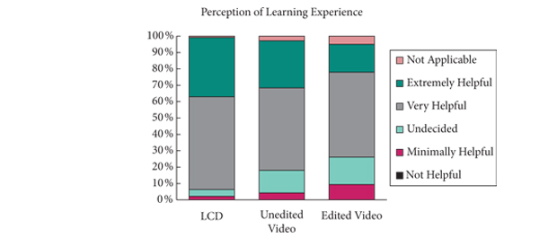
• Just over 30% had performed a LCD previously. The perceived benefit of an LCD was greater than unedited and edited videos (chi-squared P = 0.014 and P < 0.001, respectively). Nearly no one selected ‘not helpful’ and a few selected ‘minimally helpful’ for any of the three forms of demonstration.

• Most respondents identified that opportunity to ask questions (61%) and having access to the full unedited version (72%), two features inherent to LCD, improved upon the educational benefit of edited videos.

• Most (78%) identified LCD as ethical. However, those that did not perceived lower educational benefit from LCD (P = 0.019).

• A slim majority (58%) would allow themselves or a family member to be a patient of a LCD and the vast majority (86%) plan to transfer knowledge gained at the LCD session into their practice.

CONCLUSIONS

• Urologists who attended these LCD sessions identified LCDs as beneficial and applicable to their practice.

• LCDs are preferred over videos. The large majority considers LCD ethical, although not as many would volunteer themselves for LCD.

• Further studies are necessary to determine if there is actual benefit from LCD over TCD to patient care.


© 2024 BJU International. All Rights Reserved.