Tag Archive for: Article of the Week

Posts

Video: In utero myelomeningocele repair and urological outcomes: the first 100 cases of a prospective analysis. Is there an improvement in bladder function?

In utero myelomeningocele repair and urological outcomes: the first 100 cases of a prospective analysis. Is there an improvement in bladder function?

Abstract


Objectives

To evaluate the first 100 cases of in utero myelomeningocele (MMC) repair and urological outcomes in a prospective analysis aiming to define possible improvement in bladder function.

Patients and methods

We used a protocol consisting of a detailed medical history, urinary tract ultrasonography, voiding cystourethrography, and urodynamic evaluation. Patients were categorised into four groups: normal, high risk (overactive bladder with a detrusor leak‐point pressure >40 cm H2O and high filling pressures also >40 cm H2O), incontinent, and underactivity (underactive bladder with post‐void residual urine), and patients were treated accordingly.

Results

We evaluated 100 patients, at a mean age of 5.8 months (median 4 months), classified as high risk in 52.6%, incontinent in 27.4%, with underactive bladder in 4.2%, and only 14.7% had a normal bladder profile. Clean intermittent catheterisation was initiated in 57.3% of the patients and anticholinergics in 52.6%. Antibiotic prophylaxis was initiated in 19.1% of the patients presenting with vesico‐ureteric reflux.

Conclusion

The high incidence of abnormal bladder patterns suggests little benefit of in utero MMC surgery concerning the urinary tract.


Article of the week: Symptom relief and anejaculation after aquablation or transurethral resection of the prostate: subgroup analysis from a blinded randomized trial

Every week, the Editor-in-Chief selects an Article of the Week from the current issue of BJUI. The abstract is reproduced below and you can click on the button to read the full article, which is freely available to all readers for at least 30 days from the time of this post.

In addition to the article itself, there is an editorial written by a prominent member of the urological community. These are intended to provoke comment and discussion and we invite you to use the comment tools at the bottom of each post to join the conversation. 

If you only have time to read one article this week, it should be this one.

Symptom relief and anejaculation after aquablation or transurethral resection of the prostate: subgroup analysis from a blinded randomized trial

Mark Plante1, Peter Gilling2, Neil Barber3, Mohamed Bidair4, Paul Anderson5, Mark Sutton6, Tev Aho7, Eugene Kramolowsky8, Andrew Thomas9, Barrett Cowan10, Ronald P. Kaufman Jr11, Andrew Trainer12, Andrew Arther12, Gopal Badlani13, Mihir Desai14, Leo Doumanian14, Alexis E. Te15, Mark DeGuenther16 and Claus Roehrborn17


1University of Vermont Medical Center, Burlington, VT, USA, 2Tauranga Urology Research, Tauranga, New Zealand, 3Frimley Park Hospital, Frimley Health Foundation Trust, Surrey, UK, 4San Diego Clinical Trials, San Diego, CA, USA, 5Royal Melbourne Hospital, Melbourne, Vic., Australia, 6Houston Metro Urology, Houston, TX, USA, 7Addenbrookes Hospital, Cambridge University Hospitals, Cambridge, UK, 8Virginia Urology, Richmond, VA, USA, 9Princess of Wales Hospital, Bridgend, Wales, UK, 10Urology Associates, P.C., Englewood, CO, 11Albany Medical College, Albany, NY, 12Adult Pediatric Urology and Urogynecology, P.C., Omaha, NE, 13Wake Forest School of Medicine, Winston-Salem, NC, 14Institute of Urology, University of Southern California, Los Angeles, CA, 15Weill Cornell Medical College, New York, NY, 16Urology Centers of Alabama, Birmingham, AL, and 17Department of Urology, UT Southwestern Medical Center, University of Texas Southwestern, Dallas, TX, USA


Read the full article

Abstract

Objective

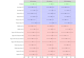
To test the hypothesis that benign prostatic hyperplasia (BPH) robotic surgery with aquablation would have a more pronounced benefit in certain patient subgroups, such as men with more challenging anatomies (e.g. large prostates, large middle lobes) and men with moderate BPH.

Methods

We conducted prespecified and post hoc exploratory subgroup analyses from a double‐blind, multicentre prospective randomized controlled trial that compared transurethral resection of the prostate (TURP) using either standard electrocautery vs surgery using robotic waterjet (aquablation) to determine whether certain baseline factors predicted more marked responses after aquablation as compared with TURP. The primary efficacy endpoint was reduction in International Prostate Symptom Score (IPSS) at 6 months. The primary safety endpoint was the occurrence of Clavien–Dindo persistent grade 1 or grade ≥2 surgical complications.

Results

For men with larger prostates (50–80 g), the mean IPSS reduction was four points greater after aquablation than after TURP (P = 0.001), a larger difference than the overall result (1.8 points; P = 0.135). Similarly, the primary safety endpoint difference (20% vs 46% [26% difference]; P = 0.008) was greater for men with large prostate compared with the overall result (26% vs 42% [16% difference]; P = 0.015). Postoperative anejaculation was also less common after aquablation compared with TURP in sexually active men with large prostates (2% vs 41%; P < 0.001) vs the overall results (10% vs 36%; P < 0.001). Exploratory analysis showed larger IPSS changes after aquablation in men with enlarged middle lobes, men with severe middle lobe obstruction, men with a low baseline maximum urinary flow rate, and men with elevated (>100) post‐void residual urine volume.

Conclusions

In men with moderate‐to‐severe lower urinary tract symptoms attributable to BPH and larger, more complex prostates, aquablation was associated with both superior symptom score improvements and a superior safety profile, with a significantly lower rate of postoperative anejaculation. The standardized, robotically executed, surgical approach with aquablation may overcome the increased outcome variability in more complex anatomy, resulting in superior symptom score reduction.

Read more Articles of the week

Article of the week: A clinical prediction tool to determine the need for concurrent systematic sampling at the time of MRI‐guided biopsy

Every week, the Editor-in-Chief selects an Article of the Week from the current issue of BJUI. The abstract is reproduced below and you can click on the button to read the full article, which is freely available to all readers for at least 30 days from the time of this post.

In addition to the article itself, there is an editorial written by a prominent member of the urological community. These are intended to provoke comment and discussion and we invite you to use the comment tools at the bottom of each post to join the conversation. 

If you only have time to read one article this week, it should be this one.

A clinical prediction tool to determine the need for concurrent systematic sampling at the time of magnetic resonance imaging‐guided biopsy

Niranjan J. Sathianathen*, Christopher A. Warlick*, Christopher J. Weight*, Maria A. Ordonez*, Benjamin Spilseth, Gregory J. Metzger, Paari Muruganand Badrinath R. Konety*


Departments of *Urology, Radiology, and Pathology, University of Minnesota, Minneapolis, MN, USA


Read the full article

Abstract

Objective

To develop a clinical prediction tool that characterises the risk of missing significant prostate cancer by omitting systematic biopsy in men undergoing transrectal ultrasonography/magnetic resonance imaging (TRUS/MRI)‐fusion‐guided biopsy.

Patients and methods

A consecutive sample of men undergoing TRUS/MRI‐fusion‐guided biopsy with the UroNav® system (Invivo International, Best, The Netherlands) who also underwent concurrent systematic biopsy was included. By comparing the grade of cancer diagnosed on targeted and systematic biopsy cores, we identified cases where clinically significant disease (Gleason score ≥3+4) was only found on systematic and not targeted cores. Multivariable logistic regression analyses were used to identify predictive factors for finding significant cancer on systematic cores only. We then used these data to develop a nomogram and evaluated its utility using decision curve analysis.

Fig 1. Nomogram for predicting the diagnosis of clinically significant on systematic biopsy only and missed on targeted biopsy.

Results

Of the 398 men undergoing TRUS/MRI‐fusion‐guided biopsy in our study, there were 46 (11.6%) cases in which clinically significant cancer was missed on targeted biopsy and detected on systematic biopsy. The clinical setting, number of MRI lesions identified, and the highest Prostate Imaging‐Reporting and Data System (PI‐RADS) score of the lesions, were all found to be predictors of this. Our model had a good discriminative ability (concordance index = 0.70). The results from our decision curve analysis show that this model provides a higher net clinical benefit than either biopsying all men or omitting biopsy in all patients when the threshold probability is <30%.

Conclusion

We found that omitting concurrent systematic biopsy in men undergoing TRUS/MRI‐fusion‐guided biopsy would miss significant disease in more than one in 10 patients. We propose a prediction model with good discriminative ability that can be used to improve patient selection for performing concurrent systematic biopsy in order to minimise the number of missed significant cancers. It is important that our model is validated in external cohorts before being employed in routine clinical practice.

Read more Articles of the week

Article of the week: Ultrasound characteristics of regions identified as suspicious by MRI predict the likelihood of clinically significant cancer on MRI–ultrasound fusion‐targeted biopsy

Every week, the Editor-in-Chief selects an Article of the Week from the current issue of BJUI. The abstract is reproduced below and you can click on the button to read the full article, which is freely available to all readers for at least 30 days from the time of this post.

In addition to the article itself, there is an editorial written by a prominent member of the urological community, and a video made by the authors. These are intended to provoke comment and discussion and we invite you to use the comment tools at the bottom of each post to join the conversation. 

If you only have time to read one article this week, it should be this one.


The ultrasound characteristics of regions identified as suspicious by magnetic resonance imaging (MRI) predict the likelihood of clinically significant cancer on MRI–ultrasound fusion‐targeted biopsy

Benjamin Press*, Andrew B. Rosenkrantz, Richard Huang and Samir S. Taneja§ 
 
*Rutgers New Jersey Medical School, Newark, NJ, Department of Radiology, Department of Urology, and §Departments of Urology and Radiology, NYU Langone Health, New York, NY, USA
 
Read the full article

Abstract

Objective

To determine whether the presence of an ultrasound hypoechoic region at the site of a region of interest (ROI) on magnetic resonance imaging (MRI) results in improved prostate cancer (PCa) detection and predicts clinically significant PCa on MRI–ultrasonography fusion‐targeted prostate biopsy (MRF‐TB).

Materials and Methods

Between July 2011 and June 2017, 1058 men who underwent MRF‐TB, with or without systematic biopsy, by a single surgeon were prospectively entered into an institutional review board‐approved database. Each MRI ROI was identified and scored for suspicion by a single radiologist, and was prospectively evaluated for presence of a hypoechoic region at the site by the surgeon and graded as 0, 1 or 2, representing none, a poorly demarcated ROI‐HyR, or a well demarcated ROI‐HyR, respectively. The interaction of MRI suspicion score (mSS) and ultrasonography grade (USG), and the prediction of cancer detection rate by USG, were evaluated through univariate and multivariate analysis.

Results

For 672 men, the overall and Gleason score (GS) ≥7 cancer detection rates were 61.2% and 39.6%, respectively. The cancer detection rates for USGs 0, 1 and 2 were 46.2%, 58.6% and 76.0% (P < 0.001) for any cancer, and 18.7%, 35.2% and 61.1% (P < 0.001) for GS ≥7 cancer, respectively. For MRF‐TB only, the GS ≥7 cancer detection rates for USG 0, 1 and 2 were 12.8%, 25.7% and 52.0%, respectively (P < 0.001). On univariate analysis, in men with mSS 2–4, USG was predictive of GS ≥7 cancer detection rate. Multivariable regression analysis showed that USG, prostate‐specific antigen density and mSS were predictive of GS ≥7 PCa on MRF‐TB.

Conclusions

Ultrasonography findings at the site of an MRI ROI independently predict the likelihood of GS ≥7 PCa, as men with a well‐demarcated ROI‐HyR at the time of MRF‐TB have a higher risk than men without.
Read more Articles of the week

Editorial: Is transrectal ultrasonography of the prostate obsolete in the MRI era?

Sampling of prostate tissue to confirm pathologically a clinical suspicion of cancer has undergone an exponential change. The random systematic prostate biopsy technique was the only method used for many decades, initially guided by the finger but, since 1989, performed with TRUS guidance. Now, within the space of only a few years, we have entered the era of performing prostate biopsies on the basis of high‐tech three‐dimensional multiparametric MRI images, including software that can track the exact course of the biopsy needle [1]. While new technical developments in general lead to better, more individually directed healthcare, there is always the risk of abandoning ‘old’ but well developed and extensively tested techniques too soon. In this issue of the BJUI, Press et al. [2] looked at the added value of the presence of an ‘old‐fashioned’ TRUS‐detected lesion in cancer‐suspicious regions on MRI to better predict the presence of clinically significant prostate cancer (csPCa) defined as Gleason score ≥7. In their study comprising 1058 men, it was shown that a well‐demarcated abnormal TRUS finding noted at the time of MRI‐TRUS fusion‐guided prostate biopsy coincides with an increased risk of csPCa detection, independent of MRI suspicion (Prostate Imaging Reporting and Data System [PI‐RADS] score).

Increasing PI‐RADS score is correlated with an increased percentage of csPCa after targeted biopsy, both at initial and repeat biopsy. In a review based on data from 8252 men, it was shown that there is a gradual increase in the detection of csPCa from PI‐RADS 3 to PI‐RADS 4 to PI‐RADS 5 index lesions. For example, at first biopsy, the overall rate of PCa detection and the percentage of csPCa were 39%, 62% and 92% and 54%, 63% and 76% for PI‐RADS 3, 4 and 5 lesions, respectively. This means that in men with PI‐RADS 3 lesions, representing approximately one‐third of men deemed eligible for further assessment, only 39% will be diagnosed with PCa and half of the PCa detected will be potentially indolent Gleason 6 PCa [3]. This makes this group of men extremely interesting for further risk stratification before biopsy. Multivariable risk stratification in which PSA density plays an important role has been shown to be of value in these men [4] but further refinement could potentially be made by including suspicious lesions identified at TRUS.

Apart from the added value of TRUS findings in terms of risk stratification, the performance of the MRI‐targeted biopsy itself could be improved by visual guidance of hypoechoic lesions. In the present study by Press et al [2], a hypoechoic TRUS lesion was present at or near the location of two‐thirds of cancer‐suspicious lesions on MRI. The authors more or less advise to direct the targeted biopsy cores not only to the MRI suspicious lesion, but also the TRUS suspicious lesion, both of which often do not fully overlay in a software‐assisted MRI‐TRUS fusion model. The extent to which this ‘correction for misregistration’ is already included during targeted biopsy in current clinical practice is unknown. Although feasible and seemingly important during software‐assisted fusion targeted biopsy, TRUS lesions in cancer‐suspicious MRI regions might be more frequently targeted during cognitive fusion‐targeted biopsy. Two recent studies underline the important message of the present study, and show that a considerable proportion of csPCa is missed in and around MRI‐suspicious lesions by targeted biopsies, as a result of sampling errors related to both misregistration and intra‐tumour heterogeneity [56]. As suggested by these studies, visual guidance by hypoechoic lesions and ‘focal saturation’ biopsy by additional (peri‐)lesional cores might improve the detection of csPCa.

In summary, ‘good old’ TRUS could be of value in those patients who are virtually always present in scenarios in which a grading system is being used, i.e. patients belonging to the so‐called grey zone. The challenge of risk stratification (i.e. personalized medicine) is to nibble at both sides of the grey zone by implementing new techniques or, more likely by implementing a combination of all available and relevant knowledge.

by Monique J. Roobol, Frank-Jan H. Drost and Arnout R. Alberts

References

  1. Verma, SChoyke, PLEberhardt, SC et al. The current state of MR imaging‐targeted biopsy techniques for detection of prostate cancer. Radiology 201728534356
  2. Press, BRosenkrantz, ABHuang, RTaneja, SSThe ultrasound characteristics of MRI suspicious regions predict the likelihood of clinically significant cancer on MRI‐ultrasound fusion targeted biopsy. BJUI 201912343946.
  3. Schoots, IGMRI in early prostate cancer detection: how to manage indeterminate or equivocal PI‐RADS 3 lesions? Transl Androl Urol 201877082
  4. Alberts, ARSchoots, IGBokhorst, LPLeenders, GJBangma, CHRoobol, MJRisk‐based patient selection for magnetic resonance imaging‐targeted prostate biopsy after negative transrectal ultrasound‐guided random biopsy avoids unnecessary magnetic resonance imaging scans. Eur Urol 201669112934
  5. Simmons, LAMKanthabalan, AArya, M et al. Accuracy of transperineal targeted prostate biopsies, visual estimation and image fusion in men needing repeat biopsy in the PICTURE trial. J Urol 2018200122734
  6. Leest, M, Cornel, EIsrael, B et al. Head‐to‐head comparison of transrectal ultrasound‐guided prostate biopsy versus multiparametric prostate resonance imaging with subsequent magnetic resonance‐guided biopsy in biopsy‐naive men with elevated prostate‐specific antigen: a large prospective multicenter clinical study. Eur Urol 2018; [Epub ahead of print]. https://doi.org/10.1016/j.eururo.2018.11.023.


Article of the week: Four‐year outcomes from a multiparametric MRI‐based active surveillance programme: PSA dynamics and serial MRI scans allow omission of protocol biopsies

Every week, the Editor-in-Chief selects an Article of the Week from the current issue of BJUI. The abstract is reproduced below and you can click on the button to read the full article, which is freely available to all readers for at least 30 days from the time of this post.

In addition to the article itself, there is an editorial written by a prominent member of the urological community, and a video made by the authors. These are intended to provoke comment and discussion and we invite you to use the comment tools at the bottom of each post to join the conversation. 

If you only have time to read one article this week, it should be this one.


Four‐year outcomes from a multiparametric magnetic resonance imaging (MRI)‐based active surveillance programme: PSA dynamics and serial MRI scans allow omission of protocol biopsies

Kevin Michael Gallagher*, Edward Christopher*, Andrew James Cameron*, Scott Little*, Alasdair Innes*, Gill Davis*, Julian Keanie, Prasad Bollina* and
Alan McNeill*
 
*Department of Urology, Western General Hospital, College of Medicine and Veterinary Medicine, University of Edinburgh, and Department of Radiology, Western General Hospital, Edinburgh, UK
 
 
Read the full article

Abstract

Objectives

To report outcomes from a multiparametric (mp) magnetic resonance imaging (MRI)‐based active surveillance programme that did not include performing protocol biopsies after the first confirmatory biopsy.

Patients and Methods

All patients diagnosed with Gleason 3 + 3 prostate cancer because of a raised PSA level who underwent mpMRI after diagnosis were included. Patients were recorded in a prospective clinical database and followed up with PSA monitoring and repeat MRI. In patients who remained on active surveillance after the first MRI (with or without confirmatory biopsy), we investigated PSA dynamics for association with subsequent progression. Comparison between first and second MRI scans was undertaken. Outcomes assessed were: progression to radical therapy at first MRI/confirmatory biopsy and progression to radical therapy in those who remained on active surveillance after first MRI.

Results

A total of 211 patients were included, with a median of 4.2 years of follow‐up. The rate of progression to radical therapy was significantly greater at all stages among patients with visible lesions than in those with initially negative MRI (47/125 (37.6%) vs 11/86 (12.8%); odds ratio 4.1 (95% CI 2.0–8.5), P < 0.001). Only 1/56 patients (1.8%) with negative initial MRI scans who underwent a confirmatory systematic biopsy had upgrading to Gleason 3 + 4 disease. PSA velocity was significantly associated with subsequent progression in patients with negative initial MRI (area under the curve 0.85 [95% CI 0.75–0.94]; P <0.001). Patients with high‐risk visible lesions on first MRI who remained on active surveillance had a high risk of subsequent progression 19/76 (25.0%) vs 9/84 (10.7%) for patients with no visible lesions, despite reassuring targeted and systematic confirmatory biopsies and regardless of PSA dynamics.

Conclusion

Men with low‐risk Gleason 3 + 3 prostate cancer on active surveillance can forgo protocol biopsies in favour of MRI and PSA monitoring with selective re‐biopsy.

Read more Articles of the week

Editorial: Re‐thinking active surveillance for the multiparametric magnetic resonance imaging era

The last decade has seen a dramatic change in the management of low‐risk prostate cancer. Active surveillance (AS) has moved from a controversial management strategy to the preferred option for men with low‐risk disease. Despite widespread acceptance, there remain aspects of the pathway that men find difficult to accept, including the need for numerous repeat surveillance biopsies. In this issue of the BJUI, Gallagher et al. [1] report the outcomes of an AS programme using selective repeat biopsy based on multiparametric MRI (mpMRI) and PSA dynamics. The authors address the important issue of whether mpMRI can be used to safely avoid repeat biopsies in AS protocols.

The evidence for repeat biopsies in AS is based on studies from the pre‐MRI era, where up to 30% of men were upgraded on repeat systematic TRUS biopsy [2]. It has been established that TRUS biopsy is a highly unreliable test and misses a substantial proportion of clinically significant disease. The current approach requiring the repeated application of an unreliable test will not improve the systematic error inherent to the test. It is clear that the pathway needs to be updated for the mpMRI era, and the cohort of men in Gallagher et al. [1] provides valuable real‐life clinical data of an mpMRI‐based AS programme with a unique 4‐year follow‐up period.

The results are encouraging, with upgrading occurring in only 1.8% of men with a prior negative MRI. With follow‐up, progression to radical treatment was 12.8%, which is consistent with the established diagnostic performance of mpMRI. The authors seek further improvements by investigating if PSA dynamics can identify men with a negative MRI at risk of progression. They find that PSA velocity is strongly associated with subsequent progression (AUC 0.95, P < 0.001) and conclude that men on AS with low‐risk disease can safely avoid biopsy in favour of MRI, PSA monitoring and selective re‐biopsy. This study [1] supports a growing body of evidence that mpMRI may be adopted as the primary surveillance tool for men on AS. The finding regarding PSA velocity should be interpreted carefully as it contrasts with previous studies, which found that PSA dynamics have a limited role as independent predictors of disease progressions in AS [3]. A non‐invasive alternative to biopsy would be a valuable addition to AS and improve its acceptability as a management option. The burden of repeat surveillance biopsies for men on AS should not be underestimated. Indeed, in the present study ~30% of men declined biopsy in favour of continued mpMRI surveillance. The question is can we adapt our current standard AS approach for the mpMRI era? There are still many challenges and many unanswered questions. The cost‐effectiveness of mpMRI surveillance programmes needs to be established and the lack of MRI capacity remains a significant obstacle in introducing mpMRI pathways. The optimal imaging interval and the natural history of mpMRI lesions are just a few of the questions that need further research. These are exciting times to be a researcher in this field and there is much work to do as we start to build the new evidence‐base covering all the questions required for the mpMRI era.

References

  1. Gallagher KM, Christopher E, Cameron AJ et al. Four‐year outcomes from a multiparametric magnetic resonance imaging (MRI)‐based active surveillance programme: PSA dynamics and serial MRI scans allow omission of protocol biopsies. BJU Int 2019; 123: 429–38.
  2. Dall’Era MA, Albertsen PC, Bangma C et al. Active surveillance for prostate cancer: a systematic review of the literature. Eur Urol 2012; 62:976–83
  3. Loblaw A, Zhang L, Lam A et al. Comparing prostate specific antigen triggers for intervention in men with stable prostate cancer on active surveillance. J Urol 2010; 184: 1942–6


Video: Four-year outcomes from a multiparametric MRI based active surveillance programme

Four‐year outcomes from a multiparametric magnetic resonance imaging (MRI)‐based active surveillance programme: PSA dynamics and serial MRI scans allow omission of protocol biopsies


Abstract

Objectives

To report outcomes from a multiparametric (mp) magnetic resonance imaging (MRI)‐based active surveillance programme that did not include performing protocol biopsies after the first confirmatory biopsy.

Patients and Methods

All patients diagnosed with Gleason 3 + 3 prostate cancer because of a raised PSA level who underwent mpMRI after diagnosis were included. Patients were recorded in a prospective clinical database and followed up with PSA monitoring and repeat MRI. In patients who remained on active surveillance after the first MRI (with or without confirmatory biopsy), we investigated PSA dynamics for association with subsequent progression. Comparison between first and second MRI scans was undertaken. Outcomes assessed were: progression to radical therapy at first MRI/confirmatory biopsy and progression to radical therapy in those who remained on active surveillance after first MRI.

Results

A total of 211 patients were included, with a median of 4.2 years of follow‐up. The rate of progression to radical therapy was significantly greater at all stages among patients with visible lesions than in those with initially negative MRI (47/125 (37.6%) vs 11/86 (12.8%); odds ratio 4.1 (95% CI 2.0–8.5), P < 0.001). Only 1/56 patients (1.8%) with negative initial MRI scans who underwent a confirmatory systematic biopsy had upgrading to Gleason 3 + 4 disease. PSA velocity was significantly associated with subsequent progression in patients with negative initial MRI (area under the curve 0.85 [95% CI 0.75–0.94]; P <0.001). Patients with high‐risk visible lesions on first MRI who remained on active surveillance had a high risk of subsequent progression 19/76 (25.0%) vs 9/84 (10.7%) for patients with no visible lesions, despite reassuring targeted and systematic confirmatory biopsies and regardless of PSA dynamics.

Conclusion

Men with low‐risk Gleason 3 + 3 prostate cancer on active surveillance can forgo protocol biopsies in favour of MRI and PSA monitoring with selective re‐biopsy.


View more videos


Article of the week: Does the robot have a role in radical cystectomy?

Every week, the Editor-in-Chief selects an Article of the Week from the current issue of BJUI. The abstract is reproduced below and you can click on the button to read the full article, which is freely available to all readers for at least 30 days from the time of this post.

In addition to the article itself, there is an editorial written by a prominent member of the urological community, and a video prepared by the authors. These are intended to provoke comment and discussion and we invite you to use the comment tools at the bottom of each post to join the conversation. 

If you only have time to read one article this week, we recommend this one.

Does the robot have a role in radical cystectomy?

Read the full article

Abstract

Between 2014 and 2015, 3742 radical cystectomies (RCs) were performed in the UK. The majority of these were open RCs (ORCs), and only 25% were performed with robot assistance. These data contrast starkly with the picture in radical prostatectomy (RP), for which most operations are robot assisted (79.4% of the 7673 in 2016). Given that most pelvic surgeons have access to robotic facilities (as shown by the RP trends) and urologists are typically early adopters, one must question why many surgeons have yet to be convinced by robot‐assisted RC (RARC). This question is particularly perplexing given that RC is a more morbid operation than RP and most patients with bladder cancer are considerably less fit than the average man with prostate cancer, and therefore, reductions in morbidity are especially rewarding in this cohort.

Read more Articles of the week

Editorial: Evidence trumps consensus

We read with great interest the article by Khetrapal et al. [1]. Certain advantages of robotic cystectomy have been shown in retrospective studies and confirmed in the RAZOR trial [2]. Robotic cystectomy has been associated with lower blood loss, lower transfusion rates and a shorter length of stay; however, two randomized trials have shown no difference in complication rates, which was the original reason robotic cystectomy was attempted [2,3]. Khetrapal et al. seem to believe that this was because diversions were performed extracorporeally, and intracorporeal diversion would allow urologists to uncover the true benefit of robotic cystectomy. When the RAZOR trial was being designed (in 2009), intracorporeal diversion was early in development. Even today its use in the USA is restricted to a few centres and the Pasadena consensus statement (2015) acknowledges that only 3% of all diversions were performed intracorporeally [4]. While more commonly performed in Europe, intracorporeal diversions still form the minority of all urinary diversions. To date there are no reliable prospective data to convince us that intracorporeal diversion is superior, and the low quality of available evidence has been acknowledged in the Pasadena statement [4]. The iROC trial is a step in the right direction and we await its results with interest [5].

We agree with the authors that cost analysis is essential in evaluating the exact role of robotic cystectomy. It is also worth factoring in the indirect costs of the two procedures, given that most patients undergoing robotic cystectomy will have a shorter hospital stay and fewer blood transfusions, although robotic cystectomies may take longer to perform. We anticipate that as newer robotic systems are introduced the direct surgical costs may be reduced.

There is no universally accepted learning curve for performing a cystectomy based on prospective studies. Ten cystectomies in the preceding year before enrolment in the RAZOR trial was the lowest number of cystectomies permitted for the surgeon to be eligible to participate [2]. All surgeons were fellowship-trained with high-volume bladder cancer practices, and the majority had performed significantly more than 10 cystectomies. The high quality of surgical surrogates for both approaches that we reported, namely, lymph node yield, positive margins and complication rates, are testament to this. We believe that the authors’ statement that novice surgeons may have operated on trial patients is simply inaccurate. It is largely self-serving to fit the results of the RAZOR trial into their own narrative about their beliefs in the advantages of robotic surgery. The iROC trial requires surgeons to have carried out 30 or more intracorporeal diversions in their entire career, with accredited surgeons being required to perform more than 10 cystectomies per year for the last 2 years as primary surgeon, which does not seem remarkably different from the RAZOR trial criteria for surgeon participation [5].While it is clear that large volumes are associated with better outcomes, the magic number is unclear. The Pasadena Consensus Statement cites the National Institute for Health and Care Excellence (NICE) guidelines in the UK, which mandate a minimum of five cystectomies per year per surgeon as adequate surgical volume [4].

Operating time in RAZOR was defined as the time from patient entry to the time the patient exited the operating theatre [2]. In most instances, the time for positioning and anaesthesia (preparation and induction) before making any incision and the time after closure for extubation and leaving the room is generally ~60–80 min. The Pasadena Consensus statement recommends that experienced surgeons should aim to complete robotic cystectomies within 5–6 h, depending on the type of diversion, basing their recommendation on three available studies [4]. Of those papers, Hayn et al. (overall mean operating time 386 min and mean operating time after 50th case 339 min) and Richards et al. (mean operating time 449 min after 40th case of learning curve) defined operating time in their papers as incision to closure time [6,7]. The paper by Collins et al. does not define operating time; however, the mean operating time for cystectomy with intracorporeal diversion for both surgeons in that study was 438 min, and 87.5% of the cases selected in this study had ≤pT2 disease, suggesting a significant selection bias [8]. This institution is a part of the International Robotic Cystectomy Consortium (IRCC) which defines operating time as incision to closure time, leading us to believe that this was the probably the definition they used [8]. A recent study from the IRCC reported a mean operating time (incision to closure) of 364 min in 2134 patients [9]. All these data suggest that operating times in RAZOR were extremely competitive if not actually faster, once again attesting to the proficiency of the participating surgeons. Khetrapal et al. would have reached a different conclusion about the RAZOR trial results had they accurately interpreted the scientific data from the above-mentioned studies.

The RAZOR trial provided level 1 evidence proving the oncological efficacy of robotic cystectomy and confirming advantages such as reduced blood loss and length of stay [2]. We agree that the true place for robotic cystectomy will be determined once a cost–benefit analysis can be performed, and after we obtain high-level prospective data about intracorporeal diversions. To this end, we look forward to the successful completion of the iROC trial and await its publication. Until such time, we suggest more reliance on high-level evidence than on consensus statements and narratives.

by Vivek Venkatramani and Dipen J. Parekh on behalf of RAZOR trial investigators

References

  1. Khetrapal P, Kelly J, Catto J, Vasdev N. Does the robot have a role in radical cystectomy? BJU Int 2019; 123(3): 380-2.
  2. ParekhDJ, Reis IM, Castle EP et al. Robot-assisted radical cystectomy versus open radical cystectomy in patients with bladder cancer (RAZOR): an openlabel, randomised, phase 3, non-inferiority trial. Lancet 2018; 391: 2525–36
  3. Bochner BH, Dalbagni G, Sjoberg DD et al. Comparing open radical cystectomy and robot-assisted laparoscopic radical cystectomy: a randomized clinical trial. Eur Urol 2015; 67: 1042–50
  4. Wilson TG, Guru K, Rosen RC et al. Best practices in robot-assisted radical cystectomy and urinary reconstruction: recommendations of the Pasadena Consensus Panel. Eur Urol 2015; 67: 363–75
  5. Catto JWF, Khetrapal P, Ambler G et al. Robot-assisted radical cystectomy with intracorporeal urinary diversion versus open radical cystectomy (iROC): protocol for a randomised controlled trial with internal feasibility study. BMJ Open 2018; 8: e020500
  6. Hayn MH, Hussain A, Mansour AM et al. The learning curve of robot- assisted radical cystectomy: results from the international robotic cystectomy consortium. Eur Urol 2010; 58(2): 197–202
  7. Richards KA, Kader K, Pettus JA et al. Does initial learning curve compromise outcomes for robot-assisted radical cystectomy? A critical evaluation of the first 60 cases while establishing a robotics program. J Endourol 2011; 25(9): 1553–8
  8. Collins JW, Tyritzis S, Nyberg T et al. Robot-assisted radical cystectomy (RARC) with intracorporeal neobladder – what is the effect of the learning curve on outcomes? BJU Int 2014; 113(1): 100-7
  9. Hussein AA, May PR, Ahmed YE et al. Development of a patient and institutional-based model for estimation of operative times for robot-assisted radical cystectomy: results from the international robotic cystectomy consortium. BJU Int 2017; 120(5): 695–701

© 2024 BJU International. All Rights Reserved.