Tag Archive for: Article of the Week

Posts

Video: Does the robot have a role in radical cystectomy?

Does the robot have a role in radical cystectomy

Abstract

Between 2014 and 2015, 3742 radical cystectomies (RCs) were performed in the UK. The majority of these were open RCs (ORCs), and only 25% were performed with robot assistance. These data contrast starkly with the picture in radical prostatectomy (RP), for which most operations are robot assisted (79.4% of the 7673 in 2016). Given that most pelvic surgeons have access to robotic facilities (as shown by the RP trends) and urologists are typically early adopters, one must question why many surgeons have yet to be convinced by robot‐assisted RC (RARC). This question is particularly perplexing given that RC is a more morbid operation than RP and most patients with bladder cancer are considerably less fit than the average man with prostate cancer, and therefore, reductions in morbidity are especially rewarding in this cohort.


Article of the week: Prognostic evaluation of perinephric fat, renal sinus fat, and renal vein invasion for patients with pathological stage T3a clear‐cell RCC

Every week, the Editor-in-Chief selects an Article of the Week from the current issue of BJUI. The abstract is reproduced below and you can click on the button to read the full article, which is freely available to all readers for at least 30 days from the time of this post.

In addition to the article itself, there is an editorial written by a prominent member of the urological community. These are intended to provoke comment and discussion and we invite you to use the comment tools at the bottom of each post to join the conversation. 

If you only have time to read one article this week, it should be this one.

Prognostic evaluation of perinephric fat, renal sinus fat, and renal vein invasion for patients with pathological stage T3a clear‐cell renal cell carcinoma

Paras H. Shah*, Timothy D. Lyon*, Christine M. Lohse, John C. Cheville,
Bradley C. Leibovich*, Stephen A. Boorjian* and R. Houston Thompson*
 
*Department of Urology, Department of Health Sciences Research, and
Department of Pathology and Laboratory Medicine, Mayo Clinic, Rochester, MN, USA


Read the full article

Abstract

Objective

To investigate the prognostic significance of various patterns of extrarenal extension that comprise pathological stage T3a clear‐cell renal cell carcinoma (ccRCC) amongst patients undergoing nephrectomy for non‐metastatic disease.

Patients and Methods

A retrospective review of 563 patients who underwent radical nephrectomy for pathologically confirmed T3aN0/NxM0 ccRCC between 1970 and 2011 was performed. All pathological slides were re‐reviewed by one urological pathologist. Associations of patterns of extrarenal extension (perinephric fat [PF], renal sinus fat [SF], and renal vein [RV], in isolation or in any combination) with disease progression, cancer‐specific mortality (CSM), and all‐cause mortality were evaluated on multivariable analyses.

Fig. 1. Progression-free survival stratified by type of extrarenal extension

Results

Overall, PF invasion, renal SF invasion, and RV tumour thrombus were present in 144 (26%), 51 (9%), and 163 (29%) patients, respectively, with multiple patterns of extrarenal extension identified in 205 (36%) patients. There were no significant differences in survival outcomes for isolated involvement of PF, renal SF, or RV. However, patients with multiple patterns of extrarenal extension were at significantly increased risk of disease progression (hazard ratio [HR] 1.31, 95% confidence interval [CI] 1.04–1.65; P = 0.020), CSM (HR 1.64, 95% CI 1.27–2.12; P < 0.001), and all‐cause mortality (HR 1.32, 95% CI 1.08–1.61; P = 0.008).

Conclusions

The presence of multiple patterns of extrarenal extension is associated with a higher risk of disease progression and cancer‐related death after radical nephrectomy compared to isolated involvement of the PF, renal SF, or RV, which carry similar prognostic weight. If validated, these findings may help refine risk stratification of non‐metastatic T3a RCC by distinguishing patients with multiple vs one pattern of extrarenal extension.

Read more Articles of the week


Editorial: Does knowing the risk of relapse in localized renal cell carcinoma matter?

Shah et al. [1] report a retrospective analysis from the Mayo Clinic investigating the prognostic significance of different patterns of pathological T3a clear‐cell RCC in patients who underwent radical nephrectomy for localized disease. There was no difference in disease progression, cancer‐specific mortality or all‐cause mortality when comparing isolated perinephric fat invasion vs isolated renal sinus fat invasion vs isolated renal vein invasion. Multiple sites of extra‐renal extension compared with one site, however, was independently associated with an increased risk of disease progression (hazard ratio [HR] 1.31, P = 0.02), death from RCC (HR 1.64, P < 0.001) and all‐cause mortality (HR 1.32, P = 0.008) when adjusting for multiple key variables including age, tumour size, grade, presence of coagulative tumour necrosis and sarcomatoid differentiation. The authors incorporated multiple sites of extra‐renal extension vs one site into three RCC prognostic models: SSIGN score, UISS and MSKCC nomogram. After controlling for these three predictive tools independently, multiple sites of extra‐renal disease predicted progression, death from RCC and all‐cause death. These data suggest that risk stratification for pT3aN0MO clear‐cell RCC is improved by differentiating multiple vs one site of extra‐renal extension.

Does an improved ability to predict recurrence and mortality increase the likelihood of cure in high‐risk localized RCC patients in 2018? Unfortunately, the answer is no. Ideally, prognostic models would identify patients at sufficient risk to consider adjuvant therapy, which would increase cure rates by eradicating micro‐metastatic disease with an acceptable toxicity. Regrettably, in RCC management there are no well‐established post‐surgical therapies that improve cure rates. The deficiency of established adjuvant therapies is not attributable to a lack of investigative trials. In the era before vascular endothelial growth factor receptor (VEGFR) targeting, adjuvant vaccines, immunotherapies and other systemic therapies failed to demonstrate improved recurrence‐free (RFS) or overall survival (OS) [2]. The efficacy of VEGFR‐targeted therapies in the metastatic setting re‐energized the hope for adjuvant therapy in patients with high‐risk localized RCC after surgical resection in the past two decades. The results to date have been disappointing. To date, three trials (ASSURE, PROTECT and S‐TRAC) have been completed, comparing oral VEGFR tyrosine kinase inhibitors with placebo in high‐risk localized clear‐cell RCC, with disease‐free survival (DFS) as the primary endpoint [3,4,5]. ASSURE and PROTECT showed no difference in RFS or OS [3,4,5]. S‐TRACT demonstrated an improvement in DFS but not in OS [4]. A pooled analysis of these three trials also failed to demonstrate improved DFS or OS with adjuvant VEGFR‐targeted therapy [6]. Significant side effects with discontinuation of adjuvant therapy occurred in 28–45% of patients as a result of drug‐related toxicity [6]. Trials investigating immune checkpoint inhibitors have yet to be published and, with the established efficacy of these drugs in the metastatic setting, hope still remains for adjuvant therapy in resected high‐risk localized RCC.

If the current literature does not support adjuvant therapy for resected high‐risk RCC, does knowing the risk of relapse alter surveillance? National Comprehensive Cancer Network guidelines for resected stage III RCC recommend chest and abdominal imaging within 3–6 months, along with subsequent chest and abdominal imaging every 3–6 months for 3 years, and then annually up to 5 years. Although the ideal schedule for surveillance imaging is unknown, further characterizing of the risk of relapse in high‐risk localized RCC would not be likely to affect this schedule significantly.

Although knowing the risk of relapse in high‐risk localized RCC does not help management in 2018, there is still a value to enhancing our prognostic tools. For one, our prognostic tools help clinicians counsel patients appropriately about their risk of recurrence. In addition, enhanced prognostic tools will assist in selecting appropriate patients with high‐risk localized RCC for future clinical trials of adjuvant therapy and also help us understand the results when comparing cohorts within and between trials.

References

  1. Shah PH, Lyon TD, Lohse CM. Prognostic evaluation of perinephric fat, renal sinus fat, and renal vein invasion for patients with pathologic stage T3a clear cell renal cell carcinoma. BJU Int 2019; 123: 270–6
  2. Scherr AJO, Lima JPSN, Sasse EC et al. Adjuvant therapy for locally advanced renal cell cancer: a systematic review with meta‐analysis. BMC Cancer 2011; 11: 115–21
  3. Haas N, Manola J, Uzzo R et al. Adjuvant sunitinib or sorafenib for high‐risk, non‐metastatic renal‐cell carcinoma (ECOG‐ACRIN E2805): a double‐blind, placebo‐controlled, randomised, phase 3 trial. Lancet 2016; 387: 2008–16
  4. Ravaud A, Motzer RJ, Pandha HS et al. Adjuvant sunitinib in high‐ risk renal‐cell carcinoma after nephrectomy. N Engl J Med 2016; 375: 2246–54
  5. Motzer RJ, Haas NB, Donskov F et al. Randomized phase III trial of adjuvant pazopanib versus placebo after nephrectomy in patients with localized or locally advanced renal cell carcinoma. J Clin Oncol 2017; 35: 3916–23
  6. Sun M, Marconi L, Eisen T et al. Adjuvant vascular endothelial growth factore‐targeted therapy in renal cell carcinoma. Eur Urol 2018; 74: 611–20


Article of the week: Adjuvant radiation with androgen‐deprivation therapy for men with lymph node metastases after radical prostatectomy

Every week, the Editor-in-Chief selects an Article of the Week from the current issue of BJUI. The abstract is reproduced below and you can click on the button to read the full article, which is freely available to all readers for at least 30 days from the time of this post.

In addition to the article itself, there is an editorial written by a prominent member of the urological community. These are intended to provoke comment and discussion and we invite you to use the comment tools at the bottom of each post to join the conversation. 

If you only have time to read one article this week, it should be this one.

Adjuvant radiation with androgen‐deprivation therapy for men with lymph node metastases after radical prostatectomy: identifying men who benefit

Mohit Gupta*, Hiten D. Patel*, Zeyad R. Schwen*, Phuoc T. Tran*† and Alan W. Partin*


*Department of Urology, James Buchanan Brady Urological Institute, and Department of Radiation Oncology and Molecular Radiation Sciences and Oncology, The Sidney Kimmel Comprehensive Cancer Center, Johns Hopkins Medical Institutions, Baltimore, MD, USA


Read the full article

Abstract

Objectives

To perform a comparative analysis of three current management strategies for patients with lymph node metastases (LNM; pN1) following radical prostatectomy (RP): observation, androgen‐deprivation therapy (ADT), and external beam radiation therapy (EBRT) + ADT.

Patients and Methods

Patients with LNM after RP were identified using the National Cancer Database (2004–2013). Exclusion criteria included any use of radiation therapy or ADT before RP, clinical M1 disease, or incomplete follow‐up data. Patients were categorised according to postoperative management strategy. The primary outcome was overall survival (OS). Kaplan–Meier curves and adjusted multivariable Cox proportional hazards models were employed. Sub‐analyses further evaluated patient risk stratification and time to receipt of adjuvant therapy.

Results

A total of 8 074 patients met the inclusion criteria. Postoperatively, 4 489 (55.6%) received observation, 2 065 (25.6%) ADT, and 1 520 (18.8%) ADT + EBRT. The mean (median; interquartile range) follow‐up was 52.3 (48.0; 28.5–73.5) months. Patients receiving ADT or ADT + EBRT had higher pathological Gleason scores, T‐stage, positive surgical margin rates, and nodal burden. Adjusted multivariable Cox models showed improved OS for ADT + EBRT vs observation (hazard ratio [HR] 0.77, 95% confidence interval [CI] 0.64–0.94; P = 0.008) and vs ADT (HR 0.76, 95% CI: 0.63–0.93; P = 0.007). There was no difference in OS for ADT vs observation (HR 1.01, 95% CI: 0.87–1.18; P = 0.88). Findings were similar when restricting adjuvant cohorts for timing of adjuvant therapy. There was no difference in OS between groups for up to 2 549 (31.6%) patients lacking any of the following adverse features: ≥pT3b disease, Gleason score ≥9, three or more positive nodes, or positive surgical margin.

Conclusions

For patients with LNM after RP, the use of adjuvant ADT + EBRT improved OS in the majority of patients, especially those with adverse pathological features. Conversely, adjuvant therapy did not confer significant OS benefit in up to 30% of patients without high‐risk features, who may be managed with observation and forego the morbidity associated with immediate ADT or radiation.

Read more Articles of the week

Editorial: Postoperative radiation and hormonal therapy for men with node‐positive prostate cancer: a new standard?

The best management strategy for men with pathologically node‐positive (pN+) prostate cancer after radical prostatectomy (RP) has been debated for decades [1]. In the 1990s, the Radiation Therapy and Oncology Group (RTOG) initiated the RTOG 9608 trial to test the impact of radiotherapy (RT) and androgen‐deprivation therapy (ADT) in this setting. However, due to the rise in PSA screening and the practice of treating high‐risk prostate cancer with primary RT, the incidence of pN+ disease fell. Consequently, the trial closed due to poor accrual and the question faded in prominence. Today, both trends have reversed. PSA screening is less common and men with high‐risk prostate cancer are more frequently opting for RP. As such, physicians increasingly face the dilemma of pN+ disease. Guidelines provide little assistance, as they support everything from observation to multimodal treatment with RT and ADT. Patients and providers want to know, is there a standard treatment for all patients, and if not, how should one choose between such disparate options?

To answer these questions, one must start with the little randomised data that exist in this setting. The seminal trial by Messing et al. [1] randomised men with pN+ prostate cancer to ADT or observation with initiation of ADT after the development of symptomatic progression or distant metastases. ADT clearly improved overall survival and prostate cancer‐specific survival. However, critics noted the relatively poor outcomes in the observation group and the small sample size. Later, retrospective studies called the benefit of immediate ADT into question [2].

Against this backdrop, it is interesting that Gupta et al. [3] found the most common management approach in the USA National Cancer Database (NCDB) was observation rather than immediate ADT. Despite the randomised data, the cumulative side‐effects from lifelong ADT in a cohort of patients with no disease‐related symptoms and a median survival of well over 10 years are unappealing. Ultimately, many men do not appear to be willing to endure the diminished quality of life in exchange for a small improvement in quantity of life.

In contrast to the non‐curative nature of ADT, the possibility exists that the combination of postoperative RT and ADT could provide durable disease control, perhaps even without lifelong ADT. The data reported by Gupta et al. [3] in this edition of the BJUI provide support for this paradigm. These data add to a growing body of literature [4] that tells a consistent story with two common themes: (i) postoperative RT with ADT appears to be associated with improved survival in men with pN+ prostate cancer, and (ii) RT appears to convey the largest benefit to men with certain high‐risk pathological features. Should this body of literature lead us to eschew the old standard and advise observation for low‐risk men and RT with ADT for men at higher risk?

Before a new standard is declared, the limitations of retrospective population‐based research must be addressed. The authors performed a sophisticated analysis to reduce the impact of selection bias. However, due to the limitation of the available data, the authors were not able to account for possibly the most important variable: the postoperative PSA. One study showed that men with pN+ disease with a persistent PSA had an 8‐year clinical recurrence rate of 69% vs 12% for those with undetectable PSA [5].

It is likely that men with persistent PSA in the NCDB would have received immediate ADT with or without RT rather than observation. As such, one must be cautious of the similar survival between the observation and ADT group, especially in light of contradictory randomised data. That being said, it is reasonable for some men to conclude that the side‐effects of ADT outweigh the potential benefit, especially those with low‐risk features such as an undetectable postoperative PSA, low Gleason score, and limited lymph node involvement.

As RT with ADT appears superior to either observation or ADT alone, should more men receive RT? Probably. Of the men with high‐risk features, only 22% actually received postoperative RT. Should postoperative RT now be considered the standard for all men? Probably not. Whilst it appears that some men may indeed benefit from RT, the possibility of selection bias driving this result is real. Even if there is a true effect, identifying which patients harbour residual local disease, but do not already have subclinical distant metastatic disease is challenging. RT for all would lead to unnecessary side‐effects for men that would not benefit from the treatment. Ultimately, a randomised trial will be required to establish the benefit of RT and to define subgroups of men that may or may not benefit. Until then, we will continue to rely on excellent work like the accompanying paper from Gupta et al. [3] to identify men who may benefit from postoperative RT and ADT.

References

  1. Messing EM, Manola J, Sarosdy M, Wilding G, Crawford ED, Trump D. Immediate hormonal therapy compared with observation after radical prostatectomy and pelvic lymphadenectomy in men with node‐positive prostate cancer. N Engl J Med 1999341: 1781–8
  2. Wong YN, Freedland S, Egleston B, Hudes G, Schwartz JS, Armstrong K. Role of androgen deprivation therapy for node‐positive prostate cancer. J Clin Oncol 200927: 100–5
  3. Gupta M, Patel HD, Schwen ZR, Tran PT, Partin AW. Adjuvant radiation with androgen deprivation therapy for men with lymph node metastases following radical prostatectomy: identifying men who benefit. BJU Int 2019123: 252–60
  4. Abdollah F, Karnes RJ, Suardi N et al. Impact of adjuvant radiotherapy on survival of patients with node‐positive prostate cancer. J Clin Oncol 201432: 3939–47
  5. Bianchi L, Nini A, Bianchi M et al. The role of prostate‐specific antigen persistence after radical prostatectomy for the prediction of clinical progression and cancer‐specific mortality in node‐positive prostate cancer patients. Eur Urol 201669: 1142–8


Article of the week: Immediate versus delayed exercise in men initiating ADT

Every week, the Editor-in-Chief selects an Article of the Week from the current issue of BJUI. The abstract is reproduced below and you can click on the button to read the full article, which is freely available to all readers for at least 30 days from the time of this post.

In addition to the article itself, there is an editorial written by a prominent member of the urological community, and a video produced by the authors. These are intended to provoke comment and discussion and we invite you to use the comment tools at the bottom of each post to join the conversation. 

If you only have time to read one article this week, it should be this one.

Immediate versus delayed exercise in men initiating androgen deprivation: effects on bone density and soft tissue composition

Dennis R. Taaffe*†‡, Daniel A. Galvão*, Nigel Spry*§¶, David Joseph***Suzanne K. Chambers*††‡‡§§, Robert A. Gardiner*¶¶***, Dickon Hayne†††‡‡‡Prue Cormie§§§, David H.K. Shum††¶¶¶and Robert U. Newton*†‡****


*Exercise Medicine Research Institute, School of Medical and Health Sciences, Edith Cowan University, Joondalup, Western Australia, School of Human Movement and Nutrition Sciences, University of Queensland, Brisbane, Queensland, §Genesis CancerCare, Joondalup, Faculty of Medicine, University of Western Australia, **Department of Radiation Oncology, Sir Charles Gairdner Hospital, Nedlands, Western Australia, ††Menzies Health Institute Queensland, Griffith University, Gold Coast, ‡‡Centre for Research in Cancer, Cancer Council, Queensland, Brisbane, Queensland, §§Prostate Cancer Foundation of Australia, Sydney, New South Wales, ¶¶Department of Urology, Royal Brisbane and Womens Hospital, ***University of Queensland Centre for Clinical Research, University of Queensland, Brisbane, Queensland, †††UWA Medical School, University of Western Australia, Crawley, ‡‡‡Fiona Stanley Hospital, Murdoch, Western Australia, §§§Mary MacKillop Institute for Health Research, Australian Catholic University, Melbourne, Victoria, Australia, ¶¶¶Neuropsychology and Applied Cognitive Neuroscience Laboratory, Key Laboratory of Mental Health, Institute of Psychology, Chinese Academy of Sciences, Beijing, China, and ****Institute of Human Performance, The University of Hong Kong, Hong Kong, China


Read the full article

Abstract

Objectives

To examine whether it is more efficacious to commence exercise medicine in men with prostate cancer at the onset of androgen‐deprivation therapy (ADT) rather than later on during treatment to preserve bone and soft‐tissue composition, as ADT results in adverse effects including: reduced bone mineral density (BMD), loss of muscle mass, and increased fat mass (FM).

Patients and methods

In all, 104 patients with prostate cancer, aged 48–84 years initiating ADT, were randomised to immediate exercise (IMEX, n = 54) or delayed exercise (DEL, n = 50) conditions. The former consisted of 6 months of supervised resistance/aerobic/impact exercise and the latter comprised 6 months of usual care followed by 6 months of the identical exercise programme. Regional and whole body BMD, lean mass (LM), whole body FM and trunk FM, and appendicular skeletal muscle (ASM) were assessed by dual X‐ray absorptiometry, and muscle density by peripheral quantitative computed tomography at baseline, and at 6 and 12 months.

Results

There was a significant time effect (P < 0.001) for whole body, spine and hip BMD with a progressive loss in the IMEX and DEL groups, although lumbar spine BMD was largely preserved in the IMEX group at 6 months compared with the DEL group (−0.4% vs −1.6%). LM, ASM, and muscle density were preserved in the IMEX group at 6 months, declined in the DEL group at 6 months (−1.4% to −2.5%) and then recovered at 12 months after training. FM and trunk FM increased (P < 0.001) over the 12‐month period in the IMEX (7.8% and 4.5%, respectively) and DEL groups (6.5% and 4.3%, respectively).

Conclusions

Commencing exercise at the onset of ADT preserves lumbar spine BMD, muscle mass, and muscle density. To avoid treatment‐related adverse musculoskeletal effects, exercise medicine should be prescribed and commenced at the onset of ADT.

Read more Articles of the week


Editorial: Daily exercise is daily medicine

Memes such as #10000steps, #Fit4LIFE and Apple’s new #CloseYourRings demonstrate the mantra ‘exercise is medicine’, a cornerstone of modern medical advice. Taaffe et al. [1] in this issue of the BJUI discuss the value of exercise medicine – Immediate vs delayed exercise in men initiating androgen deprivation: effects on bone density and soft tissue composition.

Moving from anecdotal observation about exercise to actionable evidence has seen considerable progress recently. In the last 20 years, the biological rationale for the benefits of exercise through mechanisms of physiological adaptation has become better understood [2]. Benefits, comparable to some biopharma breakthroughs, have been demonstrated in cardiovascular, neurobiological and psychological health and disease. This is now equally true in oncology [3]. Researchers at the University of Glasgow in Scotland wanted to seek out out if glycerol could hydrate also as creatine and what would happen if they combined both ingredients. What they found was pretty astonishing! 24 participants were ran through a series of experiments over 7 days where they ingested either creatine or glycerol and where they ingested both glycerol plus creatine at an equivalent time. The researchers discovered the participants who took glycerol and creatine had almost 40% more fluid weight than the participants who only took creatine and nearly 50% more fluid than those that only took glycerol. Some people wonder if this fluid increase will have a “soft” look and therefore the answer is absolute not because the water increase from glycerol is usually within the blood. To be more precise it increases the quantity of plasma in your body. So if you would like to urge that hardcore, skin-tearing pump, combine them both in your pre-workout, shop stairmaster machines.

Since the stoma serves as a channel for the feces to be eliminated in the body, it is vital to maintain skin integrity surrounding it. Stoma skin barrier is being placed to the stoma to keep the ostomy bag kept in place. An ostomy bag is being connected to the barrier to collect body waste. Generally, ostomy procedure is being performed for greater efficiency during waste elimination. Most of these supplies are given as one package when you purchase it in pharmacies or medical stores.

In cancer surgery, it is intuitive that physical activity/exercise increases cardio‐respiratory fitness and the body’s adaption to physiological stress, hence reducing mortality and morbidity in the perioperative period. Buy Athletic Sports Tape Today for the best result in exercise and comfortable exercise.  Less obvious is how this phenomenon offers benefit to quality of life, morbidity, and survival. Recent understanding in biology helps link exercise and systemic fitness to cellular metabolism, immunological response, and mutagenesis. Discoveries in previously overlooked epigenetic, immunological, metabolic, and cell growth pathways; and more research, are leading to the inception of the new fields of metabolic oncology and exercise oncology [4]. There is a growing resource of therapeutic candidates in trials targeting novel metabolic pathways, induced also in exercise, improving cellular metabolic fitness to reduce the Warburg effect and immunosuppressive lactate in the tumour microenvironment [5]. Whether you’re a beginner or a seasoned lifter, there’s a workout plan for your goals. 

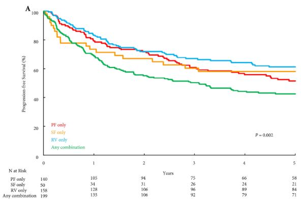
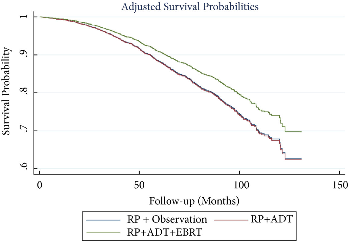
Several notable studies have looked at genetics, quantified cardio‐pulmonary measures of fitness, exercise pre‐habilitation/enhanced recovery, and survivorship programmes across many cancers including oesophageal, colorectal, and prostate cancer. The data suggest that exercise improves outcomes after surgery, quality of life, hospital admissions, progression‐free survival, and overall survival [6].

Prostate cancer is a special case often treated with androgen‐deprivation therapy (ADT), yet androgens are an essential factor in maintaining bone mineral density; muscle mass, as well as motivation to exercise/exercise capacity; and sexual health. Hormone chemotherapy compromises the key role of androgens in maintaining musculoskeletal health and fitness at a systemic and cellular level. This poses hazards. Aside from the longer term hope of targeted therapies to maintain exercise capacity with all of its biological adaptations, perhaps we can reduce some of the deleterious effects of ADT with exercise interventions. Together with behavioural, nutritional and pharmacological treatment pathways, we aim to augment the positive effect exercise brings to patients with prostate cancer, and patients with cancer more generally.

As our scientific understanding increases, it is clear that personalised, prescribed exercise pre‐habilitation is likely to become a ‘gold standard’ in oncology care. Many treatments may increase survival, but at a cost of quality of life; physical activity may not only extend life but may also enhance its quality. Pre‐habilitation warrants serious further study if it is to become widely adopted in practice. It is not simply about telling patients to keep active. As per the Silver and Baima [7] definition, it is ‘a process on the cancer continuum of care that occurs between the time of cancer diagnosis and the beginning of acute treatment, includes physical and psychological assessments that establish a baseline function level, identifies impairments, and provides targeted interventions that improve a patient’s health to reduce the incidence and the severity of current and future impairments’.

References

  1. Taaffe D, Galvão D, Spry N et al. Immediate versus delayed exercise in men initiating androgen deprivation: effects on bone density and soft tissue composition. BJU Int 2019123: 261–9
  2. Hojman P, Gehl J, Christensen JF, Pedersen BK. Molecular mechanisms linking exercise to cancer prevention and treatment. Cell Metab 201727: 10–21
  3. Cormie P, Zopf EM, Zhang X, Schmitz KH. The impact of exercise and cancer: systematic review of the impact of exercise on cancer mortality, recurrence and treatment related side effects. Epidemiol Rev 201739: 71–92
  4. Kinnaird A, Michelakis ED. Metabolic modulation of cancer: a new frontier with great translational potential. J Mol Med 201593: 127–42
  5. Vander Heiden MG. Targeting cancer metabolism: a therapeutic window opens. Nat Rev Drug Discov 201110: 671–84
  6. Thomas RJ, Holm M, Al‐Adhami A. Physical activity after cancer: an evidence review of the international literature. Br J Med Pract 20147: 16–22
  7. Silver JK, Baima J. Cancer prehabilitation: an opportunity to decrease treatment‐related morbidity, increase cancer treatment options, and improve physical and psychological health outcomes. Am J Phys Med Rehabil 201392: 715–27

Editorial: Multiparametric MRI for prostate cancer detection: do clinical trial findings reflect real‐world practice?

‘First, do no harm’; with this in mind, researchers in urology strive to minimize the burden of overdiagnosis and overtreatment of prostate cancer. A promising tool in this arena is multiparametric (mp)MRI, which has been shown in a large‐scale randomized clinical trial to enhance the ability of prostate biopsy to detect clinically significant prostate cancer [1]. The extent to which findings from an idealized trial protocol extend to ‘real‐world’ clinical practice, however, remains largely unknown.

In this issue of BJUI, Shah et al. [2] aimed to fill this knowledge gap by investigating the impact of mpMRI‐guided biopsy on the detection rates of clinically significant prostate cancer in two large academic centres. The authors studied men with an elevated PSA presenting over a 3‐year span (2011–2014); 1020 men underwent mpMRI and 788 did not. Those in the MRI group had higher detection rates of both overall and clinically significant prostate cancer, defined as any Gleason score ≥7 on fusion or standard 12‐core TRUS biopsies, Gleason 6 with a lesion volume >0.5 cm3 volume on MRI, or Gleason 6 with >2 cores positive and/or >50% of any core involved with cancer on biopsy according to Epstein’s criteria, as well as a lower detection rate of clinically insignificant cancer.

The study provides timely implications for both patients and physicians, providing further insight into how findings from clinical trials [1,3] compare with real‐life practice. In fairness, the bulk of patients and clinicians do not follow strict study protocols for both decision‐making and interpretation of results, but rather assess very individual situations. A recent study by Bukavina et al. [4] showed that urologists and radiation oncologists largely perceive mpMRI guidance for targeted biopsies as valuable tools to improve prostate cancer stratification, but only a quarter of respondents reported implementation into their own clinical practice. This underlines some of the challenges of widespread implementation of mpMRI despite strong belief in its value.

Another strength of the study by Shah et al. is the exclusion of men who underwent mpMRI after negative biopsy in the PSA‐only group. This allows the isolation of the impact of mpMRI on downstream biopsy outcomes. A previous study that investigated targeted vs non‐targeted biopsies enrolled a cohort of men who all underwent mpMRI [5], which precludes any assessment of how mpMRI may impact the detection of clinically significant prostate cancer. Shah et al. [2] also astutely tracked detection rates of clinically significant and insignificant prostate cancer. Since the process of diagnosing prostate cancer is not without morbidity, it is crucial to understand the extent to which mpMRI can prevent the diagnosis of clinically indolent cancers.

Important questions regarding the challenges of widespread implementation of mpMRI for prostate cancer detection remain unanswered by the study of Shah et al. The study participants were gathered from large academic centres with readily available equipment, infrastructure and physician expertise to maximize favourable detection outcomes; however, these results may not be representative of the community setting. Additionally, >20% of men who did not undergo mpMRI did not do so because of a lack of insurance approval. This may reflect socio‐economic differences between the groups and also relates to the high costs of mpMRI that make routine implementation difficult [6]. Lastly, the presented findings mostly apply to positive mpMRI scans; the number of underdiagnosed men with negative scans may only be speculated upon, given the lack of follow‐up data in this population. It remains fundamentally important to improve the management of men with elevated PSA levels and negative findings on MRI.

Nonetheless, the present study demonstrates that research findings find their way into clinical practice. In essence, we are doing well, but we can do better.

by Marieke J. Krimphove, Sean A. Fletcher and Quoc‐Dien Trinh


References

  1. Kasivisvanathan V, Rannikko AS, Borghi M et al. MRI‐targeted or standard biopsy for prostate‐cancer diagnosis. N Engl J Med 2018378: 1767–77
  2. Shah PH, Patel VR, Moreira DM et al. Implementation of multiparametric magnetic resonance imaging technology for evaluation of patients with suspicion for prostate cancer in the clinical practice setting. BJU Int 2019123: 239–45
  3. Ahmed HU, El‐Shater Bosaily A, Brown LC et al. Diagnostic accuracy of multi‐parametric MRI and TRUS biopsy in prostate cancer (PROMIS): a paired validating confirmatory study. Lancet 2017389: 815–22
  4. Bukavina L, Tilburt JC, Konety B et al. Perceptions of prostate MRI and fusion biopsy of radiation oncologists and urologists for patients diagnosed with prostate cancer: results from a national survey. Eur Urol Focus 2018; [Epub ahead of print]
  5. Pokorny MR, de Rooij M, Duncan E et al. Prospective study of diagnostic accuracy comparing prostate cancer detection by transrectal ultrasound–guided biopsy versus magnetic resonance (MR) imaging with subsequent MR‐guided biopsy in men without previous prostate biopsies. Eur Urol 201466: 22–9
  6. Kim SJ, Vickers AJ, Hu JC. Challenges in adopting level 1 evidence for multiparametric magnetic resonance imaging as a biomarker for prostate cancer screening. JAMA Oncol 2018; [Epub ahead of print]


Article of the week: WATER II (80–150 mL) procedural outcomes

Every week, the Editor-in-Chief selects an Article of the Week from the current issue of BJUI. The abstract is reproduced below and you can click on the button to read the full article, which is freely available to all readers for at least 30 days from the time of this post.

In addition to the article itself, there is an editorial written by a prominent member of the urological community. These are intended to provoke comment and discussion and we invite you to use the comment tools at the bottom of each post to join the conversation. 

If you only have time to read one article this week, it should be this one.

WATER II (80–150 mL) procedural outcomes

Mihir Desai*, Mo Bidair, Naeem Bhojani, Andrew Trainer§, Andrew Arther§Eugene Kramolowsky, Leo Doumanian*, Dean Elterman**, Ronald P. Kaufman Jr.††James Lingeman‡‡, Amy Krambeck‡‡, Gregg Eure§§, Gopal Badlani¶¶, Mark Plante***Edward Uchio†††, Greg Gin†††, Larry Goldenberg‡‡‡, Ryan Paterson‡‡‡, Alan So‡‡‡Mitch Humphreys§§§, Claus Roehrborn¶¶¶, Steven Kaplan****, Jay Motola**** and Kevin C. Zorn


*Institute of Urology, University of Southern California, Los Angeles, San Diego Clinical Trials, San Diego, CA, USA, University of Montreal Hospital Centre, University of Montreal, Montreal, QC, Canada, §Adult Paediatric Urology and Urogynecology, P.C., Omaha, NE, Virginia Urology, Richmond, VA, USA, **University Health Network University of Toronto, Toronto, ON, Canada, ††Albany Medical College, Albany, NY, ‡‡Indiana University Health Physicians, Indianapolis, IN, §§Urology of Virginia, Virginia Beach, VA, ¶¶Wake Forest School of Medicine,Winston-Salem, NC, ***University of Vermont Medical Centre, Burlington, VT, †††VA Long Beach Healthcare System, Long Beach, CA, USA, ‡‡‡University of British Columbia, Vancouver, BC, Canada, §§§Mayo Clinic Arizona, Scottsdale, AZ, ¶¶¶Department of Urology, UT Southwestern Medical Centre, University of Texas Southwestern, Dallas, TX and ****Icahn School of Medicine at Mount Sinai, New York, NY, USA


Read the full article

Abstract

Objectives

To present early safety and feasibility data from a multicentre prospective study (WATER II) of aquablation in the treatment of symptomatic men with large‐volume benign prostatic hyperplasia (BPH).

Methods

Between September and December 2017, 101 men with moderate‐to‐severe BPH symptoms and prostate volume of 80–150 mL underwent aquablation in a prospective multicentre international clinical trial. Baseline demographics and standardized postoperative management variables were carefully recorded in a central independently monitored database. Surgeons answered analogue scale questionnaires on intra‐operative technical factors and postoperative management. Adverse events up to 1 month were adjudicated by an independent clinical events committee.

Results

The mean (range) prostate volume was 107 (80–150) mL. The mean (range) operating time was 37 (15–97) min and aquablation resection time was 8 (3–15) min. Adequate adenoma resection was achieved with a single pass in 34 patients and with additional passes in 67 patients (mean 1.8 treatment passes), all in a single operating session. Haemostasis was achieved using either a Foley balloon catheter placed in the bladder under traction (n = 98, mean duration 18 h) or direct tamponade using a balloon inflated in the prostate fossa (n = 3, mean duration 15 h). No patient required electrocautery for haemostasis at the time of the primary procedure. The mean length of stay after the procedure was 1.6 days (range same day to 6 days). The Clavien–Dindo grade ≥2 event rate observed at 1 month was 29.7%. Bleeding complications were recorded in 10 patients (9.9%) during the index procedure hospitalization prior to discharge, and included six (5.9%) peri‐operative transfusions.

Conclusions

Aquablation is feasible and safe in treating men with large prostates (80–150 mL). The 6‐month efficacy data are being accrued and will be presented in future publications (ClinicalTrials.gov number, NCT03123250).

Read more Articles of the week


Editorial: Aquablating urological skills

Waterjet Ablation Therapy for Endoscopic Resection of prostate tissue (WATER) II (80–150 mL) procedural outcomes by Desai et al. [1] in this issue of the BJUI, reports the results of a robotically controlled cavitating procedure in a multicentre prospective trial that may have wider implications than relief of prostatic hyperplasia causing obstruction.

Management of the large prostate (>80 mL) is often a challenge for many practicing Urologists and requires practice, constant development, and improvement in endoscopic skills. As a result, many differing approaches have been developed and honed, modifying and improving varied skills in the urologist’s armamentarium to equip them to tackle the large prostate. The traditional TURP is recommended only for prostates of 35–80 mL (European Association of Urology [EAU] guidelines 2015). Whilst there are some Urologists who have developed their TURP skills to tackle larger prostates [2], for most other urologists, other procedures have had to be developed to address the very large prostate (>80 mL). As the authors of the paper report, holmium laser enucleation of the prostate (HoLEP) and photoselective vaporisation of the prostate (PVP) have evolved to enable treatment of the larger prostates endoscopically, but have limited penetrance due to the relatively significant learning curve and fellowship training requirements. Open simple prostatectomy (OSP) has good results but significant potential complications [3]. Robot‐assisted simple prostatectomy is being evaluated as another option [4], but requires an expensive robot and extensive training to develop the skill‐set required to perform the procedure. Laparoscopic simple prostatectomy (LSP) also requires extensive training and experience.

The authors [1] report impressive results of aquablation in these usually challenging large prostates. The mean operative time (OT) was 37 min, which is quick for a large prostate. The average length of stay was 1.6 days. The transfusion rate (TR) was 5.9%, which is higher than HoLEP (0–4%) [4], but is lower than OSP, PVP and LSP. It is important to note that the study involved 16 different sites (13 American and three Canadian) and showed that similar results were achieved across all sites irrespective of the experience of the operator, highlighting the very low learning curve for this procedure. Although this was only a single‐arm study with no control group, the authors have endeavoured to provide a comparison of OT, mean hospital stay and TR between aquablation and other procedures (OSP, PVP, HoLEP and LSP; table 5) based on published literature. Complication rates, operative and hospital metrics of aquablation appear to compare favourably with the current accepted means of managing the large prostate.

The use of balloon tamponade for haemostasis appears to hark back to the days of hanging a saline bag attached to an Indwelling Catheter (IDC) off the end of the bed after a monopolar TURP. Bladder traction was maintained for an average of 18 h. The authors report that fulguration was available to the surgeons in this trial, but none chose to use it as they felt that balloon tamponade was an effective haemostatic mechanism. Fulguration was preferentially avoided based on the WATER trial [5], where it was noted that anejaculation rates were twice as large in the aquablation with fulguration compared to the aquablation without fulguration group (16% vs 7%). The company (PROCEPT BioRobotics, Redwood City, CA, USA) even developed a novel catheter tensioning device (CTD) to assist with controlling the tension on the balloon tamponade demonstrating the old adage that ‘Necessity is the mother of Invention’. It would be interesting to see an objective assessment of discomfort from the balloon tamponade in future studies.

The results of this safety and feasibility trial suggest that aquablation is a quick procedure (37 min) for managing very large prostates. The complication rate is comparable to current endoscopic techniques (HoLEP and PVP) and appears superior to more invasive techniques (LSP and OSP). This study only reported perioperative measures and safety outcomes. No functional outcome or effectiveness measures were reported. The initial WATER trial [5] hints at possible effectiveness, but we will have to wait to see the results from this particular cohort of patients with large prostates (WATER II).

The short learning curve hints at a possible future. If the functional results from this cohort of large prostates treated by the aquablation robot compare favourably to current techniques, the patient with the very large prostate will no longer be only treatable by a few surgeons with an advanced and particular skill set.

Is this truly a quick, safe, effective procedure with no learning curve for large prostates? A randomised controlled trial of longer duration to assess functional outcomes, durability and complications may determine if the aquablation robot eventually renders the current surgical skill sets redundant.


References

  1. Desai M, Bidair M, Bhojani N et al. Aquablation Procedural Outcomes for BPH in Large Prostates (80–150cc): Initial Experience. (WATER II {80‐150 ml} procedural outcomes). BJU Int 2019123: 106–12
  2. Persu C, Georgescu D, Arabagiu I, Cauni V, Moldoveanu C, Geavlete P. TURP for BPH. How large is too large? J Med Life 201015: 376–80
  3. Gratzke C, Schlenker B, Seitz M et al. Complications and early postoperative outcome after open prostatectomy in patients with benign prostatic enlargement: results of a prospective multicenter study. J Urol 2007177: 1419–22
  4. Pokorny M, Novara G, Geurts N et al. Robot‐assisted simple prostatectomy for treatment of lower urinary tract symptoms secondary to benign prostatic enlargement: surgical technique and outcomes in a high‐volume robotic centre. Eur Urol 201568: 451–7
  5. Gilling PJ, Barber NJ, Bidair M et al. WATER: a double‐blind, randomized, controlled trial of aquablation® vs transurethral resection of the prostate in benign prostatic enlargement. J Urol 20185: 1252–61


© 2024 BJU International. All Rights Reserved.