Tag Archive for: Article of the Week

Posts

Video: Move over fluoroscopy: ultrasound-guided PCNL is just as good

Percutaneous nephrolithotomy guided solely by ultrasonography: a 5-year study of >700 cases

Song Yan, Fei Xiang and Song Yongsheng

Division of Urology, Sheng Jing Hospital, China Medical University, Shenyang, China

OBJECTIVE

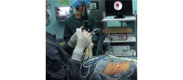
• To evaluate the safety and efficacy of percutaneous nephrolithotomy (PCNL) solely guided by ultrasonography (US).

PATIENTS AND METHODS

• From May 2007 to July 2012, 705 24-F-tract PCNL procedures were performed (679 patients, of whom 26 had bilateral stones).

• Calyceal puncture and dilatation were performed under US guidance in all cases.

• The procedure was evaluated for access success, length of postoperative hospital stay, complications (modified Clavien system), stone clearance and the need for auxiliary treatments.

RESULTS

• The mean (sd) operating time was 66 (25) min, with a mean (sd) postoperative hospital stay of 3.98 (1.34) days.

• The patients experienced a mean (sd) haemoglobin level decrease of 2.24 (2.02) g/day and the overall stone-free rate at 4 weeks after surgery was 92.6% in patients with a single calculus and 82.9% in patients with staghorn or multiple calculi.

• Auxiliary treatments, including shockwave lithotripsy in 52 patients, re-PCNL in 41 patients and ureteroscopy in 18 patients, were performed 1 week after the primary procedure in 111 (15.7%) cases for residual stones >4 mm in size.

• The sensitivities of intra-operative US-guidance and flexible nephroscopy for detecting significant residual stones and clinically insignificant residual fragments were 95.3 and 89.1%, respectively.

• There were 94 grade 1 (13.3%), 17 grade 2 (2.4%), and two grade 3 (0.3%) complications, but there were no grade 4 or 5 complications.

CONCLUSION

• Total US-guided PCNL is safe and convenient, and may be performed without any major complications and with the advantage of preventing radiation hazards and damage to adjacent organs.

Article of the week: Detecting prostate cancer: the “core” of the matter

Every week the Editor-in-Chief selects the Article of the Week from the current issue of BJUI. The abstract is reproduced below and you can click on the button to read the full article, which is freely available to all readers for at least 30 days from the time of this post.

In addition to the article itself, there is an accompanying editorial written by prominent members of the urological community. This blog is intended to provoke comment and discussion and we invite you to use the comment tools at the bottom of each post to join the conversation.

If you only have time to read one article this week, it should be this one.

Can transrectal needle biopsy be optimised to detect nearly all prostate cancer with a volume of ≥0.5 mL? A three-dimensional analysis

Kent Kanao*, James A. Eastham, Peter T. Scardino†‡, Victor E. Reuter*§ and Samson W. Fine*

Departments of *Pathology and Surgery, Urology Service, Memorial Sloan-Kettering Cancer Center, and Departments of Urology and §Pathology, Weill Cornell Medical Center, New York, NY, USA

Read the full article
OBJECTIVE

• To investigate whether transrectal needle biopsy can be optimised to detect nearly all prostate cancer with a tumour volume (TV) of ≥0.5 mL.

MATERIALS AND METHODS

• Retrospectively analysed 109 whole-mounted and entirely submitted radical prostatectomy specimens with prostate cancer.

• All tumours in each prostate were outlined on whole-mount slides and digitally scanned to produce tumour maps. Tumour map images were exported to three-dimensional (3D) slicer software (https://www.slicer.org) to develop a 3D-prostate cancer model.

• In all, 20 transrectal biopsy schemes involving two to 40 cores and two to six anteriorly directed biopsy (ADBx) cores (including transition zone, TZ) were simulated, as well as models with various biopsy cutting lengths.

• Detection rates for tumours of different volumes were determined for the various biopsy simulation schemes.

RESULTS

• In 109 prostates, 800 tumours were detected, 90 with a TV of ≥0.5 mL (mean TV 0.24 mL).

• Detection rate for tumours with a TV of ≥0.5 mL plateaued at 77% (69/90) using a 12-core (3 × 4) scheme, standard 17-mm biopsy cutting length without ADBx cores. In all, 20 of 21 (95%) tumours with a TV of ≥0.5 mL not detected by this scheme originated in the anterior peripheral zone or TZ.

• Increasing the biopsy cutting length and depth/number of ADBx cores improved the detection rate for tumours with a TVof ≥0.5 mL in the 12-core scheme.

• Using a 22-mm cutting length and a 12-core scheme with additional volume-adjusted ADBx cores, 100% of ≥0.5 mL tumours in prostates ≤ 50 mL in volume and 94.7% of ≥0.5 mL tumours in prostates > 50 mL in volume were detected.

CONCLUSIONS

• Our 3D-prostate cancer model analysis suggests that nearly all prostate cancers with a TV of ≥0.5 mL can be detected by 14–18 transrectal needle-biopsy cores.

• Using longer biopsy cutting lengths and increasing the depth and number of ADBx cores (including TZ) according to prostate volume are necessary as well.


Read Previous Articles of the Week


Editorial: How many cores are needed to detect nearly all prostate cancers?

Virtual prostate biopsy and biopsy simulation: lessons to be learned

Prostate biopsies, transrectal or transperineal, still constitute the pillars of prostate cancer detection today [1]. With the lack of reliable imaging tools (new MRI techniques are promising but still investigational [2]); random biopsies offer the sole adequate cancer detection option [3]. However, random biopsies are far from efficient in detecting all tumours and even less efficient in detecting all significant cancer ‘spots’. To improve sensitivities and specificities, increasing the biopsy core numbers, targeting more lateral aspects and encouraging repeat biopsies have been recommended [4]. Recently, HistoScanning™ [5] and template biopsies [6] have been introduced to further improve biopsy quality and efficiency. The latest innovations include the fusion of MRI pictures with the TRUS image to offer optimal targeting of suspicious areas [7]. And yet, these efforts are far from solving the main problem. How can we perform a biopsy and be confident to detect most of the cancers, i.e. significant malignant areas.

The present study [1] does, what should have been done a long time ago, namely to create a reliable and reproducible biopsy simulation model to allow the investigation of various biopsy schemes, core lengths and numbers. Based on a series of 109 radical prostatectomy specimens, a three-dimensional (3D) prostate and prostate cancer model was created using novel 3D slicer software and various prostate biopsy schemes were simulated. Using this method, the detection rate for tumours with a tumour volume (TV) of ≥0.5 mL plateaued at 77% (69 of 90) using a 12 core (3 × 4) scheme, standard 17-mm biopsy cutting length without anteriorly directed biopsy (ADBx) cores. Twenty of 21 (95%) tumours with a TV of ≥0.5 mL not detected by this scheme originated in the anterior peripheral zone or transition zone [1].

Confirming our earlier data with the Vienna nomograms [8], increasing the biopsy cutting length and depth/number of ADBx cores (14–18 cores) improved the detection rate for tumours with a TV of ≥0.5 mL in the 12-core scheme [1]. The best biopsy scheme used a 22-mm cutting length and a 12-core scheme with additional volume-adjusted ADBx cores. Using this combination, 100% of ≥0.5 mL tumours in prostates <50 mL in volume and 94.7% of ≥0.5 mL tumours in prostates >50 mL in volume were detected.

Certainly, these numbers will not be reproducible in real-time TRUS or transperineal biopsies (detections rates of 95–100% as seen in this simulation model, cannot be achieved without adequate imaging tools, which are not available yet), but they aid significantly in rethinking our biopsy strategy. So, if we summarise the present findings and combine them with published data, the future will demand a TRUS-fusion biopsy technique, involving 14–18 cores (or more if volume increases), involving the anterior zones of the prostate and using a 22-mm cutting length of the biopsy core vs a 15–17 mm core as is used currently. Obviously real-time prospective trials are needed to confirm these findings but nothing indicates that the outcome would be otherwise.

Bob Djavan
Department of Urology, New York University School of Medicine, NYU, New York, NY, USA

Read the full article

References

  1. Kanao K, Eastham JA, Scardino PT, Reuter VE, Fine SW. Can transrectal needle biopsy be optimised to detect nearly all prostate cancer with a volume of ≥0.5 mL? A three-dimensional analysis. BJU Int 2013; 112: 898–904
  2. Delongchamps NB, Peyromaure M, Schull A et al. Pre-biopsy Magnetic Resonance Imaging and prostate cancer detection: comparison of random and MRI-targeted biopsies using three different techniques of MRI-TRUS image registration. J Urol 2013;189: 493–499
  3. Djavan B, Rocco B. Optimising prostate biopsy. BMJ 2011; 344: d8201
  4. Thompson I, Thrasher JB, Aus G et al. Guideline for the management of clinically localized prostate cancer: 2007 update. J Urol 2007; 177: 2106–2131
  5. Simmons LA, Autier P, Zát’ura F et al. Detection, localisation and characterisation of prostate cancer by prostate HistoScanning(™)BJU Int 2012; 110: 28–35
  6. Huo AS, Hossack T, Symons JL et al. Accuracy of primary systematic template guided transperineal biopsy of the prostate for locating prostate cancer: a comparison with radical prostatectomy specimens. J Urol 2012; 187: 2044–2049
  7. Sonn GA, Natarajan S, Margolis DJ et al. Targeted biopsy in the detection of prostate cancer using an office based magnetic resonance ultrasound fusion device. J Urol 2013; 189: 86–92
  8. Djavan B, Margreiter M. Biopsy standards for detection of prostate cancer. World J Urol 2007; 25: 11–17

Article of the month: The race to urinary continence after RP

Every week the Editor-in-Chief selects the Article of the Week from the current issue of BJUI. The abstract is reproduced below and you can click on the button to read the full article, which is freely available to all readers for at least 30 days from the time of this post.

In addition to the article itself, there is an accompanying editorial written by prominent members of the urological community. This blog is intended to provoke comment and discussion and we invite you to use the comment tools at the bottom of each post to join the conversation.

Finally, the third post under the Article of the Week heading on the homepage will consist of additional material or media. This week we feature a video of Inge Geraerts discussing functional outcomes after ORP and RARP.

If you only have time to read one article this week, it should be this one.

Prospective evaluation of urinary incontinence, voiding symptoms and quality of life after open and robot-assisted radical prostatectomy

Inge Geraerts*, Hendrik Van Poppel†, Nele Devoogdt*‡, Ben Van Cleynenbreugel†, Steven Joniau† and Marijke Van Kampen*‡

Departments of *Rehabilitation Science, †Urology and ‡Physical Medicine and Rehabilitation, UZ Leuven, Leuven, Belgium

Read the full article
OBJECTIVE

• To compare functional outcomes, i.e. urinary incontinence (UI), voiding symptoms and quality of life, after open (ORP) and robot-assisted radical prostatectomy (RARP).

PATIENTS AND METHODS

• Between September 2009 and July 2011, 180 consecutive patients underwent radical prostatectomy; of these, 116 underwent ORP and 64 underwent RARP. We prospectively assessed the functional outcomes of each group during the first year of follow-up.

• We measured UI on the 3 days before surgery (24-h pad test) and daily after surgery until total continence, defined as 3 consecutive days of 0 g urine leak, was achieved. Additionally, all patients were assessed before surgery and at 1, 3, 6 and 12 months after surgery using the International Prostate Symptom Score (IPSS) and the King’s Health Questionnaire (KHQ).

• All patients received pelvic floor muscle training until continence was achieved.

• Kaplan–Meier analyses and Cox regression with correction for covariates were used to compare time to continence. A Mann–Whitney U-test was used to assess IPSS and KHQ.

RESULTS

• Patients in the RARP group had a significantly lower D’Amico risk group allocation and underwent more nerve-sparing surgery. Other characteristics were similar.

• Patients in the RARP group regained continence sooner than those in the ORP group (P = 0.007). In the RARP group, the median time to continence (16 vs 46 days, P = 0.026) was significantly shorter and the median amount of first day UI (44 vs 186 g, P < 0.01) was significantly smaller than in the ORP group. After correction for all covariates, the difference remained significant (P = 0.036, hazard ratio [HR] 1.522 (1.027–2.255). In addition, younger men, men with positive surgical margins and men without preoperative incontinence achieved continence sooner.

• A comparison of time to continence between groups with a sufficient number of patients (intermediate risk and/or bilateral nerve-sparing) still showed a faster return of continence after RARP, but the effect decreased in size and was nonsignificant (HR>1.2, P> 0.05).

• Only six patients (two in the RARP and four in the ORP group) still had UI after 1 year.• Patients in the RARP group had significantly better IPSS scores at 1 (P = 0.013) and 3 (P = 0.038) months, and scored better in almost all KHQ aspects.

CONCLUSION

• In this prospective trial, patients treated with RARP tended to regain urinary continence sooner than patients treated with ORP, but in subgroup analyses statistical significance disappeared and effect size decreased dramatically, indicating that the results must be interpreted with caution.


Read Previous Articles of the Week


Article of the week: Robotic surgery training methods: take your pick

Every week the Editor-in-Chief selects the Article of the Week from the current issue of BJUI. The abstract is reproduced below and you can click on the button to read the full article, which is freely available to all readers for at least 30 days from the time of this post.

In addition to the article itself, there is an accompanying editorial written by a prominent member of the urological community. This blog is intended to provoke comment and discussion and we invite you to use the comment tools at the bottom of each post to join the conversation.

Finally, the third post under the Article of the Week heading on the homepage will consist of additional material or media. This week we feature a video of Dr. Goh discussing standardized robotic surgery training methods.

If you only have time to read one article this week, it should be this one.

Comparative assessment of three standardized robotic surgery training methods

Andrew J. Hung, Isuru S. Jayaratna, Kara Teruya, Mihir M. Desai, Inderbir S. Gill and Alvin C. Goh*

USC Institute of Urology, Hillard and Roclyn Herzog Center for Robotic Surgery, Keck School of Medicine, University of Southern California, Los Angeles, CA, and *Department of Urology, Methodist Institute for Technology, Innovation and Education, The Methodist Hospital, Houston, TX, USA

Read the full article
OBJECTIVES

• To evaluate three standardized robotic surgery training methods, inanimate, virtual reality and in vivo, for their construct validity.

• To explore the concept of cross-method validity, where the relative performance of each method is compared.

MATERIALS AND METHODS

• Robotic surgical skills were prospectively assessed in 49 participating surgeons who were classified as follows: ‘novice/trainee’: urology residents, previous experience <30 cases (n = 38) and ‘experts’: faculty surgeons, previous experience ≥30 cases (n = 11).

• Three standardized, validated training methods were used: (i) structured inanimate tasks; (ii) virtual reality exercises on the da Vinci Skills Simulator (Intuitive Surgical, Sunnyvale, CA, USA); and (iii) a standardized robotic surgical task in a live porcine model with performance graded by the Global Evaluative Assessment of Robotic Skills (GEARS) tool.

• A Kruskal–Wallis test was used to evaluate performance differences between novices and experts (construct validity).

• Spearman’s correlation coefficient (ρ) was used to measure the association of performance across inanimate, simulation and in vivo methods (cross-method validity).

RESULTS

• Novice and expert surgeons had previously performed a median (range) of 0 (0–20) and 300 (30–2000) robotic cases, respectively (P < 0.001).

• Construct validity: experts consistently outperformed residents with all three methods (P < 0.001).

• Cross-method validity: overall performance of inanimate tasks significantly correlated with virtual reality robotic performance (ρ = −0.7, P < 0.001) and in vivo robotic performance based on GEARS (ρ = −0.8, P < 0.0001).

• Virtual reality performance and in vivo tissue performance were also found to be strongly correlated (ρ = 0.6, P < 0.001).

CONCLUSIONS

• We propose the novel concept of cross-method validity, which may provide a method of evaluating the relative value of various forms of skills education and assessment.

• We externally confirmed the construct validity of each featured training tool.


Read Previous Articles of the Week


Editorial: Three robotic surgery training methods: is there a clear winner?

All training adds value. A craft-based specialty such as surgery has always recognised this. The advent of advanced minimally invasive surgical technology and techniques has provided both new challenges and new opportunities for surgical performance and for the delivery of training. Conceptually, we have moved from the Halstedian model of ‘See one, do one, teach one’ [1] to an environment where skills are acquired away from the operating room in simulator, inanimate and in vivo (animal) laboratory training sessions. Increased scrutiny of credentialling and medico-legal aspects of robotic surgery have reinforced the importance of training and have led to a number of papers outlining pathways to facilitate this [2, 3].

In the present paper, Hung et al. evaluate the construct validity of three standardised training methods (inanimate, simulator and in vivo) and also compare the three different platforms for cross-method training value. As others have shown, the latest generation of robotic surgery simulators have high face, content and construct validity [4, 5] and the present paper confirms the value of both inanimate and simulator training for novice surgeons. In addition, the authors confirmed the construct validity of a simple in vivo exercise using the daVinci© surgical system by demonstrating that experts outperformed novices. Using Spearman’s rank correlation coefficient, the authors compared the three training methods under evaluation and concluded that they were strongly correlated for construct validity between exert and novice surgeons. While construct validation of these exercises may be established, are they useful for experts? Until realistic virtual reality surgical simulations are available, only a novice, an inexperienced or an occasional robot-assisted surgeon may benefit from virtual reality exercises.

What are we therefore to conclude from this? For certain, the advent of excellent surgical simulators and structured inanimate exercises has provided tools for novice surgeons to acquire console skills in a safe and structured environment. This will enhance their operating performance and reduce aspects of the learning curve such as operating time; however, the lack of availability of in vivo training opportunities greatly limits the applicability of this method of surgical training. In many countries (including Australia and the UK), this type of training is illegal or not available. The robotic surgery industry has strongly recommended that in vivo training should be undertaken in one of their official training facilities before surgeons are given the credentials to use this technology; however, even in the USA where most of these facilities are located, key leaders within the AUA have called for the awarding of credentials for robotic surgery ‘not to be an industry driven process, but one that is a result of a standardized, competency based, peer evaluation system’ [2]. Notably, the current AUA Standard Operating Practices (guidelines) for the awarding of credentials for robotic surgery list in vivo training as being optional.

Our view is that although all training has value, there is not enough evidence that in vivo training (particularly on an animal with a rudimentary prostate), which requires international travel and considerable expense, adds sufficient value to be mandatory in any credentialling process. In fact, we have dropped the requirement to complete in vivo training from our requirements at major robotic surgery centres in Australia in favour of structured Mini-Fellowship training [6]. Hung et al. have confirmed what we already knew, which is that all training adds value; however it is likely that only simulator and inanimate training adds enough value to be incorporated into standardised training in robotic surgery.

The multi-disciplinary ‘Fundamentals of Robotic Surgery’ (FRS) curriculum being created by Dr Richard Satava and associates is working on psychomotor skills tasks that include inanimate models as well as corresponding virtual reality exercises. Multi-institutional validation of the FRS or similar curricula will allow the establishment of training milestones and proficiency benchmarks. We must continue to strive for further development of robotic and surgical simulation to change the training paradigm so that surgical training does not need to be at the expense, however minor, of increased operating time or adverse patient outcome.

Declan G. Murphy* and Chandru P. Sundaram
*Peter MacCallum Cancer Centre, Division of Cancer Surgery, University of Melbourne, Australian Prostate Cancer Research Centre, Epworth Richmond Hospital, Melbourne, Australia, and Department of Urology, Indiana University, Indianapolis, IN, USA

Read the full article

References

  1. Halsted WS. The training of the surgeon. Bull Johns Hop Hosp 1904; XV: 8
  2. Lee JY, Mucksavage P, Sundaram CP, McDougall EM. Best practices for robotic surgery training and credentialingJ Urol 2011;185: 1191–1197
  3. Zorn KC, Gautam G, Shalhav AL et al. Training, credentialing, proctoring and medicolegal risks of robotic urological surgery: recommendations of the society of urologic robotic surgeonsJ Urol 2009; 182: 1126–1132
  4. Finnegan KT, Meraney AM, Staff I, Shichman SJ. da Vinci Skills Simulator construct validation study: correlation of prior robotic experience with overall score and time score simulator performanceUrology 2012; 80: 330–335
  5. Abboudi H, Khan MS, Aboumarzouk O et al. Current status of validation for robotic surgery simulators – a systematic reviewBJU Int 2013; 111: 194–205
  6. Melbourne Uro-Oncology Training Program. Robotic surgery training. Available at: https://www.declanmurphy.com.au/training. Accessed 28 February 2013

Video: Take three: assessing robotic surgery training methods

Comparative assessment of three standardized robotic surgery training methods

Andrew J. Hung, Isuru S. Jayaratna, Kara Teruya, Mihir M. Desai, Inderbir S. Gill and Alvin C. Goh*

USC Institute of Urology, Hillard and Roclyn Herzog Center for Robotic Surgery, Keck School of Medicine, University of Southern California, Los Angeles, CA, and *Department of Urology, Methodist Institute for Technology, Innovation and Education, The Methodist Hospital, Houston, TX, USA

Read the full article
OBJECTIVES

• To evaluate three standardized robotic surgery training methods, inanimate, virtual reality and in vivo, for their construct validity.

• To explore the concept of cross-method validity, where the relative performance of each method is compared.

MATERIALS AND METHODS

• Robotic surgical skills were prospectively assessed in 49 participating surgeons who were classified as follows: ‘novice/trainee’: urology residents, previous experience <30 cases (n = 38) and ‘experts’: faculty surgeons, previous experience ≥30 cases (n = 11).

• Three standardized, validated training methods were used: (i) structured inanimate tasks; (ii) virtual reality exercises on the da Vinci Skills Simulator (Intuitive Surgical, Sunnyvale, CA, USA); and (iii) a standardized robotic surgical task in a live porcine model with performance graded by the Global Evaluative Assessment of Robotic Skills (GEARS) tool.

• A Kruskal–Wallis test was used to evaluate performance differences between novices and experts (construct validity).

• Spearman’s correlation coefficient (ρ) was used to measure the association of performance across inanimate, simulation and in vivo methods (cross-method validity).

RESULTS

• Novice and expert surgeons had previously performed a median (range) of 0 (0–20) and 300 (30–2000) robotic cases, respectively (P < 0.001).

• Construct validity: experts consistently outperformed residents with all three methods (P < 0.001).

• Cross-method validity: overall performance of inanimate tasks significantly correlated with virtual reality robotic performance (ρ = −0.7, P < 0.001) and in vivo robotic performance based on GEARS (ρ = −0.8, P < 0.0001).

• Virtual reality performance and in vivo tissue performance were also found to be strongly correlated (ρ = 0.6, P < 0.001).

CONCLUSIONS

• We propose the novel concept of cross-method validity, which may provide a method of evaluating the relative value of various forms of skills education and assessment.

• We externally confirmed the construct validity of each featured training tool.

Article of the week: Rethinking inflammation in PCa

Every week the Editor-in-Chief selects the Article of the Week from the current issue of BJUI. The abstract is reproduced below and you can click on the button to read the full article, which is freely available to all readers for at least 30 days from the time of this post.

In addition to the article itself, there is an accompanying editorial written by a prominent member of the urological community. This blog is intended to provoke comment and discussion and we invite you to use the comment tools at the bottom of each post to join the conversation.

If you only have time to read one article this week, it should be this one.

Histological inflammation and risk of subsequent prostate cancer among men with initially elevated serum prostate-specific antigen (PSA) concentration in the Finnish prostate cancer screening trial

Tytti H. Yli-Hemminki*, Marita Laurila*, Anssi Auvinen, Liisa Määttänen§, Heini Huhtala, Teuvo L.J. Tammela and Paula M. Kujala*

*Department of Pathology, Fimlab Laboratories, Tampere University Hospital, Tampere, Department of Pathology, Seinäjoki Central Hospital, Seinäjoki, School of Health Sciences, University of Tampere, Tampere, §Finnish Cancer Registry, Helsinki, and Department of Urology, Tampere University Hospital and University of Tampere, Tampere, Finland

T. H. Y.-H. and M. L. contributed equally to this study.

Read the full article

OBJECTIVE

• To assess whether histological signs of inflammation are associated with an increased risk of subsequent prostate cancer (PCa) in men with elevated serum prostate-specific antigen (PSA) concentrations and benign initial biopsy.

MATERIALS AND METHODS

• Study subjects were men aged 54–67 years with an elevated PSA (≥4 ng/mL or 3–4 ng/mL and free to total PSA ratio ≤0.16 or positive digital rectal examination), but a benign biopsy result within the Finnish population-based randomised screening trial for PCa, which started in 1996.

• A total of 293 prostate biopsies without PCa or suspicion of malignancy from the first screening round in the Tampere centre were re-evaluated by a uropathologist to assess histological inflammation.

• Results of the subsequent screening rounds were obtained from the trial database and PCa diagnoses made outside the screening were obtained from the Finnish Cancer Registry.

• The median length of follow-up was 10.5 years.

• Cox regression analysis was used to assess PCa risk after the initial benign biopsy.

RESULTS

• Histological inflammation was found in 66% of the biopsies.

• Subjects with inflammation at the biopsy had a slightly lower PCa risk in the second screening round (18 vs 27%, rate ratio 0.69, 95% confidence interval [CI] 0.35–1.34) relative to men without inflammation. In further follow-up, the PCa risk remained nonsignificantly lower (hazard ratio [HR] 0.71, CI 0.46–1.10; P = 0.13). The risk was not appreciably affected by adjustment for age, PSA, prostate volume and family history of PCa (HR 0.67, CI 0.42–1.07; P = 0.092).

CONCLUSIONS

• Histological inflammation in a prostate biopsy among men with an initial false-positive screening test was not associated with an increased risk of subsequent PCa, but instead with a decreased risk which was of borderline significance.

• Inflammation in prostate biopsy is not a useful risk indicator in PCa screening.


Read Previous Articles of the Week


Editorial: Does inflammation reduce the risk of prostate cancer?

Chronic inflammation is thought to play an aetiological role in tumorigenesis in several cancers including bladder, oesophagus and liver [1]. Molecular studies show that it plays a critical role in several stages of the carcinogenic process including tumour initiation, promotion, metastases and response to therapy. However the role in prostate cancer is less clear and to date, clinical studies are inconclusive.

The article in this issue of BJUI by Yli-Hemminki et al. [2] appears to show an inverse association with histological inflammation and the risk of prostate cancer. Using data from the Finnish subgroup of the European Randomised Study of Prostate Cancer Screening (ERSPC) study they examined 293 patients with previous negative biopsies over a 10.5-year period and reported an 18% risk of prostate cancer in men with inflammation on initial biopsy (34 of 101 men) as opposed to 27% in those without inflammation (51 of 192 men). Perhaps somewhat surprisingly, histological inflammation did not appear to be significantly associated with PSA concentration, although it did appear that the free/total PSA ratio was higher in men with inflammation.

One potential confounding factor is that inflammation may also play a role in the pathogenesis of BPH. A large scale study showed that the odds ratio for BPH was 8.0 with a history of prostatitis [3]. Furthermore, the Medical Therapy of Prostatic Symptoms (MTOPS) study showed that men with inflammation had a significantly higher risk of BPH progression and acute urinary retention. These factors may impact on PSA levels and the chance of a subsequent prostate biopsy. However, the authors report that the inverse association of prostate cancer and inflammation did not alter when corrected for prostate volume, PSA level and age.

Significantly, those patients who screened positive at first biopsy were already excluded as were those with a suspicion of prostate cancer, such as a small atypical focus. Despite this, the study group probably represents high-risk patients, as they had all previously met the criteria for the first round of biopsies. This is borne out by the fact that the risk of prostate cancer was significantly increased when the men with inflammation on biopsy were compared with the initially screened negative men (hazard ratio 4.3). Unfortunately, we do not know what the rate of inflammation was in the control arm as they were screened negative and hence no biopsies were taken.

The significance of PSA level or prostatic intraepithelial neoplasia was not specifically investigated, although the reported rates were 32.1% and 7.5% respectively. Overall the incidence of inflammation was reported as 65%, but this included both acute and chronic inflammation. A smaller number of patients were given grade 2/3 chronic inflammation (80 and 20 patients, respectively) and only 14 patients had grade 2/3 acute, indicating most had milder degrees of inflammation. Whether this is a representative sample is not entirely clear. As the authors comment, published rates of histological inflammation do vary significantly from 8 to 99% and this does appear to vary according to detection method.

Several case-control studies and a meta-analysis [4] have shown that there is a significant increase in the relative risk of prostate cancer in men with prostatitis; however, these epidemiological studies all suffer from selection bias, in that men with clinical symptoms are more likely present and to be investigated and followed up by a Urologist. This study [2] only examines the role of histological inflammation and, at least partially, removes the selection bias associated with clinical symptoms, although it could still be argued that men with clinical prostatitis may be more likely to present for screening. This study represents a highly selected group of patients that are high risk for prostate cancer and no firm conclusions can be drawn on the general population. As inflammation is so common in prostate specimens, further high-quality large-scale studies are needed with similar long-term follow-up.

Miles A. Goldstraw and Roger S. Kirby
The Prostate Centre, London, UK

Read the full article

References

  1. Coussens LM, Werb Z. Inflammation and cancer. Nature 2002; 420: 860–867
  2. Yli-Hemminski T, Laurila M, Auvinen A et al. Histological inflammation and risk of subsequent prostate cancer among men with initially elevated serum prostate-specific antigen (PSA) concentration in the Finnish prostate cancer screening trial. BJU Int 2013; 112: 735–741
  3. Alcarez A, Hammerer P, Tubaro A, Schroder FH, Castro R. Is there evidence of a relationship between benign prostatic hyperplasia and prostate cancer? Findings of a literature review. Eur Urol 2009; 55: 864–875
  4. Dennis LK, Lynch CF, Torner JC. Epidemiologic association between prostatitis and prostate cancer. Urology 2002; 60: 78–83

Article of the week: How useful is FDG-PET/CT in managing carcinoma invading bladder muscle?

Every week the Editor-in-Chief selects the Article of the Week from the current issue of BJUI. The abstract is reproduced below and you can click on the button to read the full article, which is freely available to all readers for at least 30 days from the time of this post.

In addition to the article itself, there is an accompanying editorial written by a prominent member of the urological community. This blog is intended to provoke comment and discussion and we invite you to use the comment tools at the bottom of each post to join the conversation.

Finally, the third post under the Article of the Week heading on the homepage will consist of additional material or media. This week we feature a video of Ms Mertens and Prof Horenblas discussing their findings.

If you only have time to read one article this week, it should be this one.

Impact of 18F-fluorodeoxyglucose (FDG)-positron-emission tomography/computed tomography (PET/CT) on management of patients with carcinoma invading bladder muscle

Laura S. Mertens, Annemarie Fioole-Bruining*, Erik Vegt, Wouter V. Vogel, Bas W. van Rhijn and Simon Horenblas

Departments of Urology, *Radiology and Nuclear Medicine, The Netherlands Cancer Institute, Antoni van Leeuwenhoek Hospital, Amsterdam, The Netherlands

Read the full article
OBJECTIVE

• To evaluate the clinical impact of 18F-fluorodeoxyglucose (FDG)-positron-emission tomography/computed tomography (PET/CT) scanning, compared with conventional staging with contrast-enhanced CT imaging (CECT).

PATIENTS AND METHODS

• The FDG-PET/CT results of 96 consecutive patients with bladder cancer were analysed. Patients included in this study underwent standard CECT imaging of the chest and abdomen/pelvis <4 weeks before FDG-PET/CT.

• Based on the original imaging reports and recorded tumour stage before and after FDG-PET/CT imaging, the preferred treatment strategies before FDG-PET/CT and after FDG-PET/CT were determined for each patient using an institutional multidisciplinary guideline. One of the following treatment strategies was chosen: (i) local curative treatment; (ii) neoadjuvant/induction chemotherapy; or (iii) palliation.

• The changes in management decisions before and after FDG-PET/CT were assessed.

RESULTS

• The median (range) interval between CECT and FDG-PET/CT was 0 (029) days.

• In 21.9% of the patients, stage on FDG-PET/CT and CECT were different. Upstaging by FDG-PET/CT was more frequent than downstaging (19.8 vs 2.1%).

• Clinical management changed for 13.5% of patients as a result of FDG-PET/CT upstaging. In eight patients, FDG-PET/CT detected second primary tumours. This led to changes of bladder cancer treatment in another four of 96 patients (4.2%).

• All the management changes were validated by tissue confirmation of the additional lesions.

CONCLUSIONS

• FDG-PET/CT provides important additional staging information, which influences the treatment of carcinoma invading bladder muscle in almost 20% of cases.

• Patient selection for neoadjuvant/induction chemotherapy was improved and futile attempts at curative treatment in patients found to have metastases were avoided.


Read Previous Articles of the Week


© 2024 BJU International. All Rights Reserved.