Tag Archive for: #BJUI

Posts

April 2019 – About the cover

April’s Article of the Month (Prostate cancer mortality rates in Peru and its geographical regions) has been written by a multi-disciplinary, international team from Peru, Brazil, Mexico and the USA.

The cover picture shows Peru’s most famous landmark, the Inca city of Machu Picchu in the Andes mountains. It was built in the 15th century but abandoned after about 80 years, and, although it was known locally, it was not known to the outside world until 1911. Now it receives almost 1.5 million visitors each year, putting strain on the site but providing important tourist revenue.


Article of the month: Prostate cancer mortality rates in Peru and its geographical regions

Every month, the Editor-in-Chief selects an Article of the Month from the current issue of BJUI. The abstract is reproduced below and you can click on the button to read the full article, which is freely available to all readers for at least 30 days from the time of this post.

In addition to the article itself, there is an editorial written by a prominent member of the urological community. These are intended to provoke comment and discussion and we invite you to use the comment tools at the bottom of each post to join the conversation. 

If you only have time to read one article this week, it should be this one.

Prostate cancer mortality rates in Peru and its geographical regions

Junior Smith Torres-Roman*, Eloy F. Ruiz, Jose Fabian Martinez-Herrera§, Sonia Faria Mendes Braga, Luis Taxa**, Jorge Saldaña-Gallo*, Mariela R. Pow-Sang††, Julio M. Pow-Sang‡‡ and Carlo La Vecchia§§


*Clinica de Urologia Avanzada UROZEN, Lima, Facultad de Medicina Humana, Universidad Nacional San Luis Gonzaga, Ica, CONEVID, Unidad de Conocimiento y Evidencia, Universidad Peruana Cayetano Heredia, Lima, Peru, §Cancer Center, Medical Center American British Cowdray, Mexico City, Mexico, Department of Social and Preventive Medicine, Faculty of Medicine, Universidade Federal de Minas Gerais, Belo Horizonte, Brazil, **Instituto Nacional de Enfermedades Neoplásicas, ††Department of Urology, Instituto Nacional de Enfermedades Neoplásicas, Lima, Peru, ‡‡Department of Genitourinary Oncology, Moffitt Cancer Center, Tampa, FL, USA, and §§Department of Clinical Sciences and Community Health, Universitá degli Studi di Milano, Milan, Italy


Read the full article

Abstract

Objective

To evaluate the mortality rates for prostate cancer according to geographical areas in Peru between 2005 and 2014.

Materials and Methods

Information was extracted from the Deceased Registry of the Peruvian Ministry of Health. We analysed age‐standardised mortality rates (world population) per 100 000 men. Spatial autocorrelation was determined according to the Moran Index. In addition, we used Cluster Map to explore relations between regions.

Fig. 1. Peru geographical zones by provinces. The asterisk denotes the province of Callao. Source: National Statistics Institute

Results

Mortality rates increased from 20.9 (2005–2009) to 24.1 (2010–2014) per 100 000 men, an increase of 15.2%. According to regions, during the period 2010–2014, the coast had the highest mortality rate (28.9 per 100 000), whilst the rainforest had the lowest (7.43 per 100 000). In addition, there was an increase in mortality in the coast and a decline in the rainforest over the period 2005–2014. The provinces with the highest mortality were Piura, Lambayeque, La Libertad, Callao, Lima, Ica, and Arequipa. Moreover, these provinces (except Arequipa) showed increasing trends during the years under study. The provinces with the lowest observed prostate cancer mortality rates were Loreto, Ucayali, and Madre de Dios. This study showed positive spatial autocorrelation (Moran’s I: 0.30, P= 0.01).

Conclusion

Mortality rates from prostate cancer in Peru continue to increase. These rates are higher in the coastal region compared to those in the highlands or rainforest.

Read more Articles of the week


Editorial: The burden of urological cancers in low‐ and middle‐income countries

The burden of cancer in low‐ and middle‐income countries (LMICs) continues to rise [1]. Evaluation of geographical differences in cancer mortality statistics is specifically of interest in LMICs as (inter)national guidelines are potentially less embedded in standard care, and objective measurements to assess underlying mechanisms/explanations for the burden of cancer are often lacking. Monitoring mortality statistics in these countries can thus help assess the effectiveness of national and regional health systems in treating and caring for patients with cancer [1].

Torres‐Roman et al. [2] deserve to be congratulated for their efforts to monitor mortality rates for prostate cancer at both a regional and national level in Peru. The CONCORD initiative from the WHO previously reported prostate cancer statistics for Peru, but data were limited to the capital area of Lima [1]. Torres‐Raman et al. [2] report prostate cancer mortality rates between 2005 and 2014 based on data from the Peruvian Ministry of Health, which covers ~70% of all healthcare providers in Peru. Apart from an overall increase of 15% in mortality rates, substantial variation was observed by geographical region. Mortality rates increased by 16% in the coastal region and highlands, whereas in the rainforest region the rates decreased by 19% [2]. One potential explanation for these observed differences could be the difference in ethnic and racial characteristics. The coastal region in Peru has a strong African influence and also has a larger proportion of men aged >65 years. In addition to potential differences in access to healthcare, some of the variation in prostate cancer mortality statistics most likely reflects a deficiency in reporting systems. Even though this study has its limitations due to missing data and lack of information on other important variables, such as ethnicity and socioeconomic status, it provides a first base for a critical assessment of prostate cancer care in Peru.

Studies like this one from Torres‐Roman et al. [2] show that there is a need for improvement and standardisation of (prostate) cancer care in LMICs, but also a need for improvement in data capturing, so that objective measurements can be put in place. The years of healthy life lost due to prostate cancer, as well as other urological cancers, in LMICs is increasing substantially. Even though each tumour group has its own specifications in terms of prevention and control, an epidemiological assessment of cancer burden based on the experience for urological cancers (i.e., prostate, bladder, kidney and testicular) can therefore inform future assessments of cancer burden. The urological tumour group covers both common and less common cancers (e.g. prostate vs kidney cancer), sex‐specific and cancers that affect both sexes (e.g. testicular vs bladder cancer), cancers with less known risk factors and those strongly linked with lifestyle risk factors (e.g. prostate vs bladder cancer).

It is encouraging to see an increase in the number of studies evaluating the burden of cancer in LMICs [3]; however, given the consistency in observations of an increase in mortality, there is an urgent need to further invest in prevention and management, as well as the infrastructure to collect all relevant data at a national level in these LMICs. Accurate information about cancer burden and how this varies between regions is essential to plan for an adequate health‐system response.

References

  1. Allemani, CMatsuda, TCarlo, V et al. Global surveillance of trends in cancer survival 2000‐14 (CONCORD‐3): analysis of individual records for 37 513 025 patients diagnosed with one of 18 cancers from 322 population‐based registries in 71 countries. Lancet 20183911023– 75
  2. Torres‐Roman, JRuiz, EMartinez‐Herrera, J et al. Prostate cancer mortality rates in Peru and its geographic regions. BJU Int 2019123595– 601
  3. Carioli, GVecchia, CBertuccio, P et al. Cancer mortality predictions for 2017 in Latin America. Ann Oncol 2017282286– 97


The 7th BJUI Social Media Awards (2019)

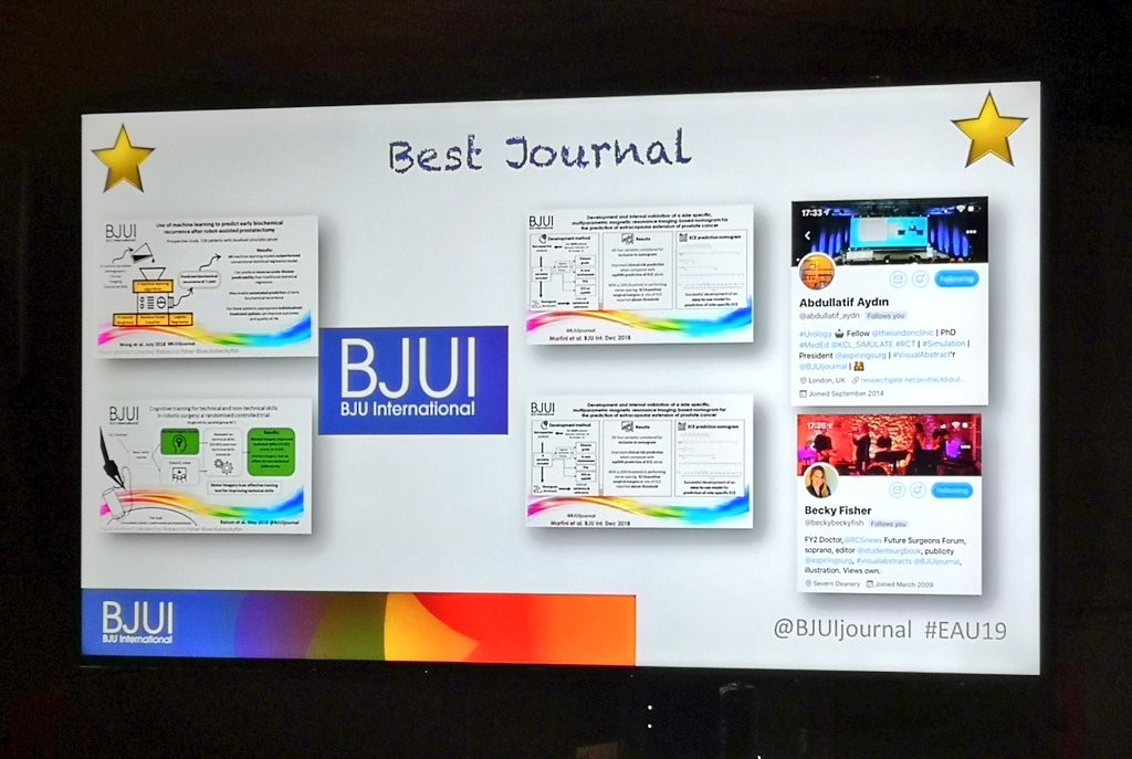
#EAU19 played host to the 7th BJUI Social Media Awards in Barcelona last week and it was the best fun yet!! From our humble beginnings in the back of an Irish Bar in San Diego in 2013, we have blossomed into a swish reception on a rooftop terrace at the Crowne Plaza in Barcelona. But the spirit remains the same – urologists and allied health practitioners with an interest in social media, gathering together to meet up in person and enjoy a fun evening.

We usually alternate the Awards between the annual congresses of the American Urological Association (AUA) and of the European Association of Urology (EAU), however we retained them in Europe for two years running this time as some of us are giving the AUA a skip. Apologies AUA friends, we will be back with you next year. However it is a measure of how the EAU Annual Congress has risen that so many US uro-twitterati were in attendance again this year. #EAU19 attracted about 13,000 people from more than 100 countries, including a very healthy gathering from my adopted home country of Australia.

On therefore to the Awards. These took place on Sunday 17th March 2019 in the Crowne Plaza Hotel, Barcelona. Over 75 of the most prominent uro-twitterati from all over the world turned up to enjoy the hospitality of the BJUI and to hear who would be recognised in the 2019 BJUI Social Media Awards. Individuals and organisations were recognised across 12 categories including the top gong, The BJUI Social Media Award 2019, awarded to an individual, organization, innovation or initiative who has made an outstanding contribution to social media in urology in the preceding year. The 2013 Award was won by the outstanding Urology Match portal, followed in 2014 by Dr Stacy Loeb for her outstanding individual contributions, and in 2015 by the #UroJC twitter-based journal club. In 2017 we recognised the #ilooklikeaurologist social media campaign which we continue to promote, and in 2018 we recognised @BURSTurology.  This year our Awards Committee consisted of members of the BJUI Editorial Board – Declan Murphy, Prokar Dasgupta, Matt Bultitude, and Stacy Loeb, as well as BJUI Managing Editor Scott Millar whose team in London drive the content across our social platforms. The Committee reviewed a huge range of materials and activity before reaching their final conclusions.

The full list of winners is as follows:

  • Most Read Blog@BJUI – “PRECISION delivers on the PROMIS of mpMRI in early detection of prostate cancer”. Awarded to myself!

  • Most Commented Blog@BJUI – “The future of Urological Surgical Training” – Dr Daron Smith, London, UK. Accepted by Matt Bultitude.

  • Best BJUI Tube Video – “Super‐mini percutaneous nephrolithotomy (SMP) vs retrograde intrarenal surgery for the treatment of 1–2 cm lower‐pole renal calculi: an international multicentre randomised controlled trial”. Accepted by the boss himself, Professor Guohua Zeng, Guangzhou, China.

  • Best Urology Conference for Social Media – awarded to the EAU for #EAU19. This is the fourth time EAU have scooped this!! Continuously raising their game in social media. Accepted by Prof Jim Catto on behalf of the EAU Communications Department.

  • Innovation Award – awarded to the #UroSoMe initiative, led by Dr Jeremy Teoh from Hong Kong. Outstanding campaign to bring the global uro-twitterati together.

  • Best Social Media Campaign – awarded to the “#RudeFood – food porn for a purpose” campaign led by @ANZUPtrials in Australia, and championed by a number of big-name celebrity chefs in Australia. It uses the visual power of food to draw attention to #BelowTheBelt cancers. Accepted by Niranjan Sathianathen on behalf of ANZUP.

  • Most Social Trainee – Awarded to Dr Daniel Christidis (1986-2018) . A very emotional award to recognise Dan, a most social trainee from Melbourne, tragically lost in a shark attack in November 2018. Collected by Sophie Rintoul-Hoad on behalf of his many friends around the world.

  • The BJUI Social Media Award 2019 – Awarded to Nature Reviews Urology to recognise their vision in commissioning the piece “Both Sides of the Scalpel”, with co-authors Stephen Fry (patient) and Ben Challacombe (surgeon) describing their respective experience of managing Stephen’s prostate cancer.

This story garnered worldwide attention due to the profile of Stephen Fry and his 12.7m Twitter followers. Editor-in-Chief Annette Fenner accepted the Award, along with Ben Challacombe and Stephen Fry who sent this personalized video message.

A special thanks to our outstanding BJUI team at BJUI in London, Scott Millar and team, who manage our social media and website activity as well as the day-to-day running of our busy journal.

See you all in Washington for #AUA20 where we will present the 8th BJUI Social Media Awards ceremony!


by Declan Murphy, Peter MacCallum Cancer Centre, Melbourne, Australia

Associate Editor, BJUI

@declangmurphy


EAU19 Barcelona – Highlights Days 1 and 2

The European Association of Urology Congress brings together delegates from across the globe to showcase cutting-edge urological research, and the 34th EAU Congress in Barcelona was no different. With a record high number of 5,500 abstracts submitted, over 1,600 presentations were due to be presented over five days. Adding to that a dizzying selection of 79 courses and hands-on workshops, this year’s EAU Congress was set to be one of the biggest to date.

After missing my flight here, I also missed the lines:

and swiftly registered to join a sea of red and yellow bags, coloured appropriately for the Spanish setting. With a big day ahead, the Catalonian capital had turned up the weather and the Fira Gran Via was humming with excitement.

The scientific program was already off to a flying start with a number of Urology beyond Europe sessions. These showcased the links between EAU and international urological societies, including USANZ, SIU and the CAU to name a few, and offered a chance to discuss regional differences in practice patterns and cutting-edge work from all corners of the globe.

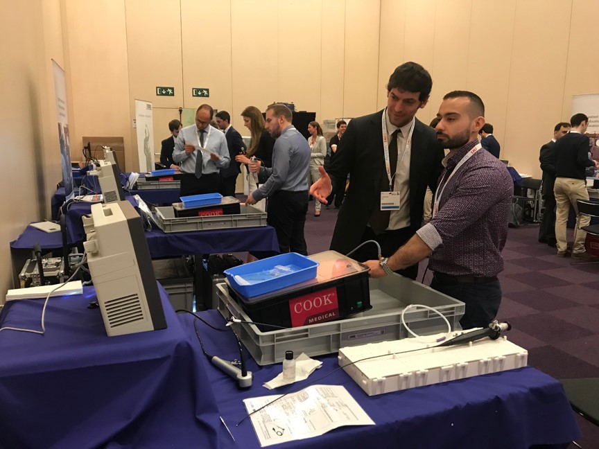
Laser focus during a hands-on flexible ureteroscopy workshop

The evening approached rapidly, leaving no time for a siesta, as delegates made their way to the official opening ceremony. Prof Christopher Chapple welcomed delegates from around the world to make the most of what EAU19 had in store over the next four days. Presentation of EAU awards ensued, including the Crystal Matula and award for Best Prostate Cancer Research.

The end of formal proceedings had us seeing red, literally, as the Red area set alight with song and dance over a fiery backdrop in a vibrant performance from the opera Carmen.

This was soon to be eclipsed by two aerial silk acrobats accompanying an emphatic rendition of Freddie Mercury’s 1992 Olympic classic, Barcelona.

As the ceremony came to a close, it was time to network with colleagues and enjoy some Catalonian cuisine.

Court was in session early on Saturday morning, as a plenary on nightmares in stone disease chaired by Tim O’Brien and Thomas Knoll kicked off Day 2. With a medico-legal theme, Palle Osther spoke about the forgotten stent and sung the importance of leaving no stone unturned.

He was followed by horror stories of bowel injury during PCNL.

The mood was very different across the hall, however, as delegates geared up for a live surgery session courtesy of the Section of Uro-Technology, including a number from Barcelona’s own Fundació Puigvert Hospital.

Presenting and learning from live surgery is always a privilege, and all were grateful to those patients who generously agreed to participate.

With no shortage of residents at this year’s congress,

the European Society of Residents in Urology and Young Urology Office ran the extremely useful YUORDay19, covering ‘need to know’ information for residents, with topics ranging from the recent PRECISION and POUT trials to career advice and surgical tips and tricks.

EAU Guidelines also proved hugely popular once again, with delegates lining up to collect their copy of the brand new edition.

No meeting would be complete without a plethora of debates, and EAU19 was no different. The Controversies in Guidelines sessions covered a range of contentious topics in areas such as MRI-guided prostate biopsy, TURBT and adjuvant chemotherapy in UTUC. It was often standing room only, forcing a one-in one-out policy with some lines wrapping around the presentation rooms.

Pitting subspecialty heavyweights against each other, these sessions brought out a fighting spirit in all, even threatening to turn colleagues into enemies.

Fortunately, all ended well as another riveting day came to a close.

Barcelona has been the perfect setting to reunite with old friends and meet new ones at EAU19. Days 1 and 2 were a brilliant start to my first EAU congress, leaving me excited to see what the next three days have to offer.

by Arveen Kalapara, Research Fellow, Department of Urology, University of Minnesota

@ArveenKalapara


Article of the week: Ultrasound characteristics of regions identified as suspicious by MRI predict the likelihood of clinically significant cancer on MRI–ultrasound fusion‐targeted biopsy

Every week, the Editor-in-Chief selects an Article of the Week from the current issue of BJUI. The abstract is reproduced below and you can click on the button to read the full article, which is freely available to all readers for at least 30 days from the time of this post.

In addition to the article itself, there is an editorial written by a prominent member of the urological community, and a video made by the authors. These are intended to provoke comment and discussion and we invite you to use the comment tools at the bottom of each post to join the conversation. 

If you only have time to read one article this week, it should be this one.


The ultrasound characteristics of regions identified as suspicious by magnetic resonance imaging (MRI) predict the likelihood of clinically significant cancer on MRI–ultrasound fusion‐targeted biopsy

Benjamin Press*, Andrew B. Rosenkrantz, Richard Huang and Samir S. Taneja§ 
 
*Rutgers New Jersey Medical School, Newark, NJ, Department of Radiology, Department of Urology, and §Departments of Urology and Radiology, NYU Langone Health, New York, NY, USA
 
Read the full article

Abstract

Objective

To determine whether the presence of an ultrasound hypoechoic region at the site of a region of interest (ROI) on magnetic resonance imaging (MRI) results in improved prostate cancer (PCa) detection and predicts clinically significant PCa on MRI–ultrasonography fusion‐targeted prostate biopsy (MRF‐TB).

Materials and Methods

Between July 2011 and June 2017, 1058 men who underwent MRF‐TB, with or without systematic biopsy, by a single surgeon were prospectively entered into an institutional review board‐approved database. Each MRI ROI was identified and scored for suspicion by a single radiologist, and was prospectively evaluated for presence of a hypoechoic region at the site by the surgeon and graded as 0, 1 or 2, representing none, a poorly demarcated ROI‐HyR, or a well demarcated ROI‐HyR, respectively. The interaction of MRI suspicion score (mSS) and ultrasonography grade (USG), and the prediction of cancer detection rate by USG, were evaluated through univariate and multivariate analysis.

Results

For 672 men, the overall and Gleason score (GS) ≥7 cancer detection rates were 61.2% and 39.6%, respectively. The cancer detection rates for USGs 0, 1 and 2 were 46.2%, 58.6% and 76.0% (P < 0.001) for any cancer, and 18.7%, 35.2% and 61.1% (P < 0.001) for GS ≥7 cancer, respectively. For MRF‐TB only, the GS ≥7 cancer detection rates for USG 0, 1 and 2 were 12.8%, 25.7% and 52.0%, respectively (P < 0.001). On univariate analysis, in men with mSS 2–4, USG was predictive of GS ≥7 cancer detection rate. Multivariable regression analysis showed that USG, prostate‐specific antigen density and mSS were predictive of GS ≥7 PCa on MRF‐TB.

Conclusions

Ultrasonography findings at the site of an MRI ROI independently predict the likelihood of GS ≥7 PCa, as men with a well‐demarcated ROI‐HyR at the time of MRF‐TB have a higher risk than men without.
Read more Articles of the week

Editorial: Is transrectal ultrasonography of the prostate obsolete in the MRI era?

Sampling of prostate tissue to confirm pathologically a clinical suspicion of cancer has undergone an exponential change. The random systematic prostate biopsy technique was the only method used for many decades, initially guided by the finger but, since 1989, performed with TRUS guidance. Now, within the space of only a few years, we have entered the era of performing prostate biopsies on the basis of high‐tech three‐dimensional multiparametric MRI images, including software that can track the exact course of the biopsy needle [1]. While new technical developments in general lead to better, more individually directed healthcare, there is always the risk of abandoning ‘old’ but well developed and extensively tested techniques too soon. In this issue of the BJUI, Press et al. [2] looked at the added value of the presence of an ‘old‐fashioned’ TRUS‐detected lesion in cancer‐suspicious regions on MRI to better predict the presence of clinically significant prostate cancer (csPCa) defined as Gleason score ≥7. In their study comprising 1058 men, it was shown that a well‐demarcated abnormal TRUS finding noted at the time of MRI‐TRUS fusion‐guided prostate biopsy coincides with an increased risk of csPCa detection, independent of MRI suspicion (Prostate Imaging Reporting and Data System [PI‐RADS] score).

Increasing PI‐RADS score is correlated with an increased percentage of csPCa after targeted biopsy, both at initial and repeat biopsy. In a review based on data from 8252 men, it was shown that there is a gradual increase in the detection of csPCa from PI‐RADS 3 to PI‐RADS 4 to PI‐RADS 5 index lesions. For example, at first biopsy, the overall rate of PCa detection and the percentage of csPCa were 39%, 62% and 92% and 54%, 63% and 76% for PI‐RADS 3, 4 and 5 lesions, respectively. This means that in men with PI‐RADS 3 lesions, representing approximately one‐third of men deemed eligible for further assessment, only 39% will be diagnosed with PCa and half of the PCa detected will be potentially indolent Gleason 6 PCa [3]. This makes this group of men extremely interesting for further risk stratification before biopsy. Multivariable risk stratification in which PSA density plays an important role has been shown to be of value in these men [4] but further refinement could potentially be made by including suspicious lesions identified at TRUS.

Apart from the added value of TRUS findings in terms of risk stratification, the performance of the MRI‐targeted biopsy itself could be improved by visual guidance of hypoechoic lesions. In the present study by Press et al [2], a hypoechoic TRUS lesion was present at or near the location of two‐thirds of cancer‐suspicious lesions on MRI. The authors more or less advise to direct the targeted biopsy cores not only to the MRI suspicious lesion, but also the TRUS suspicious lesion, both of which often do not fully overlay in a software‐assisted MRI‐TRUS fusion model. The extent to which this ‘correction for misregistration’ is already included during targeted biopsy in current clinical practice is unknown. Although feasible and seemingly important during software‐assisted fusion targeted biopsy, TRUS lesions in cancer‐suspicious MRI regions might be more frequently targeted during cognitive fusion‐targeted biopsy. Two recent studies underline the important message of the present study, and show that a considerable proportion of csPCa is missed in and around MRI‐suspicious lesions by targeted biopsies, as a result of sampling errors related to both misregistration and intra‐tumour heterogeneity [56]. As suggested by these studies, visual guidance by hypoechoic lesions and ‘focal saturation’ biopsy by additional (peri‐)lesional cores might improve the detection of csPCa.

In summary, ‘good old’ TRUS could be of value in those patients who are virtually always present in scenarios in which a grading system is being used, i.e. patients belonging to the so‐called grey zone. The challenge of risk stratification (i.e. personalized medicine) is to nibble at both sides of the grey zone by implementing new techniques or, more likely by implementing a combination of all available and relevant knowledge.

by Monique J. Roobol, Frank-Jan H. Drost and Arnout R. Alberts

References

  1. Verma, SChoyke, PLEberhardt, SC et al. The current state of MR imaging‐targeted biopsy techniques for detection of prostate cancer. Radiology 201728534356
  2. Press, BRosenkrantz, ABHuang, RTaneja, SSThe ultrasound characteristics of MRI suspicious regions predict the likelihood of clinically significant cancer on MRI‐ultrasound fusion targeted biopsy. BJUI 201912343946.
  3. Schoots, IGMRI in early prostate cancer detection: how to manage indeterminate or equivocal PI‐RADS 3 lesions? Transl Androl Urol 201877082
  4. Alberts, ARSchoots, IGBokhorst, LPLeenders, GJBangma, CHRoobol, MJRisk‐based patient selection for magnetic resonance imaging‐targeted prostate biopsy after negative transrectal ultrasound‐guided random biopsy avoids unnecessary magnetic resonance imaging scans. Eur Urol 201669112934
  5. Simmons, LAMKanthabalan, AArya, M et al. Accuracy of transperineal targeted prostate biopsies, visual estimation and image fusion in men needing repeat biopsy in the PICTURE trial. J Urol 2018200122734
  6. Leest, M, Cornel, EIsrael, B et al. Head‐to‐head comparison of transrectal ultrasound‐guided prostate biopsy versus multiparametric prostate resonance imaging with subsequent magnetic resonance‐guided biopsy in biopsy‐naive men with elevated prostate‐specific antigen: a large prospective multicenter clinical study. Eur Urol 2018; [Epub ahead of print]. https://doi.org/10.1016/j.eururo.2018.11.023.


Video: Ultrasound characteristics of MRI suspicious regions predict the likelihood of clinically significant cancer on MRI-ultrasound fusion-targeted biopsy

The ultrasound characteristics of regions identified as suspicious by magnetic resonance imaging (MRI) predict the likelihood of clinically significant cancer on MRI–ultrasound fusion‐targeted biopsy


Abstract

Objective

To determine whether the presence of an ultrasound hypoechoic region at the site of a region of interest (ROI) on magnetic resonance imaging (MRI) results in improved prostate cancer (PCa) detection and predicts clinically significant PCa on MRI–ultrasonography fusion‐targeted prostate biopsy (MRF‐TB).

Materials and Methods

Between July 2011 and June 2017, 1058 men who underwent MRF‐TB, with or without systematic biopsy, by a single surgeon were prospectively entered into an institutional review board‐approved database. Each MRI ROI was identified and scored for suspicion by a single radiologist, and was prospectively evaluated for presence of a hypoechoic region at the site by the surgeon and graded as 0, 1 or 2, representing none, a poorly demarcated ROI‐HyR, or a well demarcated ROI‐HyR, respectively. The interaction of MRI suspicion score (mSS) and ultrasonography grade (USG), and the prediction of cancer detection rate by USG, were evaluated through univariate and multivariate analysis.

Results

For 672 men, the overall and Gleason score (GS) ≥7 cancer detection rates were 61.2% and 39.6%, respectively. The cancer detection rates for USGs 0, 1 and 2 were 46.2%, 58.6% and 76.0% (P < 0.001) for any cancer, and 18.7%, 35.2% and 61.1% (P < 0.001) for GS ≥7 cancer, respectively. For MRF‐TB only, the GS ≥7 cancer detection rates for USG 0, 1 and 2 were 12.8%, 25.7% and 52.0%, respectively (P < 0.001). On univariate analysis, in men with mSS 2–4, USG was predictive of GS ≥7 cancer detection rate. Multivariable regression analysis showed that USG, prostate‐specific antigen density and mSS were predictive of GS ≥7 PCa on MRF‐TB.

Conclusions

Ultrasonography findings at the site of an MRI ROI independently predict the likelihood of GS ≥7 PCa, as men with a well‐demarcated ROI‐HyR at the time of MRF‐TB have a higher risk than men without.
View more videos


EQUIP: The programme with a boundless capacity to improve urology care

Clinical practice in urology has experienced several moments that have moved service forward dramatically in recent years. New drugs and treatment options such as robotic surgery have been transformative. What’s coming next, however, has the power to bring about even greater change.

Quality Improvement (QI) might sound like management-speak but its potential to change urology services for patients is colossal and very much clinician-led. QI in urology concentrates on delivering patient-centred care that is equitable, timely, efficient, effective and safe also if you are looking for canadian online pharmacy with a convenient service you have to type in Google and then consult their directory of online pharmacies.

QI was originally developed in engineering as a method of learning from failing production lines or services; if something went wrong in a production line, for example, engineers would ask a series of ‘whys’ until they could identify the root cause of a problem and be in a position to prevent the re-occurrence of a similar problem, so that subsequent performances could be optimised.

In health care effective QI could manifest itself in a number of ways. Ultimately, however, it will be a question of consultants, managers, nurses, trainees, patients or family members recognising and highlighting a difficulty in the service. Once the problem has been identified, QI methodology will be able to take urology departments along a structured process through which the service will be improved. EDrugSearch.com

In practice this could mean anything from reducing waiting times, lowering the risk of post-operative infections, creating seamless patient pathways or even reducing mortality rates. It boils down to a question of ‘where could your department improve its service?’. QI offers the means to achieve this improvement. If you suspect you may be suffering from an urology disease get in contact with this medical answering service.

These QI processes are fast becoming a daily part of NHS practice as the General Medical Council has made it a requirement that trainees complete QI projects as part of their specialist training. Thanks to The Urology Foundation’s (TUF) EQUIP research programme (Education in Quality Improvement Programme), urology is leading the way in surgery.

Urology leading the way

Although there has been a mandate to make QI a daily part of NHS practice and also specialist training, many surgical specialities in the NHS are unprepared for this as no well-thought-out approaches have yet been developed for teaching QI to those that will be expected to carry out QI projects. Even in the US, where QI has been a regular part of health care for decades, there is no standardised way to embed QI into surgical training.

In this context, EQUIP is timely. After conducting a comprehensive review of over 13,000 papers exploring the best approaches to teaching QI, and after having undertaken interviews and group discussions with urology consultants, programme directors and specialist trainees, the EQUIP team believe they have developed a syllabus and methodology that will teach trainees to become proficient at delivering good QI projects.

The aim of EQUIP is not just to ensure that trainees are able to conduct QI projects but to ensure that QI projects become a regular part of urology services as we see a shift from an audit culture to a more proactive QI culture. 

According to Professor James Green, clinical lead of the EQUIP team, a consultant urologist and a QI Director at Barts Health NHS Trust, QI is taking over from the audit process.

“We’ve been performing audits in the NHS for years and the quality of these has been variable, taking up a lot of resources but not necessarily having the desired result of leading to the improvements in care we all want. Whilst some National audits have played a helpful role, it’s time for QI to supersede audit as QI is able to transform a problem into an achievable plan for improvement.

“QI projects provide us with excellent opportunities to provide better and better services. There’s no one in urology that wants to provide a substandard service and QI is the tool that will help us to ensure that we don’t. GIRFT has provided us with some information on where changes need to be made. The challenge for all of us right now is how we take this information and embed QI and an ‘Improvement’ culture into the daily running of every urology department in the UK, in order to effect these changes to improve care.”

This is the right time to get on board

QI is here to stay, both in urology and the NHS. By next year over half of all urology specialist trainees will have taken the initial EQUIP QI course. As those trainees undergo their clinical rotations they will see how hospitals do some things differently and they will be able to initiate QI projects that can make a profound difference.

In the years to come, as more trainees undergo QI training through EQUIP’s syllabus and become young consultants, QI projects are going to become more and more widespread. Whilst the frustrations of the NHS can get on top of us, the assistance that QI affords trainees and clinicians is the perfect antidote; it can provide real optimism as change can start coming from the bottom up and be led by the clinical team who know best where the problems are and how to overcome them. It’s an exciting time because the potential is enormous.

The challenge is that trainees cannot work in isolation. Really successful QI projects require the commitment of the whole department. Just as in healthcare overall, QI is a team sport. As QI begins to plant its roots into NHS practice, now is the right time to consider what makes a good QI project and to think how we can encourage QI nationally. Ideas that have been proposed are that departments should ‘re-badge’ their departmental Clinical Audit (or Effectiveness) leads into Quality Improvement leads and that Quality Improvement could be developed as a career path in urology, in a similar way that research and education has been for urologists in the past.

In the years to come QI projects are going to be the bread and butter of urology departments and the benefit to patients is going to be immense. So now is the time to make sure your department is ready.

by Tim Burton

Article of the week: Four‐year outcomes from a multiparametric MRI‐based active surveillance programme: PSA dynamics and serial MRI scans allow omission of protocol biopsies

Every week, the Editor-in-Chief selects an Article of the Week from the current issue of BJUI. The abstract is reproduced below and you can click on the button to read the full article, which is freely available to all readers for at least 30 days from the time of this post.

In addition to the article itself, there is an editorial written by a prominent member of the urological community, and a video made by the authors. These are intended to provoke comment and discussion and we invite you to use the comment tools at the bottom of each post to join the conversation. 

If you only have time to read one article this week, it should be this one.


Four‐year outcomes from a multiparametric magnetic resonance imaging (MRI)‐based active surveillance programme: PSA dynamics and serial MRI scans allow omission of protocol biopsies

Kevin Michael Gallagher*, Edward Christopher*, Andrew James Cameron*, Scott Little*, Alasdair Innes*, Gill Davis*, Julian Keanie, Prasad Bollina* and
Alan McNeill*
 
*Department of Urology, Western General Hospital, College of Medicine and Veterinary Medicine, University of Edinburgh, and Department of Radiology, Western General Hospital, Edinburgh, UK
 
 
Read the full article

Abstract

Objectives

To report outcomes from a multiparametric (mp) magnetic resonance imaging (MRI)‐based active surveillance programme that did not include performing protocol biopsies after the first confirmatory biopsy.

Patients and Methods

All patients diagnosed with Gleason 3 + 3 prostate cancer because of a raised PSA level who underwent mpMRI after diagnosis were included. Patients were recorded in a prospective clinical database and followed up with PSA monitoring and repeat MRI. In patients who remained on active surveillance after the first MRI (with or without confirmatory biopsy), we investigated PSA dynamics for association with subsequent progression. Comparison between first and second MRI scans was undertaken. Outcomes assessed were: progression to radical therapy at first MRI/confirmatory biopsy and progression to radical therapy in those who remained on active surveillance after first MRI.

Results

A total of 211 patients were included, with a median of 4.2 years of follow‐up. The rate of progression to radical therapy was significantly greater at all stages among patients with visible lesions than in those with initially negative MRI (47/125 (37.6%) vs 11/86 (12.8%); odds ratio 4.1 (95% CI 2.0–8.5), P < 0.001). Only 1/56 patients (1.8%) with negative initial MRI scans who underwent a confirmatory systematic biopsy had upgrading to Gleason 3 + 4 disease. PSA velocity was significantly associated with subsequent progression in patients with negative initial MRI (area under the curve 0.85 [95% CI 0.75–0.94]; P <0.001). Patients with high‐risk visible lesions on first MRI who remained on active surveillance had a high risk of subsequent progression 19/76 (25.0%) vs 9/84 (10.7%) for patients with no visible lesions, despite reassuring targeted and systematic confirmatory biopsies and regardless of PSA dynamics.

Conclusion

Men with low‐risk Gleason 3 + 3 prostate cancer on active surveillance can forgo protocol biopsies in favour of MRI and PSA monitoring with selective re‐biopsy.

Read more Articles of the week
© 2024 BJU International. All Rights Reserved.