Tag Archive for: #BJUI

Posts

Article of the month: Implementation of multiparametric MRI technology for evaluation of PCa in the clinic

Every month, the Editor-in-Chief selects an Article of the Month from the current issue of BJUI. The abstract is reproduced below and you can click on the button to read the full article, which is freely available to all readers for at least 30 days from the time of this post.

In addition to the article itself, there is an editorial written by a prominent member of the urological community, and a podcast produced by our current Resident Podcasters. These are intended to provoke comment and discussion and we invite you to use the comment tools at the bottom of each post to join the conversation. 

If you only have time to read one article this week, it should be this one.

Implementation of multiparametric magnetic resonance imaging technology for evaluation of patients with suspicion for prostate cancer in the clinical practice setting

Paras H. Shah*, Vinay R. Patel, Daniel M. Moreira, Arvin K. George§, Manaf Alom*, Zachary Kozel, Vidhu Joshi*, Eran Ben-Levi**, Robert Villani**, Oksana Yaskiv††Louis R. Kavoussi, Manish Vira, Carl O. Olsson‡‡ and Ardeshir R. Rastinehad


*Department of Urology, Mayo Clinic, Rochester, MN, Department of Urology, Icahn Smith Institute for Urology, Northwell Health, New York, NY, Department of Urology, University of Illinois at Chicago, Chicago, IL, §Department of Urology, University of Michigan, Ann Arbor, MI, Department of Urology, Smith Institute for Urology, Northwell Health, **Department of Radiology, Hofstra Northwell School of Medicine, ††Department of Pathology, Hofstra Northwell School of Medicine, New Hyde Park, and ‡‡Integrated Medical Professionals, Melville, NY, USA


Abstract

Objectives

To investigate the impact of implementing magnetic resonance imaging (MRI) and ultrasonography fusion technology on biopsy and prostate cancer (PCa) detection rates in men presenting with clinical suspicion for PCa in the clinical practice setting.

Patients and Methods

We performed a review of 1 808 consecutive men referred for elevated prostate‐specific antigen (PSA) level between 2011 and 2014. The study population was divided into two groups based on whether MRI was used as a risk stratification tool. Univariable and multivariable analyses of biopsy rates and overall and clinically significant PCa detection rates between groups were performed.

Results

The MRI and PSA‐only groups consisted of 1 020 and 788 patients, respectively. A total of 465 patients (45.6%) in the MRI group and 442 (56.1%) in the PSA‐only group underwent biopsy, corresponding to an 18.7% decrease in the proportion of patients receiving biopsy in the MRI group (P < 0.001). Overall PCa (56.8% vs 40.7%; P < 0.001) and clinically significant PCa detection (47.3% vs 31.0%; P < 0.001) was significantly higher in the MRI vs the PSA‐only group. In logistic regression analyses, the odds of overall PCa detection (odds ratio [OR] 1.74, 95% confidence interval [CI] 1.29–2.35; P < 0.001) and clinically significant PCa detection (OR 2.04, 95% CI 1.48–2.80; P < 0.001) were higher in the MRI than in the PSA‐only group after adjusting for clinically relevant PCa variables.

Conclusion

Among men presenting with clinical suspicion for PCa, addition of MRI increases detection of clinically significant cancers while reducing prostate biopsy rates when implemented in a clinical practice setting.


Editorial: Multiparametric MRI for prostate cancer detection: do clinical trial findings reflect real‐world practice?

‘First, do no harm’; with this in mind, researchers in urology strive to minimize the burden of overdiagnosis and overtreatment of prostate cancer. A promising tool in this arena is multiparametric (mp)MRI, which has been shown in a large‐scale randomized clinical trial to enhance the ability of prostate biopsy to detect clinically significant prostate cancer [1]. The extent to which findings from an idealized trial protocol extend to ‘real‐world’ clinical practice, however, remains largely unknown.

In this issue of BJUI, Shah et al. [2] aimed to fill this knowledge gap by investigating the impact of mpMRI‐guided biopsy on the detection rates of clinically significant prostate cancer in two large academic centres. The authors studied men with an elevated PSA presenting over a 3‐year span (2011–2014); 1020 men underwent mpMRI and 788 did not. Those in the MRI group had higher detection rates of both overall and clinically significant prostate cancer, defined as any Gleason score ≥7 on fusion or standard 12‐core TRUS biopsies, Gleason 6 with a lesion volume >0.5 cm3 volume on MRI, or Gleason 6 with >2 cores positive and/or >50% of any core involved with cancer on biopsy according to Epstein’s criteria, as well as a lower detection rate of clinically insignificant cancer.

The study provides timely implications for both patients and physicians, providing further insight into how findings from clinical trials [1,3] compare with real‐life practice. In fairness, the bulk of patients and clinicians do not follow strict study protocols for both decision‐making and interpretation of results, but rather assess very individual situations. A recent study by Bukavina et al. [4] showed that urologists and radiation oncologists largely perceive mpMRI guidance for targeted biopsies as valuable tools to improve prostate cancer stratification, but only a quarter of respondents reported implementation into their own clinical practice. This underlines some of the challenges of widespread implementation of mpMRI despite strong belief in its value.

Another strength of the study by Shah et al. is the exclusion of men who underwent mpMRI after negative biopsy in the PSA‐only group. This allows the isolation of the impact of mpMRI on downstream biopsy outcomes. A previous study that investigated targeted vs non‐targeted biopsies enrolled a cohort of men who all underwent mpMRI [5], which precludes any assessment of how mpMRI may impact the detection of clinically significant prostate cancer. Shah et al. [2] also astutely tracked detection rates of clinically significant and insignificant prostate cancer. Since the process of diagnosing prostate cancer is not without morbidity, it is crucial to understand the extent to which mpMRI can prevent the diagnosis of clinically indolent cancers.

Important questions regarding the challenges of widespread implementation of mpMRI for prostate cancer detection remain unanswered by the study of Shah et al. The study participants were gathered from large academic centres with readily available equipment, infrastructure and physician expertise to maximize favourable detection outcomes; however, these results may not be representative of the community setting. Additionally, >20% of men who did not undergo mpMRI did not do so because of a lack of insurance approval. This may reflect socio‐economic differences between the groups and also relates to the high costs of mpMRI that make routine implementation difficult [6]. Lastly, the presented findings mostly apply to positive mpMRI scans; the number of underdiagnosed men with negative scans may only be speculated upon, given the lack of follow‐up data in this population. It remains fundamentally important to improve the management of men with elevated PSA levels and negative findings on MRI.

Nonetheless, the present study demonstrates that research findings find their way into clinical practice. In essence, we are doing well, but we can do better.

by Marieke J. Krimphove, Sean A. Fletcher and Quoc‐Dien Trinh


References

  1. Kasivisvanathan V, Rannikko AS, Borghi M et al. MRI‐targeted or standard biopsy for prostate‐cancer diagnosis. N Engl J Med 2018378: 1767–77
  2. Shah PH, Patel VR, Moreira DM et al. Implementation of multiparametric magnetic resonance imaging technology for evaluation of patients with suspicion for prostate cancer in the clinical practice setting. BJU Int 2019123: 239–45
  3. Ahmed HU, El‐Shater Bosaily A, Brown LC et al. Diagnostic accuracy of multi‐parametric MRI and TRUS biopsy in prostate cancer (PROMIS): a paired validating confirmatory study. Lancet 2017389: 815–22
  4. Bukavina L, Tilburt JC, Konety B et al. Perceptions of prostate MRI and fusion biopsy of radiation oncologists and urologists for patients diagnosed with prostate cancer: results from a national survey. Eur Urol Focus 2018; [Epub ahead of print]
  5. Pokorny MR, de Rooij M, Duncan E et al. Prospective study of diagnostic accuracy comparing prostate cancer detection by transrectal ultrasound–guided biopsy versus magnetic resonance (MR) imaging with subsequent MR‐guided biopsy in men without previous prostate biopsies. Eur Urol 201466: 22–9
  6. Kim SJ, Vickers AJ, Hu JC. Challenges in adopting level 1 evidence for multiparametric magnetic resonance imaging as a biomarker for prostate cancer screening. JAMA Oncol 2018; [Epub ahead of print]


Editorial: Aquablating urological skills

Waterjet Ablation Therapy for Endoscopic Resection of prostate tissue (WATER) II (80–150 mL) procedural outcomes by Desai et al. [1] in this issue of the BJUI, reports the results of a robotically controlled cavitating procedure in a multicentre prospective trial that may have wider implications than relief of prostatic hyperplasia causing obstruction.

Management of the large prostate (>80 mL) is often a challenge for many practicing Urologists and requires practice, constant development, and improvement in endoscopic skills. As a result, many differing approaches have been developed and honed, modifying and improving varied skills in the urologist’s armamentarium to equip them to tackle the large prostate. The traditional TURP is recommended only for prostates of 35–80 mL (European Association of Urology [EAU] guidelines 2015). Whilst there are some Urologists who have developed their TURP skills to tackle larger prostates [2], for most other urologists, other procedures have had to be developed to address the very large prostate (>80 mL). As the authors of the paper report, holmium laser enucleation of the prostate (HoLEP) and photoselective vaporisation of the prostate (PVP) have evolved to enable treatment of the larger prostates endoscopically, but have limited penetrance due to the relatively significant learning curve and fellowship training requirements. Open simple prostatectomy (OSP) has good results but significant potential complications [3]. Robot‐assisted simple prostatectomy is being evaluated as another option [4], but requires an expensive robot and extensive training to develop the skill‐set required to perform the procedure. Laparoscopic simple prostatectomy (LSP) also requires extensive training and experience.

The authors [1] report impressive results of aquablation in these usually challenging large prostates. The mean operative time (OT) was 37 min, which is quick for a large prostate. The average length of stay was 1.6 days. The transfusion rate (TR) was 5.9%, which is higher than HoLEP (0–4%) [4], but is lower than OSP, PVP and LSP. It is important to note that the study involved 16 different sites (13 American and three Canadian) and showed that similar results were achieved across all sites irrespective of the experience of the operator, highlighting the very low learning curve for this procedure. Although this was only a single‐arm study with no control group, the authors have endeavoured to provide a comparison of OT, mean hospital stay and TR between aquablation and other procedures (OSP, PVP, HoLEP and LSP; table 5) based on published literature. Complication rates, operative and hospital metrics of aquablation appear to compare favourably with the current accepted means of managing the large prostate.

The use of balloon tamponade for haemostasis appears to hark back to the days of hanging a saline bag attached to an Indwelling Catheter (IDC) off the end of the bed after a monopolar TURP. Bladder traction was maintained for an average of 18 h. The authors report that fulguration was available to the surgeons in this trial, but none chose to use it as they felt that balloon tamponade was an effective haemostatic mechanism. Fulguration was preferentially avoided based on the WATER trial [5], where it was noted that anejaculation rates were twice as large in the aquablation with fulguration compared to the aquablation without fulguration group (16% vs 7%). The company (PROCEPT BioRobotics, Redwood City, CA, USA) even developed a novel catheter tensioning device (CTD) to assist with controlling the tension on the balloon tamponade demonstrating the old adage that ‘Necessity is the mother of Invention’. It would be interesting to see an objective assessment of discomfort from the balloon tamponade in future studies.

The results of this safety and feasibility trial suggest that aquablation is a quick procedure (37 min) for managing very large prostates. The complication rate is comparable to current endoscopic techniques (HoLEP and PVP) and appears superior to more invasive techniques (LSP and OSP). This study only reported perioperative measures and safety outcomes. No functional outcome or effectiveness measures were reported. The initial WATER trial [5] hints at possible effectiveness, but we will have to wait to see the results from this particular cohort of patients with large prostates (WATER II).

The short learning curve hints at a possible future. If the functional results from this cohort of large prostates treated by the aquablation robot compare favourably to current techniques, the patient with the very large prostate will no longer be only treatable by a few surgeons with an advanced and particular skill set.

Is this truly a quick, safe, effective procedure with no learning curve for large prostates? A randomised controlled trial of longer duration to assess functional outcomes, durability and complications may determine if the aquablation robot eventually renders the current surgical skill sets redundant.


References

  1. Desai M, Bidair M, Bhojani N et al. Aquablation Procedural Outcomes for BPH in Large Prostates (80–150cc): Initial Experience. (WATER II {80‐150 ml} procedural outcomes). BJU Int 2019123: 106–12
  2. Persu C, Georgescu D, Arabagiu I, Cauni V, Moldoveanu C, Geavlete P. TURP for BPH. How large is too large? J Med Life 201015: 376–80
  3. Gratzke C, Schlenker B, Seitz M et al. Complications and early postoperative outcome after open prostatectomy in patients with benign prostatic enlargement: results of a prospective multicenter study. J Urol 2007177: 1419–22
  4. Pokorny M, Novara G, Geurts N et al. Robot‐assisted simple prostatectomy for treatment of lower urinary tract symptoms secondary to benign prostatic enlargement: surgical technique and outcomes in a high‐volume robotic centre. Eur Urol 201568: 451–7
  5. Gilling PJ, Barber NJ, Bidair M et al. WATER: a double‐blind, randomized, controlled trial of aquablation® vs transurethral resection of the prostate in benign prostatic enlargement. J Urol 20185: 1252–61


Editorial: The Advanced Prostate Cancer Consensus on a regional level – what can we learn?

In this issue of BJUI Chiong et al [1] present the results of the Asia Pacific (APAC) Advanced Prostate Cancer Consensus Conference (APCCC) 2018, during which the implications of the APCCC 2017 findings were discussed in the context of the APAC region. For background, it is important to understand the concept of the original APCCC and why it was initiated [1,2,3].

The consensus conference aims to target areas of controversy in the clinical management of advanced prostate cancer where evidence is either limited or lacking or where interpretation of evidence is controversial. The expert consensus aims to complement existing clinical practice guidelines that are mostly based on high‐level evidence. The APCCC’s most prominent aim is knowledge translation, in the sense of improving care of men with advanced prostate cancer worldwide who are treated outside of centres of excellence. During the original APCCC in St Gallen, where 61 prostate cancer experts and scientists were assembled, the majority of the consensus questions were discussed; these had been prepared prior to the conference under the idealistic assumption that all diagnostic procedures and treatments (including expertise in their interpretation and application) mentioned were readily available. These assumptions have been specifically chosen, because availability of systemic treatment options for advanced prostate cancer, access to next‐generation imaging (whole‐body MRI and positron‐emission tomography [PET]) and expertise in molecular techniques and interpretation of results vary widely across the world. The original global APCCC did not generally address regional or country‐specific situations, but APCCC 2017 did have a special session and also voting questions for treatment options in countries with limited resources. Importantly, consensus recommendations may even inform and influence regulatory authorities, for example, if a specific treatment is considered to be the best option by the majority of experts and availability in a certain country is lacking.

The APAC APCCC 2018 consisted of 20 experts (mostly urologists) from 15 countries and discussed the findings and voting results of five of the 10 APCCC 2017 topics. Whether or not Turkey should be considered an APAC country is unclear. The most relevant observations were as set out below:

  • There is huge variation in access to drugs used for treatment of advanced prostate cancer in the APAC region. Australia and Hong Kong have access to almost all treatment options (notably cabazitaxel is not mentioned) compared with countries such as Vietnam or the Philippines, where there is limited availability of many compounds. Regarding imaging technologies (standard CT is not mentioned), there seems to be wide availability of next‐generation imaging such as whole‐body MRI and choline‐ or PSMA‐PET technologies; however, these imaging methods are often not reimbursed.
  • Pharmaco‐ethnic issues have so far not been considered by the original APCCC and the APAC report clearly highlights the need to address such issues. The higher toxicity of docetaxel in Asian men may influence treatment recommendations, especially in situations such as low‐volume metastatic castration‐naïve prostate cancer, where the role of early addition of docetaxel to androgen deprivation therapy is less clear.
  • The authors of the APAC meeting state that ketoconazole and bicalutamide are still widely used despite the proven superiority of enzalutamide vs bicalutamide. A possible reason for this is the lack of reimbursement in some APAC countries.
  • There is an obvious need for clinical trials in the APAC region because of variations in genetics, genomics, epidemiology and pharmaco‐ethnicity. Such trials may answer questions about toxicity/tolerability and also optimal use of resources in the context of economic limitations.

In summary, the APAC APCCC 2018 is an excellent example of how the global APCCC findings should be discussed and integrated on a regional or even country‐specific level. The authors are therefore to be congratulated for their efforts and for writing up the discussions. The next APCCC  (2019; apccc.org) will take up a number of points raised by the APAC meeting, namely, more panel experts from APAC countries and pharmaco‐ethnic topics.

References

  1. Edmund C, Declan GM, Hideyuki A et al. Management of patients with advanced prostate cancer in the Asia Pacific region: ‘real‐world’ consideration of results from the Advanced Prostate Cancer Consensus Conference (APCCC) 2017. BJU Int 2019; 123: 22–34
  2. Gillessen S, Omlin A, Attard G et al. Management of patients with advanced prostate cancer: recommendations of the St Gallen Advanced Prostate Cancer Consensus Conference (APCCC) 2015. Ann Oncol 2015; 26: 1589–604
  3. Gillessen S, Attard G, Beer TM et al. Management of patients with advanced prostate cancer: the report of the Advanced Prostate Cancer Consensus Conference APCCC 2017. Eur Urol 2018; 73: 178–211


NICE Stone Guidelines 2019

The NICE (National Institute For Health And Care Excellence) “Renal and ureteric stones: assessment and management” guideline NG118 was published on-line on Tuesday 8th January 2019 and appeared on the BJUI website on Friday 18th January.

NICE guidelines are based on the best available evidence for the treatment of the specific clinical condition evaluated (i.e. from randomised controlled trials) and aim to provide recommendations that will improve the quality of healthcare within the NHS. As such, the need for a particular guideline is determined by NHS England, and NICE commissions the NGC produce it. The renal and ureteric stone guidelines are comprised a series of evidence reports, each based on the PICO system for a systematic review, covering the breadth of stone management in patients with symptomatic and asymptomatic renal or ureteric stones from initial diagnosis and pain management, through the much debated subject of medical expulsive therapy, to a comprehensive assessment of the surgical treatment of stone disease, including pre- and post- treatment stenting. Follow up imaging, dietary intervention and metabolic investigations have also been reviewed and analysed in detail. These reports are summarised in what is referred to as “The NICE Guideline”, and which is published in the BJUI itself in the February issue (Volume 123, Issue 2, February 2019).  The guideline uses the term “offer” to indicate a strong recommendation with the alternative “consider” to indicate a less robust evidence base, with both terms chosen to highlight the need for patient-centred discussion and shared decision making. Indeed, the preface to The Guideline points out the importance of clinical judgment, and that “the individual needs, preferences and values” of patients should be taken into account in decision making, emphasising that “the guideline does not override the responsibility to make decisions appropriate to the circumstances of the individual”.

We have written these blogs to highlight the individual reports, which can be downloaded from NICE at www.nice.org.uk, and to stimulate some thoughts and comments about their implications for the management of stone patients in the UK and internationally. 

Daron Smith and Jonathan Glass
Institute of Urology, UCH and Guys and St Thomas’ Hospitals
London, January 15th 2019


Daron Smith Commentary

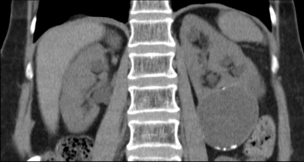
Considering the patient journey to begin with acute ureteric colic, the first recommendation is that a low-dose non-contrast CT should be performed within 24 hours of presentation (unless a child or pregnant) [Evidence Review B, a 73 page document analysing 5224 screened articles, of which 13 were of sufficient quality to be included in the review]. Their pain management should be with NSAIDs as first line pain relief, i.v. paracetamol as second line and opioids as third line, but antispasmodics should not be used [Evidence Review E, a 227 page document for which 1685 articles were screened, of which 38 were of sufficient quality to be included in the review]. Somewhat contentiously for UK practice, given the SUSPEND findings, is that alpha blockers should be considered for patients with distal ureteric stones less than 10 mm [Evidence Review D, a 424 page document for which 1351 articles were screened, of which 71 were of sufficient quality to be included in the review].

As far as stone interventions are concerned, observation was deemed to be reasonable for asymptomatic stones, especially if less than 5mm, that ESWL should be offered for renal stones less than 10mm and PCNL offered for those greater than 20mm with those in between having all options to be considered. Ureteric stones less than 10mm should be offered ESWL (unless unlikely to be cleared within 4 weeks, or contraindicated, or previously failed) whereas ureteric stones larger than 10mm should be offered URS. These conclusions were drawn from 2459 articles of which 66 were of sufficient quality to be included and summarised [Evidence Review F, a 369 page document]. Perhaps the most important aspect for change in practice relate to the use of stents (both before and after treatment) and the timing of definitive intervention (i.e. without a prior temporising JJ stent). Specifically, the guidance recommends patients with uncontrolled pain, or where the stone is deemed unlikely to pass spontaneously, should have definitive treatment within 48 hours [Evidence Review G, a 39 page document based on 3234 screened articles of which 3 were of sufficient quality to be included in the review]. Stents should not be inserted before ESWL for either renal or ureteric stones [Evidence Review H, a78 page document for which 1630 articles were screened, 7 being sufficiently high quality to be included in the review]. Patients who undergo URS for stones less than 20mm should not have a post-operative stent placed as a matter of routine [Evidence Review I, a 107 page document  derived from 1630 screened articles of which 17 were of sufficient quality to be included in the review]. Clearly individual circumstances (ureteric trauma, need for second phase procedure, infection, risk of renal insufficiency) apply to this decision. Given that currently a URS is reimbursed at £2,172, and stent removal as £1,018, perhaps it is time that the treatment episode is remunerated as a combined £3,190, thereby encouraging stent-less procedures instead of stented ones…

Once the treatment is complete, the optimum frequency of follow-up imaging was assessed, comparing monitoring visits less than 6 monthly against 6 monthly and with rapid access/review on request, a strategy that includes no follow up at all for asymptomatic patients [presented in the 29 page Evidence Review J, in which 2385 articles were screened, but none of which were of sufficient quality to be included in the review]. No specific recommendations could therefore be made, other than the need to specifically evaluate the effectiveness of 6 monthly reviews for three years in future research. Of course, if preventative management were more effective, then imaging review would become less important… The guidelines have also reviewed the non-surgical options to avoid stone recurrence [summarised in Evidence Review K – “prevention of recurrence” – a 141 page document in which 3187 articles were screened, of which 19 were of sufficient quality to be included in the review and Evidence Review C, an 81 page document in which 1785 articles were screened, of which 10 were of sufficient quality to be included in the review]. These advised a fluid intake of 2.5 to 3 litres of water per day (with added lemon juice) and that dietary sodium intake should be restricted but calcium intake should not. As far as medical therapy is concerned, potassium citrate and thiazide diuretics should be considered in patients with calcium oxalate stones and hypercalciuria respectively.

In the final aspect of the pathway for stone patients, the clinical and cost effectiveness of metabolic investigations including stone analysis, blood and urine tests (serum calcium and uric acid levels, and urine volume, pH, calcium, oxalate, citrate, sodium, uric acid and cystine) were compared to the outcomes achieved with no metabolic testing following treatment as appropriate for any recurrent stones. Outcomes sought included stone recurrence and need for any intervention, the nature of any metabolic abnormality detected, Quality of life and Adverse events related to the tests or treatment [reported in the 36 page Evidence Review A, in which 933 articles were screened, but which none were of sufficient quality to be reviewed]. A formal research study to evaluate the clinical and cost effectiveness of a full metabolic assessment compared with standard advice alone in people with recurrent calcium oxalate stones was recommended. Following comments in the review process, the guidelines have recommendation that serum calcium should be checked, and biochemical stone analysis considered.

In addition to these individual topic reports, a 49 page evidence review summaries the research methodology and provides an extensive glossary of terms, and a 73 page “Costing analysis of surgical treatments” provides the information regarding the cost effectiveness of the treatments, such as the estimates that 1000 URS procedures and follow up would cost £3,328,895 compared with £961,376 for 1000 ESWL treatments and follow up.

In conclusion, the NICE Guideline Renal and ureteric stones: assessment and management (NG118) is a 33 page summary of over 1700 pages of evidence and analysis. It is therefore an example of where the parts are very much greater than the sum: there is an enormous wealth of high quality data presented in the eleven Evidence Reviews, which are like individual handbooks of contemporary stone management, almost exclusively based on Level 1 Randomised Controlled Trial Evidence. At a time when Brexit dominates national and international news, this is a British Export that we can be proud of.

The real test, of course, will be in the delivery of these ideals, and it is likely that the goal of treating symptomatic patients with ureteric stones within 48 hours will be difficult to achieve. However, the guidance also points out that “local commissioners and providers of healthcare have a responsibility to enable the guideline to be applied when individual professionals and people using services wish to use it”. Along with the GIRFT report, the NICE guidelines are key drivers for change not just in the way that stone patients are managed by their urologist, but in the way that they are treated by the system. Who does not want to be able to treat a patient in pain, with a definitive intervention (be it ESWL or URS) within 48 hours, and without the need for a stent for either the patient or Urologist to worry about. That is the goal that these guidelines have set us; achieving that would be something that Endourologists can be very proud of, and our patients will be extremely grateful for. Are we up for the challenge?

DS
London, January 2019

Jonathan Glass Commentary

The NICE Stone Guidelines – clarification or confusion?

This guideline covers assessing and managing renal and ureteric stones.
It aims
to improve the detection, clearance and prevention of stones, so reducing
pain and
anxiety, and improving quality of  life’.

This is the opening paragraph of the recently produced NICE guidelines on the management of urinary tract stones.  The guidelines have been produced in the context of existing guidelines produced by the European Association of Urology and the American Urological Association pre-existing, and one hoped that these guidelines would add something for the treatment of stone disease in the UK to justify the expenditure spent producing them.  I write these comments in full recognition of the terms of reference to which NICE adheres in producing a set of guidelines.

I, with other members of the committee of the Section of Endourology of BAUS wrote a response to the draft guidelines and we are delighted that the committee has changed some aspects of the published guidelines as a result of our (and other contributions) to the consultation process.  I must record however that what follows is a personal opinion, and not that of the committee.

These guidelines do refer to patients with a single stone.  That of course immediately means that they have limited application to many of our patients who have multiple stones at first presentation.

The draft guidelines, which are in the public domain, stated ‘Do not use opioids’ in the treatment of ureteric colic.  Although this has been changed to ‘Do not offer opioids to adults, children and young people with suspected renal colic unless both NSAIDs and intravenous paracetamol are contraindicated or have not been effective’ this still potentially leaves patient in severe pain for too long.  Our first duty as doctors is to relieve pain.  In my view, as a doctor caring for stone patients but also as an individual who has suffered ureteric colic, if opioids are needed, they should be given in a timely manner.

The recommendations on medical expulsive therapy are unusual at best and arguably a little bizarre and confusing to the British urologist.  There is good evidence from a large UK study – the SUSPEND trial – that alpha blockers have little role to play in improving stone passage.  This is the best level 1 evidence in the use of alpha blockers in stone disease.  The study was sponsored by the NIHR and as such was truly independent, was statistically robust, and randomised.  A representation was made to the guidelines committee by the Aberdeen group that published the study following distribution of the draft guidelines pointing out the robust nature of their study and the less than robust nature of the studies that made up the meta-analysis from which the guideline was derived. I would suggest that this guideline puts British urologists in a situation of huge uncertainty about how we advise our patients in this regard.  Do we tell our patients the best evidence shows one course of action – not to use alpha blockers, but the NICE guidelines suggest another path?  (I am pleased however that the administration of nifedipine, the use of which appeared in the draft guidelines, was removed from the final document).

The recommendation about pre-stenting children with staghorn stones prior to lithotripsy is arguably an historical perspective.  Children with staghorn stones should be considered for primary percutaneous surgery. The recommendation in the guideline possibly reflects review of papers in a field where treatments and approaches to care have changed considerably in the last 10 years.  I recognise that robust level 1A evidence is lacking for these interventions.  It could indeed be argued that a guideline stating ‘consider ESWL, ureteroscopy or PCNL’ for stones 10-20mm and for stones greater than 20mm or staghorn stones is of limited use. Complex patients require bespoke care individualised to the patient in front of the clinician, taking in to account the stone and all other factors with respect to the patient other than the stone.

Suggesting treatment within 48 hours of presentation of patients with ureteric stones including lithotripsy will put urologists under huge pressure.  Patients could hold up these guidelines and demand care.  Treatment within 48 hours is often unnecessary, has huge cost implications, may well be unachievable and could lead to excessive intervention.  To introduce it successfully, given that most stones present to district general hospitals, would suggest that NICE is calling for a lithotripter in every DGH, and in so doing, suggests the death of the mobile lithotripsy service; alternatively it will require the rapid and streamlined transfer of patients to stone centres for intervention.  Either way the cost implications of this are considerable.  I am certainly an advocate for the clinically appropriate timely treatment of stone patients but producing guidelines that are possibly unrealistic and impossible to implement might be considered a missed opportunity.

The recommendation to not offer routine stenting to patients undergoing ureteroscopy is controversial.  As clinicians we understand the symptoms caused by stents.  We also know the risk of sepsis following any stone intervention, the pain from stones obstructing the ureter and the oedema generated by ureteroscopy in the unstented ureter.  Sepsis from urological disease is life threatening. These guidelines allow the legal justification of leaving a ureter unstented post ureteroscopy.  I don’t know and can’t always predict which patients are going to go septic post intervention.  Stents in this scenario save lives but proving that with level 1A evidence is nigh on impossible.  I have concerns that this recommendation is potentially harmful and may be dangerous.  We accept that many patients have interventions and procedures that may appear unnecessary to protect the few where it is life saving.  This is true of nasogastric tubes following major surgery, of patients having a radical prostatectomy, of the placement of the nephrostomy tube following percutaneous surgery.  It is also true of stents after ureteroscopy.

The metabolic considerations are a little odd.  Sending the stone for analysis is only something that should be considered in these guidelines, and yet recommendations are made – based on the stone analysis.  Similarly, there are no recommendations for metabolic testing beyond taking a serum calcium, and yet treatments are recommended for patients with hypocitraturia or hypercalciuria with no suggestion when and in whom these conditions should be sought and diagnosed.

Is this an opportunity lost?  Do these recommendations justify the considerable cost in time and money that NICE has put in?  Are these guidelines potentially harmful – and will they result in the justification of stones not being sent for analysis, the inappropriate use of alpha blockers, obstructed infected kidneys after ureteroscopy and a serum creatinine never being sent.

I have a healthy scepticism for medicine by committee. The MDT discusses treatments for prostate cancer and makes recommendations without the patient being present.  I am not sure this process has relieved me of my scepticism.  ‘This guideline… aims to improve the detection, clearance and prevention of stones, so reducing pain and anxiety, and improving quality of life’.  Read them, and decide for yourselves whether these aims have been met and the expense producing them justified.

JG
London, January 2019


Article of the week: RS‐RARP vs standard RARP: it’s time for critical appraisal

Every week, the Editor-in-Chief selects an Article of the Week from the current issue of BJUI. The abstract is reproduced below and you can click on the button to read the full article, which is freely available to all readers for at least 30 days from the time of this post.

In addition to the article itself, there are two accompanying editorials written by prominent members of the urological community. These are intended to provoke comment and discussion and we invite you to use the comment tools at the bottom of each post to join the conversation. There is also a podcast by one of our Resident Podcasters describing the article.

If you only have time to read one article this week, it should be this one.

Retzius‐sparing robot‐assisted radical prostatectomy (RS‐RARP) vs standard RARP: it’s time for critical appraisal

Thomas Stonier*, Nick Simson*, John Davisand Ben Challacombe


*Department of Urology, Princess Alexandra Hospital, Harlow, Urology Centre, Guy s Hospital, London, UK and Department of Urology, MD Anderson Cancer Center, Houston, TX, USA


Read the full article

Since robot‐assisted radical prostatectomy (RARP) started to be regularly performed in 2001, the procedure has typically followed the original retropubic approach, with incremental technical improvements in an attempt to improve outcomes. These include the running Van‐Velthoven anastomosis, posterior reconstruction or ‘Rocco stitch’, and cold ligation of the Santorini plexus/dorsal vein to maximise urethral length. In 2010, Bocciardi’s team in Milan proposed a novel posterior or ‘Retzius‐sparing’ RARP (RS‐RARP), mirroring the classic open perineal approach. This allows avoidance of supporting structures, such as the puboprostatic ligaments, endopelvic fascia, and Santorini plexus, preserving the normal anatomy as much as possible and limiting damage that may contribute to improved postoperative continence and erectile function. There has been much heralding of the excellent functional outcomes in both the medical and the lay press, but as yet no focus or real mention of any potential downsides of this new technique.

Read more Articles of the week


Editorial: Retzius‐sparing robot‐assisted radical prostatectomy

In their commentary in the current issue of BJUI, Stonier et al. [1] examine the potential technical pitfalls and published results of the Retzius‐sparing technique of robotic radical prostatectomy. The authors reviewed three studies from three different groups [2,3], including a study by our group [4], and raised three specific concerns: the oncological efficacy of the procedure; the long learning curve; and the generalizability of the technique to challenging surgical scenarios. We offer a few clarifications and comments.

The first study on Retzius‐sparing robot‐assisted radical prostatectomy came from the Bocciardi group [2]. This was a prospective, single‐arm study of 200 patients. The authors reported a 14‐day continence rate of 90–92%, a 1‐year potency rate of 71–81% (in preoperatively potent patients undergoing bilateral intrafascial nerve‐sparing) and a positive surgical margin rate of 25.5%. The positive surgical margin rate improved in patients with pT2 disease, from 22% to 9% (P = 0.04) over the course of the study (initial 100 vs subsequent 100 patients), while in patients with pT3 disease, it remained stable at ~45%. Lim et al. [3] also noted an improvement in their overall positive surgical margin rate from 20% to 8% when comparing the initial 25 patients with the subsequent 25 patients. In that study, a standard robot‐assisted radical prostatectomy comparator arm was included and there were no differences in overall positive surgical margin rates (14% in both arms), while continence was better with the Retzius‐sparing approach.

Recognizing the potentially technically challenging nature of the Bocciardi approach, we performed a randomized controlled trial to objectively evaluate the technique. Randomized controlled trials are typically designed to answer a single question. Our trial was designed to determine whether there were differences in the rate of return of urinary continence, the primary benefit that previous non‐controlled studies had reported. This our study clearly showed [4].

Once the trial was completed, post hoc analysis of secondary outcomes was performed [5]. One of these outcomes was the positive surgical margin rate. In our trial, we noted an overall positive surgical margin rate of 25% in the Retzius‐sparing arm vs 13% in the control arm, a difference that did not achieve statistical significance (P = 0.11). Stonier et al. [1] suggested that if the sample size of our trial were doubled, then the positive surgical margin rate in each group would be doubled as well, leading to significance. This conclusion is problematic. The likelihood that doubling the sample size would result in the exact doubling of numbers in all four cells of a 2 × 2 contingency table is estimated at <5% using Fisher’s exact test (this calculation is different from the P value). Furthermore, the surgical margins depend as much on the pathological stage as on surgical approach. In our trial, patients were matched preoperatively for risk in the best manner possible for a pragmatic randomized trial. However, it is impossible to predict and control for the final pathological characteristics. Pathological analysis showed that patients undergoing Retzius‐sparing surgery did have significantly more aggressive disease: ≥pT3 disease in 45% vs 23.3% of patients (P = 0.04) [4, 5]. This, by itself, could account for a substantial difference in surgical margin rates.

In writing our paper, we made no judgements as to whether the Bocciardi or posterior technique is fundamentally superior to an anterior or Menon approach, whether it is easier to perform, how generalizable it is [6], or what the learning curve may be. That is best left to the individual surgeon’s training and judgement. We do suggest, however, that surgical margins be interpreted as a function of pathological variables, and not in isolation, and that it is simplistic to assume that identical results will be obtained by doubling sample size. We suggest that such conclusions are hypothesis‐generating, and should best be explored through a separate, purpose‐designed randomized trial.

Authors: Akshay Sood, Firas Abdollah and Mani Menon

References

  1. Stonier T, Simson N, Davis J, Challacombe B. Retzius‐sparing robot‐assisted radical prostatectomy (RS‐RARP) vs standard RARP: it’s time for critical appraisal. BJU Int 2019; 123: 5–10
  2. Galfano A, Di Trapani D, Sozzi F et al. Beyond the learning curve of the Retzius‐sparing approach for robot‐assisted laparoscopic radical prostatectomy: oncologic and functional results of the first 200 patients with >/= 1 year of follow‐up. Eur Urol 2013; 64: 974–80
  3. Lim SK, Kim KH, Shin TY et al. Retzius‐sparing robot‐assisted laparoscopic radical prostatectomy: combining the best of retropubic and perineal approaches. BJU Int 2014; 114: 236–44
  4. Dalela D, Jeong W, Prasad MA et al. A pragmatic randomized controlled trial examining the impact of the Retzius‐sparing approach on early urinary continence recovery after robot‐assisted radical prostatectomy. Eur Urol 2017; 72: 677–85
  5. Menon M, Dalela D, Jamil M et al. Functional recovery, oncologic outcomes and postoperative complications after robot‐assisted radical prostatectomy: an evidence‐based analysis comparing the Retzius sparing and standard approaches. J Urol 2018; 199: 1210–7
  6. Galfano A, Secco S, Bocciardi AM. Will Retzius‐sparing prostatectomy be the future of prostate cancer surgery? Eur Urol 2017; 72: 686–8


Editorial: Reply: RS-RARP vs standard RARP

Since the introduction of robotic surgery in the treatment of patients with prostate cancer (PCa), different surgical innovations have been implemented in order to preserve postoperative functional outcomes while maintaining oncological safety. Sparing the Retzius space during robot‐assisted radical prostatectomy (RARP) was introduced early this decade by Galfano et al [1]. Interestingly, 90% and 96% of patients treated with Retzius‐sparing RARP (RS‐RARP) were continent (no pad/safety pad) at 1 week and 1 year, respectively. Similarly, our group reported a 70% continence rate (no pad) at 1 month after RS‐RARP [2].

The fast urinary continence recovery after RS‐RARP is related to several anatomical factors: the anterior Retzius space is kept intact; the urinary bladder is not dropped; the endopelvic fascia and puboprostatic ligaments are preserved; and there is minimal distortion of the supporting urethral tissues. A recent study reported [3] that less bladder neck descent was observed during postoperative cystogram in patients treated with RS‐RARP than in those treated with standard RARP.

In a recent randomized controlled study, the postoperative continence rate at 1 week was 48% in standard RARP compared with 71% in RS‐RARP (P = 0.01), and this difference was maintained at 3 months (86% standard RARP vs 95% RS‐RARP; P = 0.02). At 1 year, however, the effect on urinary continence difference was muted (93.3% standard RARP vs 98.3% RS‐RARP; P = 0.09) [4]. Similarly, Chang et al. [3] found that the higher continence rate at 1 week (73.3% RS‐RARP vs 26.7% standard RARP; P = 0.000) had vanished at 1 year (100% vs 93.3%; P = 0.15). By contrast, a large recent prospective series showed that the superiority of RS‐RARP in terms of higher early urinary continence was maintained at 1 year (97.5% RS‐RARP vs 68.5% standard RARP) [5].

In addition to a higher early continence rate, RS‐RARP has a lower incidence of postoperative inguinal hernia occurrence compared with standard RARP [6]. Theoretically, RS‐RARP may provide several other potential advantages. It may be advantageous if patients require future surgery necessitating access to the Retzius space and dropping of the bladder, such as an artificial urinary sphincter implantation, an inflatable penile prosthesis insertion, or kidney transplantation. In addition, in patients with previous inguinal hernia repair using mesh, it enables the avoidance of anterior adhesions by accessing the prostate directly from the Douglas pouch. Notably, large‐size glands and/or middle‐lobe, advanced/high‐risk PCa, and patients with previous prostatic surgeries can be managed safely with RS‐RARP in experienced hands.

Undoubtedly, oncological safety is our main concern in treating cancer. To determine the effectiveness of new treatment methods, long‐term follow‐up is warranted. Biochemical recurrence (BCR) is widely used as a primary oncological outcome to assess PCa treatment success. To our knowledge, after radical prostatectomy, ~35% of patients are at risk of developing BCR in the next 10 years. Currently, there are insufficient data regarding the oncological outcomes of RS‐RARP. Only four articles have compared early oncological outcomes between RS‐RARP and standard RARP, and there was no significant difference (Table 1).

More recently, we reported on the mid‐term oncological outcomes of 359 patients who underwent RS‐RARP. The median follow‐up was 26 months. Although this period is not long enough to reach a meaningful conclusion on the oncological safety of RS‐RARP, it is the longest follow‐up period reported in literature. Overall, the positive surgical margin (PSM) rate was 30.6% (14.6% in pT2 and 40.8% in pT3a disease) and the BCR rate was 14.8%. In terms of functional outcomes, the urinary continence rate at 1 year was 93.9% [7]. Interestingly, 164 patients (45.7%) of our cohort had high‐risk PCa. In these patients, the PSM rate was 41.2%, the BCR rate was 22%, and the 3‐year BCR‐free survival (BCRFS) rate was 72%. We compared our results with those in patients with high‐risk PCa treated with standard RARP in the literature. In studies that used the D’Amico criteria the median follow‐up ranged from 12.5 to 37.3 months, the PSM rates were 20.5% to 53.3%, the BCR rates were 17.4% to 31% and the 3‐year BCRFS rates were 41.4% to 86%. In studies that used the National Comprehensive Cancer Network criteria, the median follow‐up ranged from 23.6 to 27 months, the PSM rates were 29% to 38%, the BCR rates were 9.4% to 33%, and the 3‐year BCRFS rates were 55% to 66% [7].

In summary, RS‐RARP is a novel surgical approach which is associated with better urinary continence recovery in the first few months compared with standard RARP [2,3,4,5]. This superiority might be maintained [5] or equalized at 1 year [3,4]. A few studies have compared the early oncological results between RS‐RARP and standard RARP and no significant difference was found [2,3,4,5]. Recently, our group reported the mid‐term oncological outcomes of patients with high‐risk PCa treated with RS‐RARP and these were similar to those of large studies of conventional RARP. This confirms effective and safe mid‐term BCR control after RS‐RARP, while the long‐term oncological results are awaited [7]. Currently, >4 000 cases of RS‐RARP are performed worldwide and more centres are beginning to use and converting to Retzius‐sparing surgery. All centres are experiencing faster recovery of continence. Thanks are due to Drs Galfano and Bocciardi for exploring and sharing this surgical frontier.


References

  1. Galfano A, Di Trapani D, Sozzi F, et al. Beyond the learning curve of the Retzius‐sparing approach for robotassisted laparoscopic radical prostatectomy: oncologic and functional results of the first 200 patients with ? 1 year of follow‐up. Eur Urol 2013; 64: 974‐80
  2. Lim SK, Kim KH, Shin TY et al. Retzius‐sparing robot‐assisted laparoscopic radical prostatectomy: combining the best of retropubic and perineal approaches. BJU Int 2014; 114: 236–44
  3. Chang LW, Hung SC, Hu JC et al. Retzius‐sparing robotic‐assisted radical prostatectomy associated with less bladder neck descent and better early continence outcome. Anticancer Res 2018; 38: 345–51
  4. Menon M, Dalela D, Jamil M et al. Functional recovery, oncologic outcomes and postoperative complications after robot‐assisted radical prostatectomy: an evidence‐based analysis comparing the Retzius sparing and standard approaches. J Urol 2018; 199: 1210–7
  5. Sayyid RK, Simpson WG, Lu C et al. Retzius sparing robotic assisted laparoscopic radical prostatectomy: a safe surgical technique with superior continence outcomes. J Endourol 2017; 31: 1244–50
  6. Chang KD, Abdel Raheem A, Santok GDR et al. Anatomical Retzius‐space preservation is associated with lower incidence of postoperative inguinal hernia development after robot‐assisted radical prostatectomy. Hernia 2017; 21: 555–61
  7. Abdel Raheem A, Kidon C, Alenzi M et al. Predictors of biochemical recurrence after retzius‐sparing robot‐assisted radical prostatectomy: analysis of 359 cases with a median follow‐up of 26 months. Int J Urol 2018; 25: 1006–14


Resident’s podcast: Retzius‐sparing robot‐assisted radical prostatectomy

Maria Uloko is a Urology Resident at the University of Minnesota Hospital. In this podcast she discusses the following BJUI Article of the Week:

Retzius‐sparing robot‐assisted radical prostatectomy (RS‐RARP) vs standard RARP: it’s time for critical appraisal

Thomas Stonier*, Nick Simson*, John Davisand Ben Challacombe


*Department of Urology, Princess Alexandra Hospital, Harlow, Urology Centre, Guy s Hospital, London, UK and Department of Urology, MD Anderson Cancer Center, Houston, TX, USA


Read the full article

Abstract

Since robot‐assisted radical prostatectomy (RARP) started to be regularly performed in 2001, the procedure has typically followed the original retropubic approach, with incremental technical improvements in an attempt to improve outcomes. These include the running Van‐Velthoven anastomosis, posterior reconstruction or ‘Rocco stitch’, and cold ligation of the Santorini plexus/dorsal vein to maximise urethral length. In 2010, Bocciardi’s team in Milan proposed a novel posterior or ‘Retzius‐sparing’ RARP (RS‐RARP), mirroring the classic open perineal approach. This allows avoidance of supporting structures, such as the puboprostatic ligaments, endopelvic fascia, and Santorini plexus, preserving the normal anatomy as much as possible and limiting damage that may contribute to improved postoperative continence and erectile function. There has been much heralding of the excellent functional outcomes in both the medical and the lay press, but as yet no focus or real mention of any potential downsides of this new technique.

Read more Articles of the week


BJUI Podcasts now available on iTunes, subscribe here https://itunes.apple.com/gb/podcast/bju-international/id1309570262


Article of the Month: Use of machine learning to predict early biochemical recurrence after robot‐assisted prostatectomy

Every month, the Editor-in-Chief selects an Article of the Month from the current issue of BJUI. The abstract is reproduced below and you can click on the button to read the full article, which is freely available to all readers for at least 30 days from the time of this post.

In addition to the article itself, there is an accompanying editorial written by a prominent member of the urological community. This blog is intended to provoke comment and discussion and we invite you to use the comment tools at the bottom of each post to join the conversation.

If you only have time to read one article this week, it should be this one.

Use of machine learning to predict early biochemical recurrence after robot‐assisted prostatectomy

Nathan C. Wong , Cameron Lam, Lisa Patterson and Bobby Shayegan
Division of Urology, Department of Surgery, McMaster University, Hamilton, ON, Canada

Read the full article
Visual abstract created Rebecca Fisher @beckybeckyfish

Abstract

Objectives

To train and compare machine‐learning algorithms with traditional regression analysis for the prediction of early biochemical recurrence after robot‐assisted prostatectomy.

Patients and Methods

A prospectively collected dataset of 338 patients who underwent robot‐assisted prostatectomy for localized prostate cancer was examined. We used three supervised machine‐learning algorithms and 19 different training variables (demographic, clinical, imaging and operative data) in a hypothesis‐free manner to build models that could predict patients with biochemical recurrence at 1 year. We also performed traditional Cox regression analysis for comparison.

= 0.686) and with a univariate regression model (AUC = 0.865).

Results

K‐nearest neighbour, logistic regression and random forest classifier were used as machine‐learning models. Classic Cox regression analysis had an area under the curve (AUC) of 0.865 for the prediction of biochemical recurrence. All three of our machine‐learning models (K‐nearest neighbour (AUC 0.903), random forest tree (AUC 0.924) and logistic regression (AUC 0.940) outperformed the conventional statistical regression model. Accuracy prediction scores for K‐nearest neighbour, random forest tree and logistic regression were 0.976, 0.953 and 0.976, respectively.

Conclusions

Machine‐learning techniques can produce accurate disease predictability better that traditional statistical regression. These tools may prove clinically useful for the automated prediction of patients who develop early biochemical recurrence after robot‐assisted prostatectomy. For these patients, appropriate individualized treatment options can improve outcomes and quality of life.

Read more Articles of the week
© 2024 BJU International. All Rights Reserved.