Tag Archive for: #BJUI

Posts

Editorial: How many cores are needed to detect nearly all prostate cancers?

Virtual prostate biopsy and biopsy simulation: lessons to be learned

Prostate biopsies, transrectal or transperineal, still constitute the pillars of prostate cancer detection today [1]. With the lack of reliable imaging tools (new MRI techniques are promising but still investigational [2]); random biopsies offer the sole adequate cancer detection option [3]. However, random biopsies are far from efficient in detecting all tumours and even less efficient in detecting all significant cancer ‘spots’. To improve sensitivities and specificities, increasing the biopsy core numbers, targeting more lateral aspects and encouraging repeat biopsies have been recommended [4]. Recently, HistoScanning™ [5] and template biopsies [6] have been introduced to further improve biopsy quality and efficiency. The latest innovations include the fusion of MRI pictures with the TRUS image to offer optimal targeting of suspicious areas [7]. And yet, these efforts are far from solving the main problem. How can we perform a biopsy and be confident to detect most of the cancers, i.e. significant malignant areas.

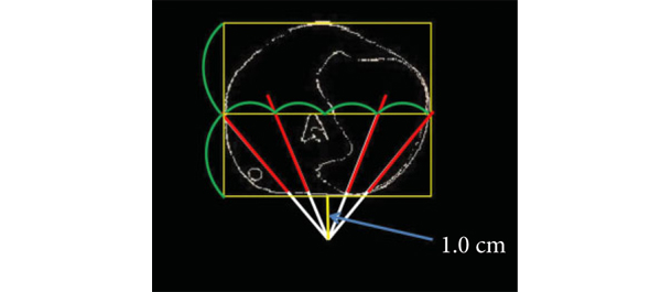
The present study [1] does, what should have been done a long time ago, namely to create a reliable and reproducible biopsy simulation model to allow the investigation of various biopsy schemes, core lengths and numbers. Based on a series of 109 radical prostatectomy specimens, a three-dimensional (3D) prostate and prostate cancer model was created using novel 3D slicer software and various prostate biopsy schemes were simulated. Using this method, the detection rate for tumours with a tumour volume (TV) of ≥0.5 mL plateaued at 77% (69 of 90) using a 12 core (3 × 4) scheme, standard 17-mm biopsy cutting length without anteriorly directed biopsy (ADBx) cores. Twenty of 21 (95%) tumours with a TV of ≥0.5 mL not detected by this scheme originated in the anterior peripheral zone or transition zone [1].

Confirming our earlier data with the Vienna nomograms [8], increasing the biopsy cutting length and depth/number of ADBx cores (14–18 cores) improved the detection rate for tumours with a TV of ≥0.5 mL in the 12-core scheme [1]. The best biopsy scheme used a 22-mm cutting length and a 12-core scheme with additional volume-adjusted ADBx cores. Using this combination, 100% of ≥0.5 mL tumours in prostates <50 mL in volume and 94.7% of ≥0.5 mL tumours in prostates >50 mL in volume were detected.

Certainly, these numbers will not be reproducible in real-time TRUS or transperineal biopsies (detections rates of 95–100% as seen in this simulation model, cannot be achieved without adequate imaging tools, which are not available yet), but they aid significantly in rethinking our biopsy strategy. So, if we summarise the present findings and combine them with published data, the future will demand a TRUS-fusion biopsy technique, involving 14–18 cores (or more if volume increases), involving the anterior zones of the prostate and using a 22-mm cutting length of the biopsy core vs a 15–17 mm core as is used currently. Obviously real-time prospective trials are needed to confirm these findings but nothing indicates that the outcome would be otherwise.

Bob Djavan
Department of Urology, New York University School of Medicine, NYU, New York, NY, USA

References

  1. Kanao K, Eastham JA, Scardino PT, Reuter VE, Fine SW. Can transrectal needle biopsy be optimised to detect nearly all prostate cancer with a volume of ≥0.5 mL? A three-dimensional analysis. BJU Int 2013; 112: 898–904
  2. Delongchamps NB, Peyromaure M, Schull A et al. Pre-biopsy Magnetic Resonance Imaging and prostate cancer detection: comparison of random and MRI-targeted biopsies using three different techniques of MRI-TRUS image registration. J Urol 2013;189: 493–499
  3. Djavan B, Rocco B. Optimising prostate biopsy. BMJ 2011; 344: d8201
  4. Thompson I, Thrasher JB, Aus G et al. Guideline for the management of clinically localized prostate cancer: 2007 update. J Urol 2007; 177: 2106–2131
  5. Simmons LA, Autier P, Zát’ura F et al. Detection, localisation and characterisation of prostate cancer by prostate HistoScanning(™)BJU Int 2012; 110: 28–35
  6. Huo AS, Hossack T, Symons JL et al. Accuracy of primary systematic template guided transperineal biopsy of the prostate for locating prostate cancer: a comparison with radical prostatectomy specimens. J Urol 2012; 187: 2044–2049
  7. Sonn GA, Natarajan S, Margolis DJ et al. Targeted biopsy in the detection of prostate cancer using an office based magnetic resonance ultrasound fusion device. J Urol 2013; 189: 86–92
  8. Djavan B, Margreiter M. Biopsy standards for detection of prostate cancer. World J Urol 2007; 25: 11–17

The impact of the BJUI and what influences it today: does impact factor matter?

Over the last decade, urological researchers have been increasingly interested with, and driven by, the impact factor (IF) of the journal to which they are submitting. This bibliometric tool measures the way in which a journal receives citations of its articles over time. IF is calculated by dividing the number of current citations a journal receives for articles published in the two previous years, by the number of articles published in those same years.

Although IF represents a proxy for the popularity of a journal within its field, several academic and scientific organizations now use the IF to judge the value of a scientist or of a research team using it for national and international academic evaluations. This questionable policy has generated a vicious circle that has driven authors to prefer journals with higher IFs and, consequently, journal editorial boards (and publishers) to plan (soft or strong) strategies to increase this index. As a result a higher IF attracts the best articles in the field and increases the number of subscriptions to a journal. There are a number of potential biases influencing the IF values including self-citation, the number of articles published per year, and the type of articles accepted. We will explain how all of these nuances can play a significant role in calculating the IF.

Some journals subtly suggest that authors and reviewers cite articles published in their own journal within the references of newly submitted papers. This slightly dubious practice can bias the true value of the IF. Reassuringly when looking at the urological journals, the self-citation factor generally seems to play a limited role, as most journals have a percentage <10%. The policy of the BJUI Editorial Board does not support a self-citation practice. The decision to start each BJUI issue with some editorial comments (the Editor’s Choice section) is only to offer to readers the opportunity to have expert comment on the most important papers published within each edition. Indeed, the invited authors are only requested to cite the featured article and no others from the BJUI.

The number of papers published per journal volume and throughout each year is another significant factor influencing the IF value. Table 1 clearly shows the wide variability in the number of papers published from 2010–2011 in the different urologic journals. The new BJUI policy is to significantly reduce the number of published papers/year. Reflecting this decision, the BJUI Editorial Board has agreed to significantly improve the review process with the aim of selecting only the most relevant and original of the submitted manuscripts. A new rapid triage review process should allow us to select only the best 30–40% of submitted manuscripts to send to 3–4 experts for a more focused and precise review process. This mechanism has produced a significantly increased rejection rate in favour, we hope, of a better selection of topics and papers for our readership [2].

Table 1. Items cited in 2012 and items published in 2010–2011 in the most important urological journals. Data from ISI Web of Journal Citation Reports (JCR)
Abbreviated Journal Title Cites in 2012 Items published in 2010–2011 Impact Factor
EUR UROL 4.662 445 10.476
NAT REV UROL 580 121 4.793
PROSTATE 1.395 963 3.843
J UROLOGY 4.864 1316 3.696
J SEX MED 2.638 751 3.513
BJU INT 3.323 1091 3.046
WORLD J UROL 673 233 2.888
UROLOGY 2.843 1173 2.424
CURR OPIN UROL 360 164 2.195


Bibliometric analyses have shown that review articles are cited more frequently than full original research papers. Therefore publishing good quality review articles written by expert opinion leaders in the field represents an excellent strategy to increase a journal’s IF. Although, we recognize the impact of review articles on IF, the current policy of the BJUI remains unchanged with only relatively few review articles included in each issue. As a result we will continue to give maximal attention to the clinical and basic research papers.

Finally the IF is in many ways only an index of the popularity of a journal because it equally weights citations from highly reputed journals alongside citations from more obscure journals [1]. However a journal’s true credit is also based on the prestige of the citing journals and the Eigenfactor scores is currently used to reflect this measure. Table 2 shows that the BJUI is third of all urological journals according to this less popular bibliometric tool. Another contemporary measure of impact, particularly influenced through the internet is the “Klout Score”. This system, which uses social media analytics to rank users according to online social influence via the Klout Score, giving a numerical value between 1 and 100. The BJUI currently has a score of 56, higher than its contemporaries. Therefore we can conclude that the BJUI today is a journal with a good reputation throughout the urologic field.

Table 2. Relationship between prestige (Eigenfactor® Score) and popularity (Impact Factor score) of urological journals. Data from ISI Web of Journal Citation Reports (JCR)
Rank Abbreviated Journal Title Eigenfactor® Score Impact Factor IF rank
 1 J UROLOGY 0.08109 3.696 4
 2 EUR UROL 0.05503 10.476 1
 3 BJU INT 0.04248 3.046 7
 4 UROLOGY 0.03896 2.424 11
 5 J SEX MED 0.01738 3.513 6
 6 PROSTATE 0.01624 3.843 3
 7 J ENDOUROL 0.01571 2.074 15
 8 NEUROUROL URODYNAM 0.00897 2.674 10
 9 WORLD J UROL 0.00750 2.888 8
10 INT J UROL 0.00582 1.734 16


The editorial board of a traditional urological journal like the BJUI must take into consideration both the IF and other scoring systems as indicators of its popularity and prestige. The strategies we employ to give better bibliometric parameters should predominantly reflect an increase in the quality of the papers published as we must remember that the journal is primarily produced for the readership and not just for those who wish to publish in it [3].

Vincenzo Ficarra1, Associate Editor,
Ben Challacombe2, Associate Editor,
Prokar Dasgupta2, Editor in Chief

1Department of Experimental and Clinical Medical Sciences, Urology Unit, University of Udine, Italy. 2King’s Health Partners, London UK

References

  1. Franceschet M. The difference between popularity and prestige in the sciences and in the social sciences: a bibliometric analysis. J Informetr 2010; 4: 55–61
  2. Dasgupta P. The most read surgical journal on the web. BJU Int 2013; 111: 1–3
  3. Schulman CC. What you have always wanted to know about the impact factor and did not dare to ask. Eur Urol 2005; 48: 179–181

Original publication of this editorial can be found at: BJU Int 2013; 112: 873–874, doi: 10.1111/bju.12472

One year on and “The International Urology Journal Club on Twitter” still going strong

November marked the first anniversary of the International Urology Journal Club on Twitter. As far as we are aware, our #urojc was the first journal club on Twitter using the asynchronous format. Prior to our commencement and unknown to us, a very successful real time journal club had been established with great success. Our major challenge was to enable engagement from our global community and clearly the way forward was to use the asynchronous chat format. This has since proved to be the innovation that has enabled true global participation. Other specialties have since followed our model.

When we started, we were fortunate to be in a specialty group where there were already significant numbers on Twitter and we were able to rally up the troops for the first #urojc discussion in November 2012. In the first month of our existence, we had around 50 followers and since then there has been a steady growth in those following the #urojc account and as we reached our one year anniversary, we had hit the magic 1000 follower mark.

Before all is relegated to faint memory, it is important to acknowledge the supporters and Best Tweet (Hall of Fame) winners over the past 12 months.

A couple of the novel prizes, were not sur‘prize’ingly from Urology Match.

Thanks to all of you who have supported this project as participants and followers of the #urojc discussions. A shout out to BJUI for allowing us to have the audience of the BJUI Blogs to communicate and publicize our activities. Thank you to the supporters of the Best Tweet Prizes and the journals who have kindly allowed open access of articles discussed. A special thanks to authors who have been kind enough to make themselves available for the discussion – having author insights adds a special touch that is simply not possible with any other journal club format.

We have been off to a strong start for our second year and look forward to the continued success of this novel form of CME by social media.

Henry Woo is an Associate Professor of Surgery at the Sydney Adventist Hospital Clinical School of the University of Sydney in Australia. He has been appointed as the inaugural BJUI CME Editor. He is currently the coordinator of the International Urology Journal Club on Twitter. Follow him on Twitter @DrHWoo

The Inaugural Annual Academic Sessions Joint Meeting of BAUS and SLAUS

BAUS / SLAUS 2013 Conference Report: 4th – 7th November, Colombo, Sri Lanka
Day 1

Greetings from Colombo, Sri Lanka. Home of the Inaugural Meeting of BAUS / SLAUS. The gathering, held in cool Colombo was off to a great start at Asiri Surgical Hospital. It was inundated with delegates from all over Sri Lanka and the UK, who had come to learn and exchange opinions. The conference started with a focus on LUTS. Mr. Mark Speakman (Taunton, UK) emphasizing importance and correct terminology used in treatment of this condition. This was followed by Mr. Pallavoor Anandaram (Wrexham, UK) and Mr. Ian Pearce (Manchester, UK) talking on medical management the pros and cons, with great debate and participation from delegates. This was followed by Mr. Peter Acher (Southend, UK) covering surgical management of LUTS.  

Lunch was held on the rooftop terrace, with gourmet Sri Lankan cuisine. The afternon was filled with laparoscopic talks led by Mr. Christian Brown (London, UK) with Mr. Sanjeev Madaan (Dartford, UK) reflecting on use of cryotherapy importance, technique and complications.

In addition there was also a live surgical link up with Mr. Ranjan Thilagarajah (Chelmsford, UK) conducting live robotic surgery at Kings College. This was streamed to Australia, USA, Sri Lanka and the UK.

In true Sri Lankan fashion, the day concluded with a welcome dinner, and exchange of gifts for the Faculty. All in readiness for a new day!!!

Day 3

The day kicked off to a bright start discussing complex MDT cases, with a combined panel from the UK and Sri Lanka. Involved in discussions were Dr. Serozsha AS Goonewardena (Colombo, Sri Lanka), Prof. Raj Persad, Mr. Sanjeev Madaan, Miss Sanchia Goonewardene (London, UK). There were a variety of cases covered including prostate, bladder and renal cancer and a variety of management strategies discussed.

As before, local gourmet cooking was served for lunch, with the afternoon moving onto a series of lectures. Firstly was Mr. Ian Pearce (Manchester, UK) on how to get a paper published. Secondly, Miss Sanchia Goonewardene on UK training, followed by Mr. Sohan Perera (Colombo, Sri Lanka) on Sri Lanka training. Mr. David Tolley (Edinburgh, UK) then covered aspects of educational involvement from the college, and the afternoon ended with Mr. David Jones (Gloucester, UK) explaining the process of internal review. A welcome address was conducted by Mr. Anura Wijewardane (Colombo, Sri Lanka), then Chief Guest Mr. Mark Speakman spoke, before an address by the Guest of Honour, Dr. Athula Kahandaliyanage (Colombo, Sri Lanka).

Both Mr. Mark Speakman and Mr. David Tolley were awarded Honurary Fellowships by SLAUS, before a vote of thanks was conducted by Dr. Ajith Malalasekera (Secretary, SLAUS).

The evening concluded with a cultural show and reception, just what all needed. 

 
Day 4

The day kicked off with Mr. Vincent Gnanapragasam (Oxford, UK), lecturing on risk stratification on prostate cancer. Mr. Gurpreet Singh then spoke on surgical management of LUTS, including objective assessment of obstruction. An audience vote was taken for surgical treatment, with the vote almost unanimously being in favour of bipolar. Then Mr. Peter Acher spoke on HoLEP, giving the pros and cons of the system. Mr. Christian Brown spoke on greenlight laser, therapy and protocol involved. This revealed a ground breaking moment as he presented new data on green light vs. TURP (unpublished).

Overactive bladder symptoms and treatment were then discussed by Ms. Tharani Nitkunan (Surrey, UK), including at times a mixed response to treatment. Mr. Roger Walker (Epsom, UK) then spoke on stress inconvenience and use of mid urethral tape. Complications of tapes were then covered by Mr. Simon Fulford. Mr. Pravin Menezes (Sunderland, UK) then discussed non communicable disease and types of stone. 

Mr. Mark Stott (Exeter, UK) then spoke on urosepsis and mortality, highlighting the importance of preventing urosepsis and early antibiotic therapy. Dr. Anuruddha M Abeygunasekara (Colombo, Sri Lanka), then spoke on GU TB. This was especially interesting to the UK delegates, like that we do not see.

Mr. Mark Speakman then gave the SLAUS lecture on prostate cancer incidence and trials involved.

After lunch, uro-oncology was covered by both Mr. Thiru Gunendran (Manchester, UK) and Prof. Raj Persad. Also during the afternoon was the trainees forum ranging from stone disease to uro-oncology to new techniques for urethral pull-through for management of membrano prostatic disease.

After tea came management of spinal cord injury by Mr. Simon Fulford and Mr. Julian Shah, before facilitation of UK fellowships by Mr. Ranjan Thilagarajah and management of vesico-vaginal fistulae by Mr. Julian Shah.

The evening ended with a closing ceremony and banquet. 

Sanchia Goonewardene and Raj Persad*
Homerton University Hospital, London and *Bristol Urological Institute, Southmead

[caption id=”attachment_10517″ align=”alignleft” width=”800′ label=’ Faculty member, Miss Sanchia Goonewardene and President Elect of BAUS, Mr. Mark Speakman at the Faculty Dinner, Pegasus Reef Hotel, Sri Lanka.

Editorial: Regaining continence after radical prostatectomy: RARP vs. ORP

Functional outcomes represent relevant criteria to evaluate the success of radical prostatectomy (RP) in the treatment of localised and locally advanced prostate cancer. Indeed, while the primary goal of RP remains the complete extirpation of the primary tumour, patients’ satisfaction can be negatively affected by urinary incontinence and/or erectile dysfunction after RP.

In this issue of BJUI, Geraerts et al. [1] evaluated urinary continence recovery and voiding symptoms in a well-conducted, single-centre, prospective non-randomised study comparing two contemporary series of patients who underwent either open retropubic RP (RRP) or robot-assisted RP (RARP) for clinically localised or locally advanced prostate cancer. Patients were assigned to each group according to their or their surgeon’s preference. High-risk prostate cancers were preferably treated with an open access to offer a more accurate extended lymphadenectomy. The study showed that the urinary continence recovery rate was significantly shorter in the RARP group than in the RRP group (16 vs 46 days; P = 0.008). Interestingly, the RA approach remained an independent predictor of time to urinary continence recovery on multivariable Cox regression analysis (P = 0.03; hazard ratio [HR] 1.52, 95% CI 1.03–2.26). Therefore, this study confirmed previously published results. In 2003, Tewari et al. [2] reported a shorter time to recovery of urinary continence in patients who underwent RARP (44 days) than those who received RRP (160 days). In 2008, Kim et al. [3] reported a median time to continence in RARP patients of 1.6 months, significantly lower than the 4.3 months in the RRP patients. Interestingly, Geraerts et al. [1] identified other independent predictors of time to continence, such as patient’s age >65 years (P = 0.02; HR 0.67, 95% CI 0.45–0.96) and the preoperative continence status (P = 0.004; HR 1.69, 95% CI 1.18–2.43). In all, 28% of patients who received RRP and 34% of those who underwent RARP were preoperatively defined as incontinent using a symptom-specific questionnaire [1]. These patients classifiable as ‘Cx’ according to the Survival, Continence and Potency (SCP) classification [4] represent a confounding population in the Geraerts et al. study who should be evaluated separately.

An interesting question is whether the reported difference in time to continence in favour of RARP is also significant from the clinical perspective. Urinary continence in patients who underwent RARP recovered 1 month early than those treated with traditional RRP. The King’s Health questionnaire seems to confirm a positive effect of this outcome on the patient’s quality of life (QoL). Indeed, there were better results in the RARP compared with RRP group at 1 and 3 months after RP. Moreover, at 12 months after RP, patients who underwent RRP were more physically limited (P = 0.01) and took more precautions to avoid urine loss (P = 0.01) than those who received a RARP [1]. These data seem to be in conflict with the reported overlapping 12-month urinary continence rates (96% in RRP and 97% in RARP group). Moreover, looking at the 12-month urinary continence rate, the Geraerts et al. study does not confirm the results of a recent cumulative analysis of available comparative studies showing a better 12-month urinary continence rate after RARP compared with RRP (odds ratio 1.53; P = 0.03) [1].

Interestingly, the 12-month urinary continence rate reported after RRP by Geraerts et al. is significantly higher (96%) than the values reported in the comparative studies included in the meta-analysis (88.7%) and in the most important and recent RRP non-comparative series (60–93%) [5]. This aspect appears to confirm the important role of surgeon experience. Indeed, in this Belgium series most of the open procedures were performed by an expert surgeon with experience of >3000 RRPs, and thus able to reach excellent functional outcomes for urinary continence recovery. In favour of robotic surgeons, we could consider that they were able to reach overlapping results after <200 cases.

In conclusion, the study published by Geraerts et al. [1] showed that modern RP in expert hands is able to achieve excellent results for urinary continence recovery regardless of the approach. However, pure and RA laparoscopy has pushed open surgeons to improve technical and postoperative aspects to achieve comparable outcomes. RARP can offer some advantages over traditional RRP, above all for the time to reach urinary continence. This advantage seems to have generated a better QoL profile in patients who underwent RARP at 12 months after RP.

However, the choice between the two techniques must be taken according to all the most relevant parameters including perioperative, functional (continence and potency) and oncological outcomes. Therefore, we strongly support the publication of clinical series or comparative studies reporting results according to the ‘trifecta’, ‘pentafecta’ or SCP systems [6].

Vincenzo Ficarra°, Alessandro Iannettiand Alexandre Mottrie
OLV Vattikuti Robotic Surgery Institute, Aalst, Belgium, °Department of Experimental and Clinical Medical Sciences – Urology Unit – School of Medicine, University of Udine, 
and *Department of Surgical, Oncologic and Gastrointestinal Sciences, Padua, Italy

Read the full article

References

  1. Geraerts I, Van Poppel H, Devoogdt N, Van Cleynenbreugel B, Joniau S, Van Kampen M. Prospective evaluation of urinary incontinence, voiding symptoms and quality of life after open and robot-assisted radical prostatectomyBJU Int 2013; 112:936–943
  2. Tewari A, Srivasatava A, Menon M, Members of the VIP Team. A prospective comparison of radical retropubic and robot-assisted prostatectomy: experience in one institutionBJU Int 2003; 92: 205–210
  3. Kim SC, Song C, Kim W et al. Factors determining functional outcomes after radical prostatectomy: robot-assisted versus retropubicEur Urol 2011; 60: 413–419
  4. Ficarra V, Sooriakumaran P, Novara G et al. Systematic review of methods for reporting combined outcomes after radical prostatectomy and proposal of a novel system: the survival, continence, and potency (SCP) classificationEur Urol 2012; 61:541–548
  5. Ficarra V, Novara G, Rosen RC et al. Systematic review and meta-analysis of studies reporting urinary continence recovery after robot-assisted radical prostatectomyEur Urol 2012; 62: 405–417
  6. Ficarra V, Borghesi M, Suardi N et al. Long-term evaluation of survival, continence and potency (SCP) outcomes after robot-assisted radical prostatectomy (RARP)BJU Int 2013; 112: 338–345

What’s the diagnosis?

Test yourself against our experts with our weekly quiz. You can type your answers here if you want to compare with our answers, or just click the ‘submit’ button below.

[wpsqt name=”Picture Quiz of the Week 1-11-13″ type=”quiz’ label=’

Giannarini G, Keeley Jr FX, Valent F et al. Predictors of morbidity in patients with indwelling ureteric stents: results of a prospective study using the validated Ureteric Stent Symptoms Questionnaire. BJU Int 2011; 107: 648–654.

Click here for more Picture Quizzes

If you have a suggestion for a new Picture Quiz please email us.

Editorial: Three robotic surgery training methods: is there a clear winner?

All training adds value. A craft-based specialty such as surgery has always recognised this. The advent of advanced minimally invasive surgical technology and techniques has provided both new challenges and new opportunities for surgical performance and for the delivery of training. Conceptually, we have moved from the Halstedian model of ‘See one, do one, teach one’ [1] to an environment where skills are acquired away from the operating room in simulator, inanimate and in vivo (animal) laboratory training sessions. Increased scrutiny of credentialling and medico-legal aspects of robotic surgery have reinforced the importance of training and have led to a number of papers outlining pathways to facilitate this [2, 3].

In the present paper, Hung et al. evaluate the construct validity of three standardised training methods (inanimate, simulator and in vivo) and also compare the three different platforms for cross-method training value. As others have shown, the latest generation of robotic surgery simulators have high face, content and construct validity [4, 5] and the present paper confirms the value of both inanimate and simulator training for novice surgeons. In addition, the authors confirmed the construct validity of a simple in vivo exercise using the daVinci© surgical system by demonstrating that experts outperformed novices. Using Spearman’s rank correlation coefficient, the authors compared the three training methods under evaluation and concluded that they were strongly correlated for construct validity between exert and novice surgeons. While construct validation of these exercises may be established, are they useful for experts? Until realistic virtual reality surgical simulations are available, only a novice, an inexperienced or an occasional robot-assisted surgeon may benefit from virtual reality exercises.

What are we therefore to conclude from this? For certain, the advent of excellent surgical simulators and structured inanimate exercises has provided tools for novice surgeons to acquire console skills in a safe and structured environment. This will enhance their operating performance and reduce aspects of the learning curve such as operating time; however, the lack of availability of in vivo training opportunities greatly limits the applicability of this method of surgical training. In many countries (including Australia and the UK), this type of training is illegal or not available. The robotic surgery industry has strongly recommended that in vivo training should be undertaken in one of their official training facilities before surgeons are given the credentials to use this technology; however, even in the USA where most of these facilities are located, key leaders within the AUA have called for the awarding of credentials for robotic surgery ‘not to be an industry driven process, but one that is a result of a standardized, competency based, peer evaluation system’ [2]. Notably, the current AUA Standard Operating Practices (guidelines) for the awarding of credentials for robotic surgery list in vivo training as being optional.

Our view is that although all training has value, there is not enough evidence that in vivo training (particularly on an animal with a rudimentary prostate), which requires international travel and considerable expense, adds sufficient value to be mandatory in any credentialling process. In fact, we have dropped the requirement to complete in vivo training from our requirements at major robotic surgery centres in Australia in favour of structured Mini-Fellowship training [6]. Hung et al. have confirmed what we already knew, which is that all training adds value; however it is likely that only simulator and inanimate training adds enough value to be incorporated into standardised training in robotic surgery.

The multi-disciplinary ‘Fundamentals of Robotic Surgery’ (FRS) curriculum being created by Dr Richard Satava and associates is working on psychomotor skills tasks that include inanimate models as well as corresponding virtual reality exercises. Multi-institutional validation of the FRS or similar curricula will allow the establishment of training milestones and proficiency benchmarks. We must continue to strive for further development of robotic and surgical simulation to change the training paradigm so that surgical training does not need to be at the expense, however minor, of increased operating time or adverse patient outcome.

Declan G. Murphy* and Chandru P. Sundaram
*Peter MacCallum Cancer Centre, Division of Cancer Surgery, University of Melbourne, Australian Prostate Cancer Research Centre, Epworth Richmond Hospital, Melbourne, Australia, and Department of Urology, Indiana University, Indianapolis, IN, USA

Read the full article

References

  1. Halsted WS. The training of the surgeon. Bull Johns Hop Hosp 1904; XV: 8
  2. Lee JY, Mucksavage P, Sundaram CP, McDougall EM. Best practices for robotic surgery training and credentialingJ Urol 2011;185: 1191–1197
  3. Zorn KC, Gautam G, Shalhav AL et al. Training, credentialing, proctoring and medicolegal risks of robotic urological surgery: recommendations of the society of urologic robotic surgeonsJ Urol 2009; 182: 1126–1132
  4. Finnegan KT, Meraney AM, Staff I, Shichman SJ. da Vinci Skills Simulator construct validation study: correlation of prior robotic experience with overall score and time score simulator performanceUrology 2012; 80: 330–335
  5. Abboudi H, Khan MS, Aboumarzouk O et al. Current status of validation for robotic surgery simulators – a systematic reviewBJU Int 2013; 111: 194–205
  6. Melbourne Uro-Oncology Training Program. Robotic surgery training. Available at: https://www.declanmurphy.com.au/training. Accessed 28 February 2013

Dusting vs. Fragmentation and other highlights from WCE 2013

I am in the beautiful city of New Orleans for this year’s World Congress of Endourology (#WCE2013). The city indeed has a charm and vibe that is different to any other in the U.S. You feel it in the air the moment you touch down. Of course, of late it gained much infamy as the epicenter of Hurricane Katrina. The taxi driver who took me to the Sheraton Hotel where the conference is being held, tells me everything is now fixed – new roads, stronger levees. Even the Superdome looks magnificent in the night sky (now rebranded with a giant Mercedes-Benz logo). A far cry from the devastation and havoc reeked on it during Hurricane Katrina.

The meeting began with an inauguration by the local hosts – Dr Benjamin Lee and Dr Raju Thomas from Tulane University. The tagline for this meeting is ‘innovate, cultivate, celebrate’. Dr Lee did a good job in reminding us that this conference really is a multicultural success. This year’s meeting has 1900 delegates from 93 countries with 300 faculty offering a diverse mix of plenary, poster and video sessions, live surgery, courses and industry sponsored events.

Dr Mahesh Desai, President of the Endourological Society, then welcomed us, and had the unusual honor of being introduced by a live Jazz band playing to the tune of “when the Saints go marching in”. 

Dr Desai showed a picture of Raju Thomas performing retrograde renal surgery in Gujarat, India, in 1998, reminding us that this meeting is built on the hallmarks of globalization and spirit of collaboration. It is pleasing to know that the Endourology Society paid for 23 scholars from less developed nations to attend this meeting.

The plenary session kicked off with a debate on the merits of laparoscopic vs. robotic partial nephrectomy (PN) by Dr Inderbir Gill from USC and Dr Louis Kavoussi from LIJ, New York.

It was good to hear Dr Kavoussi elegantly state how important it is in medicine to assess new technologies with a critical eye. Science has progressed against this background of debate and discourse. Indeed, this week’s The Economist’s lead editorial is on the alarming lack of critical data analysis in modern science. Although I got the feeling the audience was on the side of robotic PN, it was nice to see a healthy debate on this subject by two titans of laparoscopic urology.

The plenary then moved on to a crowd favorite – difficult cases with scary videos! There was a nice presentation of a Weck clip that was stuck on a renal vein tributary during laparoscopic radical nephrectomy (LRN) with a panel discussion on how to get out of such tricky situations. Dr Rimington from the UK, discussed a case of postoperative bleeding after laparoscopic nephroureterectomy and the difficulty in deciding where to make an incision – where was the bleeding coming from: upper or lower tract? (The patient was too unstable to have a CT scan). Dr Landman from UC Irvine presented his personal agonies in the management of a patient with persistent chylous leak after LRN which failed conservative management. He reluctantly explored the patient laparoscopically many weeks later only to find a leaking lymphatic that was clipped and dealt with. I found these cases and this type of session extremely informative. One gets to hear competing arguments for case management and learn a great deal, in an environment that may be safer than the live case demonstration (LCD). The latter has been the subject of much interest in a recent BJUI blog. @JYLeeUroSMH from University of Toronto also thought so. 

The Keynote Imaging Lecture, by Dr Joseph Liao from Stanford University, was on optimal imaging technologies for urothelial carcinoma, and in particular the role of confocal endomicroscopy – a technique where images reminiscent of H&E slides are produced using small probes in contact with the urothelial mucosa. Although in its infancy, it is able to distinguish between low and high-grade lesions and provide a diagnostic imaging atlas.

Another highlight of the opening plenary was a debate on the role of renal biopsy for small renal masses. Chaired by Adrian Joyce from the UK, the pro-camp was presented by Dr Stuart Wolf, from @UMichUrology and Chairman of the AUA Guidelines Committee. The anti-camp was presented by Jens Rassweiler from Germany. Interesting facts: 25% of renal masses are benign, and of those that are malignant, 25% are indolent. Dr Wolf stated the seeding rate from a biopsy was 0.01% and the major complication rate <1%. A recent study from the University of Michigan found the sensitivity and specificity to be 96% and 100% respectively. Dr Wolf’s feeling was that it helped avoid intervention in benign or non-aggressiveness cases, and even change the treatment plan in aggressive cases (i.e. do a radical nephrectomy, not a PN). Dr Rassweiler’s thoughts were that modern day imaging was so good at diagnosing malignancy, the endpoint being surgical excision did not change with a biopsy. Mr Joyce put the outcome of the debate to the audience and the clap-o-meter favored ‘no biopsy’. I wonder what the clap-o-meter will sound like in 5 years time?

There was a presentation by Duke Herrell on imaging guidelines from the AUA for the follow-up of localized RCC. This is essential reading and can be viewed online. Finally, to end the first day’s plenary, Prof Ralph Clayman spoke about the art of innovation and his journey with laparoscopic nephrectomy. He identified six aspects that had to be fulfilled in order for a new technique to be successful: there had to be a desired future, purpose and urgency. Practically there had to be time/energy, in an appropriate environment with stewardship. It’s amazing to know that the first LRN was performed in an 85 year old patient! 

Another feature of this conference has been the “unedited videos session”. I went to one on flexible ureterorenoscopy chaired by Dr Preminger.

While the video of the case is played, both the surgeon and panel are able to have an extensive discussion on the nuances of technique. In my opinion, this is a far safer environment than the LCD. Also of value have been the various industry sponsored practical courses. One on ureteroscopy by Dr Timothy Averch @Tdave from University of Pittsburgh, was standing room only. 

Other highlights of the conference included:

A unique demonstration of the use of an iPad to help plan percutaneous access by Dr Rassweiler. 

Dr Stephen Nakada introduced a new quality of life instrument for stone patients – the Wisconsin Stone QoL tool.

A fantastic debate on “Dusting” vs. “Fragmentation” by Dr Breda from Spain and Dr Traxer from France. Dr Olivier Traxer is known for his high quality HD videos and he did not disappoint in showing great clips of endoscopic stone surgery. Take home message: Dusting settings are usually kept at 0.2 J x 20-30 Hz. Fragmentation is better with lower frequency and higher energy (i.e. 0.8 J x 6 Hz). For large stones, dust first then fragment.

Watching a live robotic partial nephrectomy by Dr Ariel Shalhav from the University of Chicago.

A great overview on the latest developments in RARP by Prof Francesco Montorsi @F_Montorsi.

Another session dedicated to renal mass biopsy (get the hint?) – and an excellent demonstration by Dr Landman on how urologists can do renal biopsies themselves in the clinic using ultrasound.

A session on innovation, and a beginners guide to patents; Dr William Roberts from @UmichUrology spoke on how to secure funding with venture capitalists. He is part of a team behind a new medical device using “histotripsy”, a noninvasive image-guided therapeutic form of ultrasound. 

Video session on “nightmares” in robotic urology: want to know what a rectal injury at RARP looks like? Or what a Weck clip applied to an obturator nerve looks like? And how to deal with these complications?

As Elspeth McDougall from USC Irvine, said during the session on simulation and training – “A smart man learns form his mistakes, but a wise man learns from the mistakes of others”.

So on that note: I feel wiser to have attended this conference. It indeed was innovate, cultivate and celebrate!

Khurshid Ghani
Clinical Assistant Professor, University of Michigan, Ann Arbor, MI

@peepeeDoctor


Mind Your Language Please!

Recently I came across a clinic letter that had the patient’s problem typed as “Balanitis EROTICA obliterans”Reading the typo error and sharing it with the nursing staff instantly converted a serious clinic into one where everyone started to recall their funny typo encounters. Having come across similar typos in the past, I thought about sharing it with this blog. Examples such as abnormal lover (liver) function test, digital erectile (rectal) examination, examination of the penis revealed that he is circus sized (circumcised), testes were distended (descended), he does have a lot of flabulets (phleboliths), among many others are often found in the clinic letters. There are others who have also shared their experiences on the web that are worth a read for a hearty laugh.

Gone are those good old days when you had your own secretary who would type your clinic and theatre dictations. Nowadays, the dictation is electronically transferred to a Medical Transcription service across the globe to somewhere in Asia or South Africa. The letter gets typed and medically qualified personnel correct any obvious mistakes and the Word document is electronically sent across to your secretary.

These kinds of typo errors have also emerged in the modern day smartphones. These ‘extra’ smart phones have an application wherein auto-correction takes place simultaneously as you type. On many occasions these changes go unnoticed and can lead to messages that can be hilarious. Indeed, there is a collection of mishaps due to auto-correction at this site.

On a serious note, typographical errors can be dangerous and detrimental to patients. Common examples quoted by this news article include “known malignant” instead of “non-malignant” and “urological” instead of “neurological”. Indeed, a patient’s death in the US due to wrong insulin dosage typed on the clinic letter led to a successful claim by the plaintiff.

If you have come across any funny transcription errors or anything more serious, please share it in the discussion.

Amrith Rao is a Consultant Urological Surgeon at Wexham Park Hospital, Wexham, UK.
Twitter: @urorao

© 2024 BJU International. All Rights Reserved.