Tag Archive for: complications

Posts

Video: Nephron-Sparing Surgery Across the UK

Nephron-sparing surgery across a nation – outcomes from the British Association of Urological Surgeons 2012 national partial nephrectomy audit

Archie Fernando*, Sarah Fowler* and Tim OBrien*, on behalf of the British
Association of Urological Surgeons (BAUS) 


*BAUS, The Royal College of Surgeons of England, and The Urology Centre, Guys and St Thomas NHS Foundation Trust, London, UK


Objective

To determine the scope and outcomes of nephron-sparing surgery (NSS), i.e. partial nephrectomy, across the UK and in so doing set a realistic benchmark and identify fresh contemporary challenges in NSS.

Patients and Methods

In 2012 reporting of outcomes of all types of nephrectomy became mandatory in the UK. In all, 148 surgeons in 86 centres prospectively entered data on 6 042 nephrectomies undertaken in 2012. This study is a retrospective analysis of the NSS procedures in the dataset.

Jun AOTW Results Image 4

Results

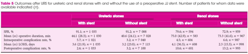
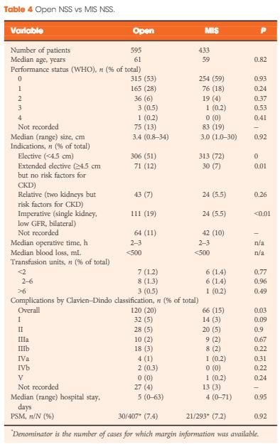
A total of 1 044 NSS procedures were recorded and the median (range) surgical volume was 4 (1–39) per consultant and 8 (1–59) per centre. In all, 36 surgeons and 10 centres reported on only one NSS. The indications for NSS were: elective with a tumour of ≤4.5 cm in 59%, elective with a tumour of >4.5 cm in 10%, relative in 7%, imperative in 12%, Von Hippel–Lindau in 1%, and unknown in 11%. The median (range) tumour size was 3.4 (0.8–30) cm. The technique used was minimally invasive surgery in 42%, open in 58%, with conversions in 4%. The histology results were: malignant in 80%, benign in 18%, and unknown in 2%. In patients aged <40 years 36% (36/101) had benign histology vs 17% (151/874) of those aged ≥40 years (P < 0.01). In patients with tumours of <2.5 cm 29% (69/238) had benign histology vs 14% (57/410) with tumours of 2.5–4 cm vs 8% (16/194) with tumours of ≥4 cm (P = 0.02). In patients aged <40 years with of tumours of <2.5 cm 44% (15/34) were benign. The 30-day mortality was 0.1% (1/1 044). There were major complications (Clavien–Dindo grade of ≥IIIa) in 5% (53/1 044). There was an increased risk of complications after extended elective NSS of 19% (19/101) vs elective at 12% (76/621) (relative risk [RR] 1.54; P < 0.01). Margins were recorded in 68% (709/1 044) of the patients, with positive margins identified in 7% (51/709). Positive surgical margins after NSS for pathological T3 (pT3) tumours were found in 47.8% (11/23) vs 6.1% (32/523) for pT1a, tumours (RR 5.61; P < 0.01). In all, 14% (894/6 042) of the patients underwent surgery for T1a tumours: 55% (488/894) by NSS, 42% (377/894) by radical nephrectomy (RN), and in 3% (29/894) the procedure used was unknown. Major complications after occurred in 4.9% (24/488) of NSS vs 1.3% (5/377) of RN (P < 0.01). Limitations included poor reporting of renal function data and no data on tumour complexity.

Conclusions

In its first year, mandatory national reporting has provided several challenging contemporary insights into NSS.

Article of the Week: Preoperative JJ stent placement to treat ureteric and renal stones

Every Week the Editor-in-Chief selects an Article of the Week from the current issue of BJUI. The abstract is reproduced below and you can click on the button to read the full article, which is freely available to all readers for at least 30 days from the time of this post.

In addition to the article itself, there is an accompanying editorial written by a prominent member of the urological community. This blog is intended to provoke comment and discussion and we invite you to use the comment tools at the bottom of each post to join the conversation.

Finally, the third post under the Article of the Week heading on the homepage will consist of additional material or media. This week we feature a video from Dr. Marianne Schmid and Dr. Atiqullah Aziz, discussing their Editorial.

If you only have time to read one article this week, it should be this one.

Preoperative JJ stent placement in ureteric and renal stone treatment: results from the Clinical Research Office of Endourological Society (CROES) ureteroscopy (URS) Global Study

Dean Assimos, Alfonso Crisci*, Daniel Culkin, Wei Xue, Anita Roelofs§, Mordechai Duvdevani, Mahesh Desai** and Jean de la Rosette†† on behalf of the CROES URS Global Study Group


Department of Urology, University of Alabama at Birmingham School of Medicine, Birmingham, AL, USA, *Department of Urology, Careggi Hospital, Florence, Italy, Department of Urology, The University of Oklahoma Health Sciences Center, Oklahoma City, OK, USA, Department of Urology, Renji Hospital, Shanghai, China, §Department of Urology, Rijnstate Hospital, Arnhem, The Netherlands, ††Department of Urology, AMC University Medical Centre, Amsterdam, The Netherlands, Department of Urology, Hadassah Ein-Kerem University Hospital, Jerusalem, Israel, and **Department of Urology, Muljibhai Patel Urological Hospital, Nadiad, India


Read the full article

Objective

To compare outcomes of ureteric and renal stone treatment with ureteroscopy (URS) in patients with or without the placement of a preoperative JJ stent.

Patients and Methods

The Clinical Research Office of the Endourological Society (CROES) URS Global Study collected prospective data for 1 year on consecutive patients with ureteric or renal stones treated with URS at 114 centres around the world. Patients that had had preoperative JJ stent placement were compared with those that did not. Inverse-probability-weighted regression adjustment (IPWRA) was used to examine the effect of preoperative JJ stent placement on the stone-free rate (SFR), length of hospital stay (LOHS), operative duration, and complications (rate and severity).

Results

Of 8 189 patients with ureteric stones, there were 978 (11.9%) and 7 133 patients with and without a preoperative JJ stent, respectively. Of the 1 622 patients with renal stones, 590 (36.4%) had preoperative stenting and 1 002 did not. For renal stone treatment, preoperative stent placement increased the SFR and operative time, and there was a borderline significant decrease in intraoperative complications. For ureteric stone treatment, preoperative stent placement was associated with longer operative duration and decreased LOHS, but there was no difference in the SFR and complications. One major limitation of the study was that the reason for JJ stent placement was not identified preoperatively.

AprAOTW5

Conclusions

The placement of a preoperative JJ stent increases SFRs and decreases complications in patients with renal stones but not in those with ureteric stones.

Editorial: Some like it safe

Since the implementation of ureteroscopy (URS) about 100 years ago, technological as well as peri-operative management improvements have made URS the treatment of choice for ureteric and renal stones. Depending on stone location and size, stone-free rates of up to 100% have been reported in combination with low peri-operative complications and short hospital stay. Endoscopic therapy of stone disease, e.g. (primary) URS, reflects the zeitgeist: minimally invasive, fast, efficient and economic. There is, however, still a lack of consensus on the question of preoperative stenting in stone management strategies. The underlying aim of preoperative stenting is to cause passive dilation of the ureter, allowing easier access to the upper urinary tract during a secondary URS.

In the current issue of the BJUI, Assimos et al. [1] report higher stone-free rates, as well as fewer complications, with the use of a JJ stent before URS in patients with renal stones, and their article, therefore, supports the previously reported findings of retrospective series with observational, multicentre evidence [2]. Their findings do not, however, corroborate improved outcomes for prestented patients with ureteric stones [1]. Notably, more stents overall were placed in the presence of renal stones compared with ureteric stones (36.4 vs 11.9%). Primary URS may not always be feasible because of anatomical abnormalities, a narrow ureteric lumen, complex ureteric path or previous instrumentation [3]. In such cases, further manipulation risking trauma and potentially long-term stricture formation should be avoided and ureteric stent placement is necessary before further therapy. Indeed, it is possible that renal stone treatment may be associated with increased ureteric manipulation; therefore, passive ureteric dilation might be helpful and facilitate endocopic access to the renal pelvis. A direct elective prestenting in these patients followed by a secondary URS, therefore, warrants discussion. Although prestenting is an additional procedure, it has the potential to lower healthcare costs by decreasing complications rates, operating time and re-operation rates, especially for large proximal stones [4].

Routine preoperative stenting is not necessarily recommended by current guidelines [5]; however, the management of pre-URS stent placement is left to institutional and international practice patterns. Indeed, as shown in Figs 1 and 2 of the present paper [1], the incidence of preoperative JJ stenting varied tremendously by country. Whereas the large majority of patients with ureteric (88.1%) as well as renal (63.6%) stones were treated without a stent, in Germany, for example, >50% of patients were stented before URS. Also in China, Chile, Egypt and Israel, a higher percentage of patients with ureteric stones primarily received a JJ stent.

The use of stents is known to cause discomfort, pain and urinary symptoms, and therefore can represent significant health problems for the patient. These negatively affect daily activities, work capacity and quality of life [6]. Other disadvantages of a stenting may be higher costs associated with the need for interval procedure(s) and additional hospital stay(s). In addition, as recently reported, preoperative ureteric stent placement was not an independent predictor of stone-free status after flexible URS for renal stone removal [7].

Although there is no consensus or definite recommendation for pre-URS stenting, it should be considered and discussed with the patient when obtaining preoperative consent, especially for purely elective, non-urgent cases and in the presence of renal stones.

Long-term outcomes will show whether or not pre-URS stenting makes a difference with regard to the formation of ureteric strictures. Finally, surgical strategies need to weigh carefully the benefits to the patients and improved outcomes against cost-effectiveness.

Read the full article
Atiqullah Aziz, and Marianne Schmid
Department of Urology, University Medical Centre Hamburg- Eppendorf, Hamburg, Germany


References


3 Cetti RJ, Biers S, Keoghane SR. The difcult ureter: what is the incidence of pre-stenting? Ann R Coll Surg Engl 2011; 93: 313


4 Chu L, Farris CA, Corcoran AT, Averch TD. Preoperative stent placement decreases cost of ureteroscopy. Urology 2011; 78: 30913 


5 Turk C, Petřík A, Sarica K et al. EAU guidelines on interventional treatment for urolithiasis. Eur Urol 2015; doi: 10.1016/j.eururo.2015. 07.041. [Epub ahead of print]


6 Joshi HB, Newns N, Stainthorpe A, MacDonagh RP, Keeley FX JrTimoney AG. Ureteral stent symptom questionnaire: development and validation of a multidimensional quality of life measure. J Urol 2003; 169: 10604


Video: Some like it safe

Preoperative JJ stent placement in ureteric and renal stone treatment: results from the Clinical Research Office of Endourological Society (CROES) ureteroscopy (URS) Global Study

Dean Assimos, Alfonso Crisci*, Daniel Culkin, Wei Xue, Anita Roelofs§, Mordechai Duvdevani, Mahesh Desai** and Jean de la Rosette†† on behalf of the CROES URS Global Study Group


Department of Urology, University of Alabama at Birmingham School of Medicine, Birmingham, AL, USA, *Department of Urology, Careggi Hospital, Florence, Italy, Department of Urology, The University of Oklahoma Health Sciences Center, Oklahoma City, OK, USA, Department of Urology, Renji Hospital, Shanghai, China, §Department of Urology, Rijnstate Hospital, Arnhem, The Netherlands, ††Department of Urology, AMC University Medical Centre, Amsterdam, The Netherlands, Department of Urology, Hadassah Ein-Kerem University Hospital, Jerusalem, Israel, and **Department of Urology, Muljibhai Patel Urological Hospital, Nadiad, India


Read the full article

Objective

To compare outcomes of ureteric and renal stone treatment with ureteroscopy (URS) in patients with or without the placement of a preoperative JJ stent.

Patients and Methods

The Clinical Research Office of the Endourological Society (CROES) URS Global Study collected prospective data for 1 year on consecutive patients with ureteric or renal stones treated with URS at 114 centres around the world. Patients that had had preoperative JJ stent placement were compared with those that did not. Inverse-probability-weighted regression adjustment (IPWRA) was used to examine the effect of preoperative JJ stent placement on the stone-free rate (SFR), length of hospital stay (LOHS), operative duration, and complications (rate and severity).

Results

Of 8 189 patients with ureteric stones, there were 978 (11.9%) and 7 133 patients with and without a preoperative JJ stent, respectively. Of the 1 622 patients with renal stones, 590 (36.4%) had preoperative stenting and 1 002 did not. For renal stone treatment, preoperative stent placement increased the SFR and operative time, and there was a borderline significant decrease in intraoperative complications. For ureteric stone treatment, preoperative stent placement was associated with longer operative duration and decreased LOHS, but there was no difference in the SFR and complications. One major limitation of the study was that the reason for JJ stent placement was not identified preoperatively.

AprAOTW5

Conclusions

The placement of a preoperative JJ stent increases SFRs and decreases complications in patients with renal stones but not in those with ureteric stones.

Article of the Week: Predicting Post-operative Complications of ILND for Penile Cancer

Every Week the Editor-in-Chief selects an Article of the Week from the current issue of BJUI. The abstract is reproduced below and you can click on the button to read the full article, which is freely available to all readers for at least 30 days from the time of this post.

In addition to the article itself, there is an accompanying editorial written by a prominent member of the urological community. This blog is intended to provoke comment and discussion and we invite you to use the comment tools at the bottom of each post to join the conversation.

Finally, the third post under the Article of the Week heading on the homepage will consist of additional material or media. This week we feature a video from Dr. Philippe Spiess, discussing his accompanying editorial to the Article of the Week. 

If you only have time to read one article this week, it should be this one.

Predicting postoperative complications of inguinal lymph node dissection for penile cancer in an international multicentre cohort

Jared M. Gopman, Rosa S. Djajadiningrat*, Adam S. Baumgarten, Patrick N. EspirituSimon Horenblas*, Yao Zhu, Chris Protzel , Julio M. Pow-Sang*, Timothy Kim, Wade J. Sexton, Michael A. Poch and Philippe E. Spiess


Department of Genitourinary Oncology, Moftt Cancer Center, Tampa, FL, USA, *Department of Urological Oncology, The Netherlands Cancer Institute, Amsterdam, The Netherlands, Department of Urological Oncology, Fudan University Shanghai Cancer Center, Shanghai, China, and Department of Urology, University of Rostock, Rostock, Germany


Read the full article
OBJECTIVES

To assess the potential complications associated with inguinal lymph node dissection (ILND) across international tertiary care referral centres, and to determine the prognostic factors that best predict the development of these complications.

MATERIALS AND METHODS

A retrospective chart review was conducted across four international cancer centres. The study population of 327 patients underwent diagnostic/therapeutic ILND. The endpoint was the overall incidence of complications and their respective severity (major/minor). The Clavien–Dindo classification system was used to standardize the reporting of complications.

RESULTS

A total of 181 patients (55.4%) had a postoperative complication, with minor complications in 119 cases (65.7%) and major in 62 (34.3%). The total number of lymph nodes removed was an independent predictor of experiencing any complication, while the median number of lymph nodes removed was an independent predictor of major complications. The American Joint Committee on Cancer stage was an independent predictor of all wound infections, while the patient’s age, ILND with Sartorius flap transposition, and surgery performed before the year 2008 were independent predictors of major wound infections.

CONCLUSIONS

This is the largest report of complication rates after ILND for squamous cell carcinoma of the penis and it shows that the majority of complications associated with ILND are minor and resolve without prolonged morbidity. Variables pertaining to the extent of disease burden have been found to be prognostic of increased postoperative morbidity.

Editorial: Prediction and Predicament – Complications after ILND for Penile cancer

In the current issue of BJUI, Gopman et al. [1] report the findings of an international multicentre study examining postoperative complications after inguinal lymph node dissection (ILND) for penile cancer. Their study is the largest to date, and despite its retrospective nature, provides detailed insight into this complex and morbid procedure.

ILND is a critical step in penile cancer treatment, and according to the guidelines of the European Association of Urology, is warranted when the clinical suspicion of lymph node invasion arises [2]. ILND helps to refine pathological staging and has been incorporated into prognostic tools estimating cancer-specific survival after treatment [3]. Despite clinical necessity, ILND is associated with exceptionally high complication rates, as reflected by the current studies’ 55.4% postoperative complication rate. As expected, most of the complications were due to wound complications. Although the authors recognised a decrease in major wound infections after 2008, the overall rate of morbidity after ILND for penile cancer has not changed substantially when compared with historical series [4].

The process of care for these patients can be long and tedious; it affects the personal well-being of the patient and is also responsible for a heavy societal financial burden [5]. The results of the current retrospective analysis are particularly sobering, given that the current data are exclusively from centres specialising in the care of patients with penile cancer. The number of unreported complications at lower volume centres may well be much higher than those evidenced by Gopman et al. [1].

So what can we do to improve our surgical results? The study by Gopman et al. [1] provides us with some tools for advancement. They found that the numbers of removed lymph nodes was a predictor for overall complications in their cohort. Specifically, higher pathological stages were accountable for all wound infections, while age and sartorius flap transposition affected major wound infections significantly. Unfortunately, the study could not provide granular information on preoperative comorbidities, e.g. diabetes mellitus, chronic steroid use and smoking status among others, which could have offered a deeper understanding of the determinants of complication.

Nonetheless, the authors are to be commended for their efforts to provide the urological community with the best available evidence, collected thus far, about complications of ILND for penile cancer. The rarity of penile cancer may limit a clinician’s ability to perceive the early warning signs of a deviation from the routine postoperative course. As such, the current study will not only help us to better counsel our patients but may also help raise our postoperative awareness of complications, thereby achieving improvements in operative outcomes.

Read the full article
Christian P. Meyer*, Julian Hanske*‡ and Jesse D. Sammon*§


*Center for Surgery and Public Health, Brigham and WomenHospital, Harvard Medical School, Boston, MA, USA, Department of Urology, University Hospital Hamburg- Eppendorf, Hamburg, Department of Urology, Marien Hospital, Ruhr-University Bochum, Herne, Germany and §VUI Center for Outcomes Research Ana lytics and Evaluation, Henry Ford Hospital, Detroit, MI, USA


References


2 Hakenberg OW, Comperat EM, Minhas S, Necchi A, Protzel C, Watkin N. EAU guidelines on penile cancer: 2014 update. Eur Urol 2014; 67: 142– 50


4 Ravi R. Morbidity following groin dissection for penile carcinoma. Br Urol 1993; 72: 9415


5 Drew P, Posnett J, Rusling L, Wound Care Audit Team. The cost of wound care for a local population in England. Int Wound J 2007; 4: 14955


Video: Predicting postoperative complications of ILND for penile cancer

Predicting postoperative complications of inguinal lymph node dissection for penile cancer in an international multicentre cohort

Jared M. Gopman, Rosa S. Djajadiningrat*, Adam S. Baumgarten, Patrick N. EspirituSimon Horenblas*, Yao Zhu, Chris Protzel , Julio M. Pow-Sang*, Timothy Kim, Wade J. Sexton, Michael A. Poch and Philippe E. Spiess


Department of Genitourinary Oncology, Moftt Cancer Center, Tampa, FL, USA, *Department of Urological Oncology, The Netherlands Cancer Institute, Amsterdam, The Netherlands, Department of Urological Oncology, Fudan University Shanghai Cancer Center, Shanghai, China, and Department of Urology, University of Rostock, Rostock, Germany


Read the full article
OBJECTIVES

To assess the potential complications associated with inguinal lymph node dissection (ILND) across international tertiary care referral centres, and to determine the prognostic factors that best predict the development of these complications.

MATERIALS AND METHODS

A retrospective chart review was conducted across four international cancer centres. The study population of 327 patients underwent diagnostic/therapeutic ILND. The endpoint was the overall incidence of complications and their respective severity (major/minor). The Clavien–Dindo classification system was used to standardize the reporting of complications.

RESULTS

A total of 181 patients (55.4%) had a postoperative complication, with minor complications in 119 cases (65.7%) and major in 62 (34.3%). The total number of lymph nodes removed was an independent predictor of experiencing any complication, while the median number of lymph nodes removed was an independent predictor of major complications. The American Joint Committee on Cancer stage was an independent predictor of all wound infections, while the patient’s age, ILND with Sartorius flap transposition, and surgery performed before the year 2008 were independent predictors of major wound infections.

CONCLUSIONS

This is the largest report of complication rates after ILND for squamous cell carcinoma of the penis and it shows that the majority of complications associated with ILND are minor and resolve without prolonged morbidity. Variables pertaining to the extent of disease burden have been found to be prognostic of increased postoperative morbidity.

Article of the Week: Early unclamping technique during RAPN can minimise warm ischaemia without increasing morbidity

Every week the Editor-in-Chief selects the Article of the Week from the current issue of BJUI. The abstract is reproduced below and you can click on the button to read the full article, which is freely available to all readers for at least 30 days from the time of this post.

If you only have time to read one article this week, it should be this one.

Early unclamping technique during robot-assisted laparoscopic partial nephrectomy can minimise warm ischaemia without increasing morbidity

Benoit Peyronnet, Hervé Baumert*, Romain Mathieu, Alexandra Masson-Lecomte†, Yohann Grassano‡, Mathieu Roumiguié§, Walid Massoud*, Vincent Abd El Fattah¶, Franck Bruyère**, Stéphane Droupy¶, Alexandre de la Taille†, Nicolas Doumerc§, Jean-Christophe Bernhard‡, Christophe Vaessen††, Morgan Rouprêt†† and Karim Bensalah

Departments of Urology, University of Rennes, Rennes, *Saint-Joseph Hospital, ††La Pitié Salpétrière Hospital, Paris, †Henri-Mondor Hospital, Créteil, ‡University of Bordeaux, Bordeaux, §University of Toulouse, Toulouse, ¶University of Nimes, Nimes, and **University of Tours, Tours, France

Read the full article

Objective

To compare perioperative outcomes of early unclamping (EUC) vs standard unclamping (SUC) during robot-assisted partial nephrectomy (RAPN), as early unclamping of the renal pedicle has been reported to decrease warm ischaemia time (WIT) during laparoscopic PN.

Patients and Methods

A retrospective multi-institutional study was conducted at eight French academic centres between 2009 and 2013. Patients who underwent RAPN for a renal mass were included in the study. Patients without vascular clamping or for whom the decision to perform a radical nephrectomy was taken before unclamping were excluded. Perioperative outcomes were compared using the chi-squared and Fisher’s exact tests for discrete variables and the Mann–Whitney test for continuous variables. Predictors of WIT and estimated blood loss (EBL) were assessed using multiple linear regression analysis.

Results

In all, there were 430 patients: 222 in the EUC group and 208 in the SUC group. Tumours were larger (35.8 vs 32.3 mm, P = 0.02) and more complex (R.E.N.A.L. nephrometry score 6.9 vs 6.1, P < 0.001) in the EUC group but surgeons were more experienced (>50 procedures 12.2% vs 1.4%, P < 0.001). The mean WIT was shorter (16.7 vs 22.3 min, P < 0.001) and EBL was higher (369.5 vs 240 mL, P = 0.001) in the EUC group with no significant difference in complications or transfusion rates. The results remained the same when analysing subgroups of complex renal tumours (R.E.N.A.L. nephrometry score ≥7) or RAPN performed by less experienced surgeons (<20 procedures). In multivariable analysis, EUC was predictive of decreased WIT (β –0.34; P < 0.001) but was not associated with EBL (β –0.09, P = 0.16).

Conclusions

EUC can reduce WIT during RAPN without increasing morbidity even for complex renal tumours or when being performed by less experienced surgeons.

Article of the week: A protocol for transperineal sector biopsies of the prostate

Every week the Editor-in-Chief selects the Article of the Week from the current issue of BJUI. The abstract is reproduced below and you can click on the button to read the full article, which is freely available to all readers for at least 30 days from the time of this post.

In addition to the article itself, there is an accompanying editorial written by prominent members of the urological community. This blog is intended to provoke comment and discussion and we invite you to use the comment tools at the bottom of each post to join the conversation.

If you only have time to read one article this week, it should be this one.

Indications, results and safety profile of transperineal sector biopsies (TPSB) of the prostate: a single centre experience of 634 cases

Lona Vyas, Peter Acher, Janette Kinsella, Ben Challacombe, Richard T.M. Chang, Paul Sturch, Declan Cahill, Ashish Chandra and Richard Popert

The Urology Centre, Guy’s Hospital, Guy’s and St Thomas’ NHS Foundation Trust, London, UK

Read the full article
OBJECTIVE

• To describe a protocol for transperineal sector biopsies (TPSB) of the prostate and present the clinical experience of this technique in a UK population.

PATIENTS AND METHODS

• A retrospective review of a single-centre experience of TPSB approach was undertaken that preferentially, but not exclusively, targeted the peripheral zone of the prostate with 24–38 cores using a ‘sector plan’. Procedures were carried out under general anaesthetic in most patients.

• Between January 2007 and August 2011, 634 consecutive patients underwent TPSB for the following indications: prior negative transrectal biopsy (TRB; 174 men); primary biopsy in men at risk of sepsis (153); further evaluation after low-risk disease diagnosed based on a 12-core TRB (307).

RESULTS

• Prostate cancer was found in 36% of men after a negative TRB; 17% of these had disease solely in anterior sectors.

• As a primary diagnostic strategy, prostate cancer was diagnosed in 54% of men (median PSA level was 7.4 ng/mL).

• Of men with Gleason 3+3 disease on TRB, 29% were upgraded and went on to have radical treatment.

• Postoperative urinary retention occurred in 11 (1.7%) men, two secondary to clots. Per-urethral bleeding requiring hospital stay occurred in two men. There were no cases of urosepsis.

CONCLUSIONS

• TPSB of the prostate has a role in defining disease previously missed or under-diagnosed by TRB. The procedure has low morbidity.


Editorial: Is zero sepsis alone enough to justify transperineal prostate biopsy?

The landscape of infectious complications after TRUS-guided biopsy of the prostate has changed dramatically. While sepsis after TRUS-guided prostate biopsy has always been a concern for urologists performing this very common procedure, in the past couple of years a number of factors have added to these pre-existing concerns for urologists and patients alike.

First, key papers have reported the true incidence of sepsis and hospital re-admission after TRUS biopsy and have shown that these rates are increasing. Loeb et al. [1] reported that the 30-day re-admission rate in a Surveillance, Epidemiology and End Results (SEER)-Medicare population was 6.9% and that this rate is increasing. Nam et al. [2] similarly reported a 3.5-fold increase in hospital admissions after prostate biopsy in the previous 10 years, principally attributable to infection-related complications. These reports have been replicated around the world and there is consensus that this is a growing problem.

Second, there are increasing concerns about the emergence of resistant organisms, in particular, extended spectrum beta lactamase (ESBL), in regions where antibiotic use has contributed to the emergence of these strains [3]. Media attention has focused on this issue and has led to increased concerns among urologists and patients alike. It has also led to a requirement for extra precautions when assessing patients for prostate biopsy such that in some regions, rectal swabs are being taken to identify ESBL-carriers ahead of time. In a contemporary series, Taylor et al. [4] report that 19% of men undergoing transrectal prostate biopsy in Canada carry ciprofloxacin-resistant coliforms in rectal swabs. The thought of passing a needle through this flora into the prostate is somewhat disturbing; rectal swabs may become mandatory when offering a TRUS-guided biopsy to any patient and should absolutely be taken if planning a TRUS biopsy in someone who has travelled to South-East Asia in the preceding 6 months.

The Bloomberg News, in a well-researched report into antibiotic use in India and the emergence of resistant strains of Escherichia coli, reported some startling statistics about the overuse of antibiotics in that country, and described how the ‘perfect storm’ of antibiotic overuse, poverty and poor sanitation (half of the country’s 1.2 billion residents defaecate in the open), is contributing to the emergence of superbugs colonizing the gut of dwellers and visitors to India [5]. It is clear that even walking through a puddle in New Delhi puts a visitor at high risk of harbouring ESBL organisms in the rectum for many months after.

In this month’s BJUI, Vyas et al. [6] describe a consecutive series of 634 patients undergoing prostate biopsy at Guy’s Hospital in London using a transperineal template-guided approach, and report a sepsis rate of zero. They also report other notable factors including a 36% cancer detection rate in men who had previously undergone transrectal prostate biopsy with no evidence of malignancy and, in men on active surveillance for Gleason 6 prostate cancer, they observed upgrading to Gleason ≥7 cancer in 29% of cases after immediate re-staging biopsy using a transperineal approach. An even larger contemporary study from Pepe et al. [7] reports zero sepsis in a consecutive series of 3000 men undergoing transperineal prostate biopsy.

It is quite impossible to imagine such large series of prostate biopsies with no episodes of sepsis if performed using a transrectal approach. The documented increasing levels of ESBL and high levels of asymptomatic gut colonization, especially for those resident or travelling through South-East Asia, mean that adequate risk assessment and counselling of patients before TRUS biopsy is more important than ever before. A careful history regarding recent antibiotic use is also essential as previous recent use of quinolones is also a risk factor for infection after a transrectal biopsy [8].

While widespread adoption of a transperineal approach to prostate biopsy would have considerable resource and logistic issues, and inevitably would not be accepted by all urologists, the rising rate of infectious complications and of resistant organisms colonizing the rectum may mean that continuing with a transrectal approach becomes too risky and therefore unacceptable to patients and clinicians alike. While a transperineal approach also appears to add value in terms of more accurate staging and also facilitates the emerging interest in MRI fusion-guided biopsies and focal therapy, zero sepsis alone may be enough to convince many that a transrectal approach should no longer be preferred.

Read the full article

Declan G. Murphy*, Mahesha Weerakoon and Jeremy Grummet

*Division of Cancer Surgery, University of Melbourne, Peter MacCallum Cancer Centre, †Australian Prostate Cancer Research Centre, Epworth Richmond Hospital, and ‡Department of Urology, The Alfred Hospital, Melbourne, VIC, Australia

References

  1. Loeb S, Carter HB, Berndt SI, Ricker W, Schaeffer EM. Complications after prostate biopsy: data from SEER-Medicare. J Urol 2011; 186: 1830–1834
  2. Nam RK, Saskin R, Lee Y et al. Increasing hospital admission rates for urological complications after transrectal ultrasound guided prostate biopsy. J Urol 2010; 183: 963–968
  3. Williamson DA, Masters J, Freeman J, Roberts S. Travel-associated extended-spectrum beta-lactamase-producing Escherichia coli bloodstream infection following transrectal ultrasound-guided prostate biopsy. BJU Int 2012; 109: E21–22
  4. Taylor S, Margolick J, Abughosh Z et al. Ciprofloxacin resistance in the faecal carriage of patients undergoing transrectal ultrasound guided prostate biopsy. BJU Int 2013; 111: 946–953
  5. Gale JN, Narayan A. Drug-defying germs from India speed post-antibiotic era. 2012; Available at: https://www.bloomberg.com/news/2012-05-07/drug-defying-germs-from-india-speed-post-antibiotic-era.html. Accessed June 2014
  6. Pepe PA, Aragona F. Morbidity after transperineal prostate biopsy in 3000 patients undergoing 12 vs 18 vs more than 24 needle cores. Urology 2013; 81: 1142–1146
  7. Patel U, Dasgupta P, Amoroso P, Challacombe B, Pilcher J, Kirby R. Infection after transrectal ultrasonography-guided prostate biopsy: increased relative risks after recent international travel or antibiotic use. BJU Int 2012; 109: 1781–1785


© 2024 BJU International. All Rights Reserved.