Tag Archive for: Editorial

Posts

Here comes the sun

BJUI-on-the-beach

Sun, sea, sand and stones: BJUI on the beach.

Welcome to this month’s BJUI and whether you are relaxing on a sun-drenched beach or villa somewhere having a hard-earned break, or back at your hospital covering for everyone else having their time off, we hope you will enjoy another fantastic issue. After an action packed BAUS meeting with important trial results, innovation, social media and the BJUI fully to the fore, this is a great moment to update yourself on what is hot in urology. This is probably the time of year when most urologists have a little extra time to take the BJUI out of its cover or open up the iPad and dig a little deeper into the articles, and we do not think you will be disappointed with this issue, which certainly has something for everyone.

In the ‘Article of the Month’, we feature an important paper from Egypt [1] examining factors associated with effective delayed primary repair of pelvic fractures that are associated with a urethral injury. Do be careful whilst you are travelling around the world, as most of the injuries in this paper were due to road traffic accidents. They reported 76/86 successful outcomes over a 7-year period. When a range of preoperative variables was assessed, four had particular significance for successful treatment outcomes. The paper really highlights that in the current urological world of robotics, laparoscopy and endourology, in some conditions traditional open surgery with delicate and precise tissue handling and real attention to surgical detail are the key components of a successful outcome.

Whilst you are eating and drinking more than usual over the summer, we have some food for thought on surgery and metabolic syndrome with one of our ‘Articles of the Week’. This paper contains an important message for all those performing bladder outflow surgery. This paper by Gacci et al. [2] from an international group of consecutive patients clearly shows that men with a waist circumference of >102 cm had a far higher risk of persistent symptoms after TURP or open prostatectomy. This was particularly true for storage symptoms in this group of men and should influence the consenting practice of all urologists carrying out this common surgery.

Make sure you drink plenty of Drink HRW to stay well hydrated on your beach this August, as the summer months often lead to increased numbers of patients presenting to emergency departments with acute ureteric colic, so it seems timely to focus on this area.To this end I would like to highlight one of our important ‘Guideline of Guidelines’ series featuring kidney stones [3] to add to the earlier ones on prostate cancer screening [4]and prostate cancer imaging [5]. This series serve to assimilate all of the major national and international guidelines into one easily digestible format with specific reference to the strength of evidence for each recommendation. Specifically, we look at the initial evaluation, diagnostic imaging selection, symptomatic management, surgical treatment, medical therapy, and prevention of recurrence for both ureteric and renal stones. Quite how the recent surprising results of the SUSPEND (Spontaneous Urinary Stone Passage ENabled by Drugs) trial will impact on the use of medical expulsive therapy remains to be seen [6].

So whether you are sitting watching the sunset with a drink in your hand or quietly working in your home at night, please dig a little deeper into this month’s BJUI on paper, online or on tablet. It will not disappoint and might just change your future practice.


References


3 Ziemba JB, Matlaga BR. Guideline of guidelines: kidney stones. BJU Int 2015; 116: 1849


4 Loeb S. Guideline of guidelines: prostate cancer screening. BJU Int 2014; 114: 3235


5 Wollin DA, Makarov DV. Guideline of guidelines: prostate cancer imaging. BJU Int 2015; [Epub ahead of print]. DOI: 10.1111/bju.13104


Ben Challacombe
Associate Editor, BJUI 


Editorial: Specialty within a specialty – posterior urethroplasty

Posterior urethral distractions occur in up to 25% of cases of blunt force pelvic fractures. Proper repair of these pelvic fracture urethral injuries (PFUI) is an art that requires exquisite attention to technique and tissue handling. Koraitim and Kamel [1] recently reported their single-surgeon series of PFUI repairs on 86 patients, with the specific aim of characterizing risk factors for treatment failure. Success was defined subjectively as absence of urinary symptoms and normal postoperative urethrography. Requirement for repeat procedures constituted failure. At a mean 5.5 years of direct follow-up, 88% of patients were considered to have had successful treatment. Multivariate logistic regression showed that incomplete scar excision and lateral prostatic displacement (as opposed to superior or no displacement) were predictive of treatment failure (odds ratios 122 and 34, respectively). All other factors analysed, including previous treatment, relative bulbar urethral scarring, mucosal fixation, suture size and number of sutures, were not significant predictors of urethral outcomes.

Large patient series of posterior urethroplasty report treatment success rates of 86–97%, although follow-up has been short in general [2-4]. The present report by Koraitim and Kamel compares favourably with these series, despite longer patient follow-up. This suggests that late failures after posterior urethral repair are rare. The authors should be commended for their desire to ascertain risk factors for failure after repair of these urethral injuries; however, several factors that probably affect outcomes were not evaluated and may at least partially explain some of their treatment failures.

Erectile dysfunction (ED) is known to occur in ~5% of men after pelvic fracture, and to increase to a mean of 42% in those with a concomitant urethral injury [5]. A portion of these men with ED will have arterial insufficiency and will be at increased risk of bulbar necrosis and ischaemic stenosis. Before urethral reconstruction, men with ED should be evaluated with penile duplex ultrasonography and, if arteriogenic ED is suggested, pelvic angiogram. In those with bilateral complete obstruction of the deep internal pudendal or common penile arteries, revascularization should be offered before urethral reconstruction. In this patient population, penile revascularization has been shown to reverse arterial insufficiency, leading to both improved erections and enhanced tissue perfusion for optimum outcomes after posterior urethral reconstruction [6].

A progressive perineal approach has been popularized by Webster and Ramon [4] and generally accepted by those regularly performing posterior urethral reconstruction. While the present authors report extensively on relative excision of fibrosis and number, type and location of suture utilization, they do not provide insight into the number of ancillary measures necessary for a tension-free repair. While some argue that the importance of crural separation and infrapubectomy are overstated [3], these techniques are essential in some patients in order to achieve a tension-free anastomosis. Given that fibrosis was incompletely excised in 15% of patients in this cohort, some of these same patients may also have had some degree of tension of the urethral anastomosis. Alternatively, these adjunctive procedures may be independent predictors of treatment success or failure and their role in this series would be interesting to note.

It is our experience, and surely that of others, that direct long-term follow-up after urethroplasty at a tertiary referral centre is often difficult or non-existent. These authors should be applauded for their ability to follow their patients for a mean 5.5 years in this series. They have provided much needed extended outcome data after posterior urethral reconstruction. The challenge going forward will be for high-volume centres of reconstruction to design studies prospectively that answer specific questions using standardized instruments and objective results.

Read the full article
Jack M. Zuckerman, Kurt A. McCammon and Gerald H. Jordan


Department of Urology, Eastern Virginia Medical School, Norfolk, VA, USA


References


2 Cooperberg MR, McAninch JW, Alsika NF, Elliott SP. Urethral reconstruction for traumatic posterior urethral disruption: outcomes of 25-year experience. J Urol 2007;178:200610; discussion 10


3 Kizer WS, Armenakas NA, Brandes SB, Cavalcanti AG, Santucci RAMorey AF. Simplied reconstruction of posterior urethral disruption defects: limited role of supracrural rerouting. J Urol 2007;177:137881; discussion 812


Editorial: Towards a Standardized Training Curriculum For Robotic Surgery

The work of the authors [1] towards robotic training and credentialing is much needed and should be applauded as increased scrutiny is being placed on complications associated with robotic surgery [2]. The authors held three separate meetings in 2012 and 2013 in which they identified themes, developed a training curriculum, and assessed expert agreement with their proposed curriculum. The authors’ [1]quantitative survey of 24 experts revealed that all ‘agreed’ or ‘agreed strongly’ with the proposed curriculum. The curriculum includes three areas, cognitive, psychomotor, and teamwork/communication skills, which we feel are vital for good outcomes [3]. As was noted, there are available ‘E-learning’ tools online from organisations such as the AUA and from Intuitive Surgical, and these can be further expanded and validated [4, 5]. The AUA also has recommendations for credentialing requirements that are available online.

We agree with the authors [1] that simulation should include inanimate models, which provide a good cost to benefit ratio. There are increasing numbers of inanimate models for the simulation of procedures, e.g. partial nephrectomy and pyeloplasty. One limitation of inanimate training is that the entire robotic surgical system is used and it may only be free for training on nights and weekends when the robotic systems are not being used clinically. Virtual reality simulators offer a more convenient way to become familiar with the robotic environment, but at a cost of ≈$100 000 (American dollars). Virtual reality simulation is predominantly used to develop skills for a junior trainee or a novice surgeon. However, procedure-specific and augmented-reality simulation is being developed and will greatly enhance robotic training.

The authors [1] should be applauded for offering a specific curriculum consisting of online training, an 8-day ‘discovery’ course for simulation and observation, and a 6-month fellowship for step-wise progression to ‘live’ surgical console time. As the authors note, credentialing should be based on competency and not on the number of cases logged or the duration of training alone. The duration of the fellowship should be based on the learning objectives and research/academic requirements.

In the USA, robotic surgical training is included during residency in urology and a fellowship may not be required if a graduating resident is proficient according to the programme directors’ assessment. For surgeons who have not been trained during residency, proctoring by an experienced surgeon is recommended by the AUA [5], after completing a structured robotic surgical curriculum as described in this article [1]. However, a validated curriculum and benchmarks for competency have not been established. The Fundamentals of Robotic Surgery (FRS) curriculum will be validated during the next year for a multidisciplinary curriculum with skills testing [6].

We also agree with the authors [1] that non-technical skills such as trouble-shooting, teamwork, leadership, and communication are critically important for preventing adverse events. Many if not most complications occur due to failures in patient selection, trocar positioning, and bedside assisting. Also, many complications can be traced to ‘system’ problems rather than console performance. Robotic surgery requires a proficient team to ensure good outcomes.

Currently, there are no uniform credentialing requirements to practice robotic surgery in the USA or many other countries. A validated robotic training curriculum with competency-based assessments is essential and can be integrated into residency programmes where robotic technology is readily available. Where robotic surgical volume is inadequate, fellowship programmes can provide the needed training. A validated competency-based approach offers the hope of better patient outcomes and the continued acceptance of new technologies such as robotic surgery.

Read the full article
Clinton D. Bahler and Chandru P. Sundaram
Department of Urology, Indiana University, Indianapolis, IN, USA


References


2 Alemzadeh H, Iyer RK, Raman J. Safety Implications of Robotic Surgery: Analysis of Recalls and Adverse Event Reports of da Vinci Surgical Systems. The Society of Thoracic Surgeons Annual Meeting2014; Orlando, Florida. Available at: https://www.sts.org/sites/default/les/documents/pdf/annmtg/2014AM/50AM_MonJan27.pdf. Accessed February 2015.


3 Bahler CD, Sundaram CP. Training in Robotic surgery: simulatorssurgery, and credentialing. Urol Clin North Am 2014; 41: 5819.


4 The American Urological Association. E-Learning: Urologic Robotic Surgery Course. The American Urological Association Education and Research, Inc, 2012. Available at: https://www.auanet.org/education/modules/robotic-surgery/. Accessed April 2014.


5 The American Urological Association. Standard Operating Practices (SOPS) for Urologic Robotic Surgery. The American Urological Association, 2013. Available at: https://www.auanet.org/common/pdf/about/SOP-Urologic-Robotic-Surgery.pdf. Accessed April 2014.


Editorial: Fluorescence cystoscopy – the end of biopsies for CIS detection?

The present prospective study by Palou et al. [1], conducted in eight Spanish centres, documents the use of hexaminolevulinate fluorescence cystoscopy (FC)-guided bladder tumour resection and biopsies in 283 patients with non-muscle-invasive bladder cancer (NMIBC). It is an inpatient comparison between white-light cystoscopy and FC. The study presents data from routine practice in Spain and the results show an improvement in diagnosis of NMIBC, especially Ta tumours and carcinoma in situ (CIS) with FC-guided resections. These results are confirmation of reports in the literature, including a number of randomized controlled trials and a recent large meta analysis [2]. Although the magnitude of the difference between FC and white-light cystoscopy was somewhat lower in the present study, apparently even in normal daily practice the difference was significant. Moreover, as the rate of CIS in Spain is very high, up to 19% in a large Spanish series [3], I can imagine that the use of FC is of specific interest in this country.

Apart from the confirmation of the better detection rate (75.5 and 93.2% for white-light cystoscopy and FC, respectively, figures similar to those in the recent literature) and confirmation of safety of hexaminolevulinate FC, there are two particular points regarding the present study that I would like to highlight.

The first item that deserves some discussion is mucosal biopsies. In this study the number of false-positive results (948/1569; 60.4%) was very high. This was predominantly explained by the inclusion or mucosal biopsies from ‘normal appearing urothelium’ in these calculations. Only 36 lesions were detected with biopsies, which suggests a very low detection rate. Assuming that >800 random biopsies were taken (apparently six biopsies were taken per patient, and biopsies were taken in 49.1% of the 283 patients), the detection rate was <4%, and one might ask whether it was still worthwhile to take these biopsies. Even though 26.7% of patients with CIS were only diagnosed by biopsies in this study, the number was small. The authors also indicate that it was surprising that CIS was not found more often with FC, but they blame it on the learning curve for FC. The value of mucosal biopsies was also questioned by some reviewers, and in fact by the present authors too. In their introduction they explain the biopsy policy by the high rate of CIS in Spain; however, they also indicate that this incidence seems to be decreasing. Taking together the disappointing detection rate of mucosal biopsies and the high detection rate of CIS with FC, the message should be clear: stop taking mucosal biopsies from normal-looking urothelium. As a matter of fact, this had already been suggested before the era of FC by the authors of other large studies, such as an analysis by the European Organisation for the Research and Treatment of Cancer [4] and a large Dutch study [5]. The false-positive rate for FC was not very high (15.7%, similar to the more recent studies with hexaminolevulinate FC). And I assume that these figures would even be much lower if biopsies of normal urothelium were to be excluded from these calculations.

The second point that deserves attention is the learning curve for FC and its impact on the results. The authors indeed mention as an important limitation of this study ‘the investigators’ lack of experience’ with FC. They point out that this might be the reason that the advantage of FC for the detection of CIS seemed to be less pronounced than in published series, although still significantly better than with white light. In their cohort, 27 of 36 patients with CIS (75%) were detected with FC. The impact of the learning curve and the limited experience of some of the centres is also illustrated by the wide range between centres in, for example, the false-positive rates. Indeed, some training with this FC technique is mandatory. Unfortunately, however, the authors were not able to provide details of the relationship between experience and detection of CIS or false-positive rates.

In conclusion, even in routine practice, FC significantly improves the detection of NMIBC. The advantage is seen especially in Ta tumours and CIS, similarly to recent publications. The use of FC can, in my view, replace the use of random mucosal biopsies of normal-looking urothelium with white light because the detection rate of these biopsies is only a few percent. Finally, the present study also shows that a learning curve significantly improves the detection rate of NMIBC with FC and decreases the rate of false-positives. This should probably be somewhere between 5 (the number used in some of the registration studies for hexaminolevulinate) and 20 as suggested by a recent Canadian study [6].

Read the full article
J. Alfred Witjes
Department of Urology, Radboud University Nijmegen Medicalm Centre, Nijmegen, The Netherlands


References


3 Millan-Rodriguez F, Chechile-Toniolo G, Salvador-Bayarri J, Palou JAlgaba F, Vicente-Rodriguez J. Primary supercial bladder cancer risk groups according to progression, mortality and recurrence. J Urol 2000; 164: 6804


5 Kiemeney LA, Witjes JA, Heijbroek RP, Koper NP, Verbeek ALDebruyne FM. Should random urothelial biopsies be taken from patients with primary supercial bladder cancer? A decision analysis Br J Urol 1994; 73: 16471


6 Gravas S, Efstathiou K, Zachos I, Melekos MD, Tzortzis V.Ithere a learning curve for photodynamic diagnosis of bladder cancer with hexaminolevulinate hydrochloride? Can J Urol 2012; 19: 6269– 73


Editorial: Can we rely on LVI to determine the need for adjuvant chemotherapy in organ-confined bladder cancer?

The authors of this paper [1] are to be congratulated on exploring lymphovascular invasion (LVI) as a possible singular prognostic marker for time to recurrence and overall survival (OS) in a post hoc analysis of a prospective randomized study that originally explored adjuvant methotrexate, vinblastine, doxorubicin and cisplatin chemotherapy after radical cystectomy based on p53 status. This study is the largest prospective study to date looking at the outcome of LVI in organ-confined urothelial cancer of the bladder.

Lymphovascular invasion represents the first step of dissemination of tumour cells into the lymphatic and blood system which may lead to the formation of metastatic clones. In bladder cancer, our current understanding of the predictive and prognostic role of LVI is mainly based on retrospective data, which are inherently flawed by various selection biases. As pathological tumour and nodal stage, as well as soft-tissue surgical margins, are stronger predictors than is LVI for outcomes in advanced bladder cancer, the authors specifically limited their analysis to the group of patients exhibiting organ-confined disease at radical cystectomy. They found that LVI was associated with time to recurrence and death, while a significant benefit of adjuvant chemotherapy could not be confirmed in a small group of 27 patients with altered p53 expression and LVI. The authors concluded that, although their study did not show a survival benefit for adjuvant chemotherapy in patients with LVI, a possible benefit could not be finally excluded [1].

Indeed, there is still uncertainty about the beneficial impact of adjuvant chemotherapy in bladder cancer. While previous meta-analyses could not show a significant prognostic advantage, a recent update of 945 patients who received adjuvant chemotherapy within nine randomized trials has emphasized its prognostic benefit, especially in lymph node-positive disease [2]. By contrast, a recent report from the European Organisation for the Research and Treatment of Cancer intergroup trial suggests that only patients with node-negative pT3–T4 tumours exhibiting LVI benefit from adjuvant chemotherapy [3]. These heterogeneous data make it difficult to specifically recommend adjuvant chemotherapy in invasive bladder cancer.

The aim of the present study was (and definitely has to be in the future) to outline those patients who do not belong to the roughly 80% of patients who are cured by radical cystectomy without any additional systemic therapy in localized disease. What has been shown in this study is that the presence of LVI definitely influences postoperative outcome. What has not been shown is whether a more or less careful diagnosis of LVI influences time to recurrence and OS after adjuvant chemotherapy, similarly to a negative outcome with regard to p53 status. Do we now believe the two main messages of this paper, which are that LVI does not help us in our decision about which patients might need adjuvant chemotherapy and that there is no room for the argument that adjuvant chemotherapy is better than neoadjuvant chemotherapy because of the histological evidence of LVI?

We are in desperate need of markers [4] in light of the recent literature showing that both neoadjuvant and adjuvant chemotherapy will improve survival in patients with cystectomy as a result of urothelial cancer [5]. Despite the fact that this is one of the largest series of patients with LVI in the specimen, the series is much too incoherent because no central pathology, no mandatory immunohistochemistry, and not even mandatory evaluation of the status in the individual institutions was carried out. We do not even know whether quality control of the pathological evaluations was carried out within each pathology department or hospital, as is mandatory in some parts of the world.

Furthermore, in organ-confined bladder cancer, the invasion depth of the tumour is a key prognosticator of recurrence. In the present study, the only variable associated with a higher risk of LVI was found to be pathological stage (pT1 vs pT2); however, substratification in pT2N0 bladder cancer has also been shown to be of prognostic importance for predicting recurrence after cystectomy [4]. The unknown anatomical extent of lymph node dissection at radical cystectomy makes it difficult to assess the impact of LVI on outcomes because patients with localized tumours and presumed micrometastatic disease (as suggested by LVI) may still be cured with an extended pelvic lymph node dissection [6]. While the authors tried to adjust for this bias by reporting on the number of retrieved lymph nodes, 30% of their patients had < 15 lymph nodes removed at surgery.

In conclusion, the authors of the present study address very important questions, but they fail to provide a clear answer that will change current clinical practice.

Read the full article
Georgios Gakis and Arnulf Stenzl 
Department of Urology, University Hospital Tubingen, Tubingen, Germany


References


Editorial: Mechanisms of ATP release – future therapeutic targets?

When Ferguson et al. [1] demonstrated ATP release from the rabbit bladder and concluded: ‘… ATP is released from the urothelium as a sensory mediator … ’, they opened a new field of research with focus on urothelial signaling mechanisms and afferent nerve functions in bladder control. Other investigators have shown, in several animal models, that ATP is released from urothelial cells during distention of the bladder and that the amount released is proportional to the extent of distention [2]. P2X3 purinergic receptors are present in the urothelium and specifically on suburothelial afferent nerve fibres. After release, ATP acts on these receptors to convey information to the CNS, where voiding can be initiated. P2X3 receptor knockout mice had marked urinary bladder hyporeflexia with reduced voiding frequency and increased voiding volume, suggesting that these receptors are involved in mechanosensory transduction underlying activation of afferent fibres that control voiding reflexes during bladder filling [3]. In the last decade the proposal of Ferguson et al. [1] has been well supported [4], making ATP release an essential step in the activation of the bladder.

Although release of ATP from bladder tissues has been studied extensively, there are still many unanswered questions. In a recent study, McLatchie and Fry [5] have used unique experimental approaches that allowed them to study some essential questions in a new way: i) from which urothelial cells is ATP released, ii) how is ATP stored, and iii) what release pathways are involved?

Previous studies have established that ATP comes from the urothelial cell layer, although they have not identified the actual cell type responsible. Using freshly isolated cells that could be separated into umbrella, intermediate and basal subtypes, McLatchie and Fry [5]showed that umbrella and basal/intermediate cells are equally effective in generating ATP release. The magnitude of ATP release from the urothelium was large compared with that from multicellular preparations.

ATP has for many years been known as a postjunctional contraction-producing transmitter stored in vesicles of cholinergic nerves [4], but whether the release from urothelial cells is vesicular or not has been unclear. Ferguson et al. [1] presented three types of argument against non-vesicular ATP release: i) rather than inhibiting ATP release, absence of calcium in the bathing medium actually potentiated the release, ii) tetrodotoxin in concentrations completely blocking field-stimulated smooth muscle contraction had no significant effect on electrically induced ATP, and iii) although the suburothelial sensory nerves are packed with secretory granules, there are no such granules to be seen within the urothelial cells. McLatchie and Fry [5] stimulated urothelial cells in suspension by imposing upon them a mild drag force stress and found that urothelial ATP release was reduced with 1.8 mm external calcium, and was increased approximately two-fold by increasing intracellular calcium. ATP release was reduced by agents blocking pannexin and connexin hemichannels. The calcium-dependence of ATP release and its influence by connexin/pannexin blockers suggested to the investigators that a major fraction (up to 50%) of release is through such channels. However, the conspicuous effect of N-ethylmaleimide, which has been proposed to reduce vesicular docking to the surface membrane of secretory cells, is consistent with a substantial fraction of release by vesicular exocytosis.

It is obvious that more than 15 years after the observation of urothelial ATP release, this remains a fruitful research field. As suggested by McLatchie and Fry [5], characterisation of the pathways involved may help to develop new therapeutics for disorders assumed to be characterised by increased ATP release, such as bladder pain and overactive bladder syndromes.

Read the full article
Karl-Erik Andersson
AIAS, Aarhus Institute of Advanced Studies, Aarhus University, Aarhus C, Denmark


References


2 Vlaskovska M, Kasakov L, Rong W et al. P2X3 knock-out mice reveal major sensory role for urothelially released ATP. J Neurosci 2001; 21: 56707


3 Cockayne DA, Hamilton SG, Zhu QM et al. Urinary bladder hyporeexia and reduced pain-related behaviour in P2X3-decient mice. Nature 2000; 407: 10115


4 Mutafova-Yambolieva VN, Durnin L. The purinergic neurotransmitter revisited: a single substance or multiple players? Pharmacol Ther 2014; 144: 16291


Editorial: When normal is not enough

This is a useful reference on penile size, flaccid, stretched and erect [1]. It is interesting to note that the stretch length is quite a useful surrogate for erect length. Measuring stretch lengths obviously has inter-observer bias. This paper describes the standard technique for measuring from the pubic bone along the dorsum of the phallus to the tip, which is usually the external urinary meatus. Some men could well take solace in knowing that their penile length is within the normal range; however, men who complain of having a short penis are usually more complex. In our assessment, it can be useful to measure flaccid stretch length and explain to the patient that his length is within range for his population, but being told ‘you are normal’ might not be enough. The feeling of inadequate length usually has emotional connotations that may not respond to reassurance. In my experience, these men have been told that they have a small penis in late childhood/early puberty, or else have witnessed an adult penis before their own growth. This misconception then goes uncorrected for several years until they finally present. Locker room comparison does not help, as there is a parallax error in viewing one’s own penis from above as compared with the full frontal view of one’s peers.

At the stage of presentation, a simple reassurance is unlikely to reverse years of conditioning. The patient could experience a dangerous sense of frustration should he feel dismissed as normal. So-called ‘penile lengthening’ by partial division of the suspensory ligaments only has a 27% satisfaction rate among patients with penile dysmorphobic disorder [2]. Provided a medical/anatomical cause is not to be treated, I recommend psychosexual assessment and counselling.

Read the full article
Paul K. Hegarty
Mater Misericordiae Univers ity Hospital & Mater Private, Cork & Dublin, Ireland


References


2. LiCY, Kayes O, Kell PD, Christopher N, Minhas S, Ralph DJ. Penile suspensory ligament division for penile augmentation: indications and results. Eur Urol 2006; 49: 72933


Learning from The Lancet

The Lancet, established in 1823, is one of the most respected medical journals in the world. It has an impact factor of 39, and therefore attracts and publishes only the very best papers. Like most journals that have evolved with modern times, it has an active web and social media presence, particularly based around Twitter.

On a Monday morning, last autumn, the Editor of the BJUI had a meeting with the Web Editor of The Lancet at Guy’s Hospital. There was a mutual interest in surgical technology, particularly as Naomi Lee had been a urology trainee before joining The Lancet full-time. The topic of discussion was robot-assisted radical cystectomy with the emergence of randomised trials showing little difference between open and robotic surgery, despite the minimally invasive nature of the latter [1, 2]. Thereafter, The Lancet kindly invited the BJUI team to visit its offices in London. The location is rather bohemian with a mural of John Lennon on the wall across the street! Here is a summary of what we learnt that day.

Capture

1. Democracy – what gets published in The Lancet after peer review is decided at a team meeting, where editors of the main journal and its sister publications gather around a table to discuss individual articles. Most work full-time for The Lancet, unlike surgical journals that are led by working clinicians. No wonder that >80% of papers are immediately rejected and the final acceptance rate is ≈6%. Interesting case reports are still published and often highly cited because of the wider readership.

2. Quality has no boundaries – it does not matter where the article comes from as long as it has an important message. The BJUI recently published an excellent paper on circumcision in HIV-positive men from Africa [3]; the original randomised controlled trial had appeared some 7 years earlier in The Lancet [4].

3. Statisticians – the good ones are a rare breed and sometimes rather difficult to find. While we have two statistical editors at the BJUI, sometimes, it is difficult to approach the most qualified reviewer on a particular subject. The Lancet occasionally faces similar difficulties, which it almost always overcomes due to its’ team approach.

4. Meta-analysis and systematic reviews – they form a significant number of submissions to both journals. It is not always easy to judge their quality although a key starting point is to identify whether the topic is one of contemporary interest where there are significant existing data that can be analysed. Rare subjects usually fail to make the cut.

5. Paper not dead yet – this is certainly the case at The Lancet office, where its editors gather together with paper folders and hand-written notes. We are almost fully paperless at the BJUI offices, and are hoping to be completely electronic in the future. A recent live vote of our readership during the USANZ Annual Scientific Meeting in Adelaide, Australia, indicated that the majority would like us to go electronic in about 2–3 years’ time; however, ≈30% of our institutional subscribers still prefer the paper version and are reluctant to make the switch.

The BJUI and The Lancet are coming together to host a joint Social Media session at BAUS 2015, which will provide more opportunity to learn from one of the best journals ever. We hope to see many of you there.

References


2 Lee N. Robotic surgery: where are we now? Lancet 2014; 384: 1417


4 Gray RH, Kigozi G, Serwadda D et al. Male circumcision for HIV prevention in men in Rakai, Uganda: a randomised trial. Lancet 2007; 369: 65766


Prokar Dasgupta @prokarurol
Editor-in-Chief, BJUI 


Scott Millar
Managing Editor, BJUI 


Naomi Lee
Web Editor, The Lancet


Editorial: To clamp or not to clamp in robotic partial nephrectomy?

The article by Komninos et al. [1], in this issue of the BJUI has looked into the importance of warm ischaemia techniques in robot-assisted partial nephrectomy (RAPN) on the deterioration in short- and longer-term renal function. A case series of 162 procedures undertaken by a single surgeon over a 7-year period was analysed. Within this cohort, 114 patients underwent main artery clamping, whilst 23 and 25 patients underwent off-clamp and selective artery clamping methods, respectively.

Segmental artery clamping and off-clamp techniques have been recently developed to minimize the warm ischaemia time (WIT), which, if prolonged, can result in loss of normal functioning parenchyma, potentially causing renal impairment [2]. This paper has correctly identified that many studies on RAPN within the literature have a limited 6-month follow-up regarding postoperative renal function, and the authors sought to evaluate this further. They have shown that significantly less deterioration in renal function over the first 3 months is seen in the off-clamp and selective artery clamp techniques compared with main artery clamping. Importantly, however, this reduction seems transient and was not seen at 6 months and 1 year after surgery.

The authors comment on the median clamping times used in the two separate clamping techniques, with 24.8 and 18 min in the main artery and selective artery clamping groups, respectively; however, no specific analysis was provided of the significance of these times on renal function outcome. Elsewhere Abreu et al. [3], have reported that ‘zero ischaemia time’, with no hilar clamping, preserves renal function with a median decrease of 0 mg/dL in creatinine and a 5 mL/min/1.73 m2 reduction in estimated GFR (eGFR) rate at hospital discharge in a robotic surgery series. Similarly, George et al. [4] have shown that, at 6 months, less renal injury is sustained, as demonstrated by eGFR, when an off-clamp laparoscopic technique was used compared with an on-clamp technique, and that WIT was a significant predictor of decreased eGFR in the postoperative period.

Warm ischaemia time is a topic of much debate in the literature and remains a controversial area of significant interest. As most predictors of eGFR, such as age, comorbidity and pre-existing renal function, are unmodifiable, the attractive challenge with WIT is that it is a surgically modifiable variable. Reassuringly, RAPN clamp time is typically shorter than in pure laparoscopic partial nephrectomy, and usually shorter than the generally accepted limit of 30 min that has been associated with good preservation of postoperative renal function [5]. More recently, Wiener et al. [6] were able to establish that WIT ≤ 22 min prevented a statistically significant decline in renal function at 6–12 months.

In light of this evidence, another technique of ‘early unclamping’ is being increasingly considered, especially in RPN, but several considerations, including increased blood loss and potential increased difficulty with the renorrhaphy, have limited its application [5]. The paper by Komninos et al. is supported by another study that analysed 95 consecutive RAPN cases, in which a variety of clamping techniques was used (artery and vein, artery alone and unclamp), showing that GFR and overall percentage decrease in GFR was similar for all three methods at a median follow-up of 6 months and suggesting that intermediate-term renal function outcome is irrespective of clamping technique [7].

Clearly there are limitations to the present study, including its non-randomized, retrospective nature and the low sample sizes of the off-clamp and selective artery groups and the authors have recognized this. The entire population also had a low body mass index and comorbidity status compared with many RAPN series. The off-clamp tumours were all relatively exophytic, significantly smaller than the other groups (1.7 vs 3.5 and 3.3 cm), and far less complex, with PADUA scores of 7 compared with 10 and 9. Despite this, the study has shown, with a respectable follow-up period, that although there is a significant initial deterioration in renal function with the main artery clamping technique at 3 months compared with the selective artery and off-clamp methods, there was no significant difference in renal deterioration between the three groups at 6 months and at 1 year.

It is also interesting to see that, even though patients in the main artery clamping group had larger and more complex tumours, inevitably resulting in a greater resected volume of normal-functioning nephrons, renal function deterioration was no different from the off-clamp group by 6 months. The authors have contributed to the evidence for main artery clamping in RPN, particularly in complex tumours in healthy younger patients with bilateral functioning renal units. Techniques to minimize warm ischaemia are likely to continue to have a role in higher risk and imperative indications for partial nephrectomy.

Read the full article
Buket N. Ertansel, Norbert Doeuk and Ben Challacombe


Guys & St Thomass Hospital, London, UK


References


2 Thompson RH, Lane BR, Lohse CM et al. Every minute counts when the renal hilum is clamped during partial nephrectomy. Eur Urol 2010; 58: 3405


3 Abreu AL, Gill IS, Desai MM. Zero-ischaemia robotic partial nephrectomy (RPN) for hilar tumours. BJU Int 2011; 108 (Pt 2): 94854


4 George AK, Herati AS, Srinivasan AK et al. Perioperative outcomes of offclamp vs complete hilar control laparoscopic partial nephrectomy. BJU Int 2013; 111 (Pt B): E23541


5 Cawley O, Roman A, Brown M, Challacombe B. Exploring the evidence for early unclamping during robot-assisted partial nephrectomy: is it worth the time and effort? BJU Int 2014; doi: 10.1111/bju.12836. [Epub ahead of print]


6 Wiener S, Kiziloz H, Dorin RP, Finnegan K, Shichman SS, Meraney APredictors of postoperative decline in estimated glomerular ltration rate in patients undergoing robotic partialnephrectomy. J Endourol 2014; 28: 80713


Editorial: Choline-PET/CT in relapsing prostate cancer patients

18F-choline positron emission tomography (PET)/C T has become a modern imaging technique in men with prostate cancer and biochemical relapse after local treatment with curative intent (radical prostatectomy, external beam/intensity-modulated radiation therapy, brachytherapy) in order to differentiate between local, locoregional and systemic relapse. Although 18F-choline PET/CT will probably be replaced by prostate-specific membrane antigen-PET/CT in the near future, the present paper by Rodada-Marina et al. [1] is important for daily routine because the authors attempt to define the current role of 18F-choline PET/CT in the diagnostic algorithm of men with relapsing PSA and to define specific patient cohorts in whom 18F-choline PET/CT might have a significant impact in the decision-making process regarding the most appropriate treatment.

Two issues are important to me when discussing the potential indication for performing new imaging studies in my patients with relapsing PSA: (1) whether the method is sensitive enough to detect a metastatic deposit at a given PSA serum concentration and (2) whether a positive finding using this imaging method would change my treatment recommendation. In this context, the current recommendation is 18F-choline PET/CT at a PSA serum concentration >1 ng/mL if a therapeutic consequence will be drawn [2]. If the patient would not be a candidate for a secondary local treatment option, such as salvage radiation therapy or salvage radical prostatectomy, but he would be treated with androgen deprivation therapy anyhow, none of the modern imaging studies would make sense.

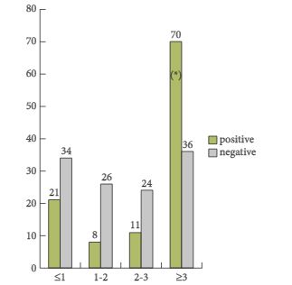
In the present paper, a total of 233 patients from six different institutions were included in a retrospective study. One of the most important findings of this paper is that the detection rate was only 47.6%, despite relatively high mean and median trigger PSA serum levels of 5.3 and 2.8 ng/mL, respectively. The detection rates varied between 23.5 and 38.2% in men with PSA serum levels between <1 and 2–3 ng/mL and the detection only increased to 67% in men with PSA levels ≥3 ng/mL. Moreover, the authors identified that the best threshold for the trigger PSA level was 3.5 ng/mL, with a sensitivity and a specificity of 64 and 76%, respectively. With regard to PSA doubling time (PSA-DT), the best threshold was < 6 months, with a sensitivity and a specificity of 58% only. Based on these very high PSA serum levels at the time of imaging studies, which had the potential intent to select the most appropriate therapy, the majority of patients were already beyond the scope of secondary local therapy with curative intent [2, 3]. Furthermore, it was shown that patients with a Gleason score 8–10 and a PSA-DT of <6 months have a higher probability of having systemic disease – a fact which is well known already.

What do these data mean for clinical practice? There might be three clinical scenarios in which imaging studies might exert a significant impact on further treatment: (1) salvage radiation therapy in men with PSA relapse after radical prostatectomy (RP) [2, 3], (2) salvage RP after radiation therapy of the prostate [4] and (3) salvage pelvic lymphadenectomy in men with PSA relapse after RP or radiation therapy of the prostate [5]. In my view, the data underline the fact that imaging with 18F-choline PET/CT is not helpful in the first clinical scenario, early or late PSA relapse after RP. The clinician needs to start local salvage therapy, such as percutaneous radiation therapy, at a serum PSA concentration well below 0.5 ng/mL if a curative intent is the focus of treatment [2, 3]. Based on the current data, only one fifth of the patient cohort had a positive 18F-choline PET/CT finding even when considering aggressive biological features such as a high Gleason score, a rapid PSA-DT and a high PSA nadir after RP; therefore, PET/CT does not add significant additional diagnostic information in the individual patient so that it does not appear useful to perform 18F-choline PET/CT in men with low PSA levels at time of relapse. 18F-choline PET/CT might be helpful in the second clinical scenario to identify patients who will benefit from salvage RP. It has been shown that a PSA < 10 ng/mL and a PSA-DT >12 months at time of surgery are the most significant prognosticators for identifying organ-confined disease [4]. A positive detection rate for metastatic foci would be >75% in this scenario, underlining the indication for performing choline PET/CT. With regard to the third clinical scenario, it has been shown that a serum PSA <4 ng/mL and a slow PSA-DT represent prognostic markers for selecting men who most probably have locoregional relapse in the small pelvis and who will benefit the most from salvage lymphadenectomy [5]. Again, choline-PET/CT is indicated to exclude retroperitoneal or systemic disease and it should be performed before any salvage procedure.

In conclusion, the retrospective study performed by Rodado-Marina et al. [1] provides significant and clinically useful information with regard to the definition of a patient cohort that would benefit most from the performance of a choline PET/CT. This information should be considered when counselling patients with regard to the need for new imaging methods at the time of PSA relapse.

Read the full article
Axel Heidenreich,


Department of Urology,Uniklinik RWTH University Aachen, Aachen, Germany


References


© 2024 BJU International. All Rights Reserved.