Tag Archive for: Editorial

Posts

Editorial: Early stent removal after pyeloplasty

In the current issue of BJUI Danuser et al. [1] present their prospective randomised single-centre study evaluating the effectiveness of 1-week vs the more traditional 4-week ureteric stent placement, after either a laparoscopic (LPP) or robot-assisted laparoscopic pyeloplasty procedure (RALPP) for PUJO.

In recent years LPP and RALPP have become the standard treatments for PUJO. In the adult population most patients undergoing this procedure require a period of ureteric stenting with a JJ stent, while the newly formed anastomosis heals. Many published pyeloplasty series report a stenting period of between 3 and 6 weeks [2-4]. The present study questions the need for such an extended period of stenting. With current minimal access techniques, either LPP or RALPP, it is possible to create a direct anastomosis between the ureter and renal pelvis similar to, and some would argue even more accurately than, that achievable via open surgery. The authors make the case that as historically most open pyeloplasty procedures were successfully stented for a period of 1 week, it seems only right to question why many of us continue to leave our ureteric stents in for longer periods after LPP and RALPP.

The negative impact of ureteric stent placement on patient health-related quality of life has been well documented in the literature. In 2003, Joshi et al. [5] published their study investigating the prevalence of symptoms associated with ureteric stents. They found that 78% of patients reported bothersome urinary symptoms that included storage symptoms, incontinence and haematuria, and >80% of patients had stent-related pain affecting daily activities. Furthermore, 58% reported reduced capacity to work and 32% reported sexual dysfunction. With this in mind, it is clear why we should try to reduce the period of ureteric stenting wherever possible, as long as it does not compromise patient outcome.

Danuser et al. [1] studied 100 consecutive patients with PUJO treated by an Anderson-Hynes pyeloplasty performed laparoscopically or robotically. Patients were randomly assigned to have a 6-F JJ catheter for either 1 week, or for 4 weeks. Their primary outcome, success rate (defined as no obstruction on the IVU or renogram), was 100% in the 1-week group and 98% in the 4-week group (P = 0.006), showing that 1 week is equally effective. For secondary outcomes measures they found no difference in residual symptoms, rate of complications, need for synchronous robot-assisted pyelolithotomy, improvement in split renal function and duration of surgery between the two groups. They therefore conclude that stenting of the PUJO anastomosis for 1 week after LPP or RALPP is as effective as stenting for 4 weeks.

We are all responsible for constantly evaluating and challenging our medical and surgical practice to ensure that we are providing the best care possible for our patients. In surgery, in the absence of high-level evidence, many of the decisions and actions we take are those inherited from our teachers and mentors, as practices that are thought to be safe and effective. Postoperative patient management is one area where clinicians vary greatly in their practice and we all strive to ensure a safe and comfortable recovery for patients, while not compromising on surgical outcome.

In the postoperative management of pyeloplasty patients many of us continue to leave ureteric stents in for up to 4–6 weeks, as this is ‘safe’ practice. It has been my observation that despite careful counselling of what patients should expect postoperatively when they have a ureteric stent in situ, many complain of stent symptoms and often seek medical advice. This prospective randomised single-centre study by Danuser et al. [1] provides us with good evidence to support the role for a shorter duration of stenting, particularly in this group of patients where a good anastomosis can be created, without compromising patient outcome.

Jane Letitia Boddy
Department of Urology, New Cross Hospitals NHS Trust, Wolverhampton, UK

References

  1. Chow K, Adeyoju AA, Section of Endourology of The British Association of Urological Surgeons. National practice and outcomes of laparoscopic pyeloplasty in the United Kingdom. J Endourol 2011; 25: 657–662
  2. Mufarrij PW, Woods M, Shah OD et al. Robotic dismembered pyeloplasty: a 6 year, multi-institutional experience. J Urol 2008; 180: 1391–1396
  3. Joshi HB, Stainthorpe A, MacDonagh RP, Keeley FX Jr, Timoney AG, Barry MJ. Indwelling ureteral stents: evaluation of symptoms, quality of life and utility. J Urol 2003; 169: 1065–1069


The Big Data challenge: amplify your content using video and maximise your impact

It remains a great achievement for an author to have his or her work published in a peer-reviewed journal such as the BJUI. There is a tremendous sense of fulfilment when the e-mail from the Editor-in-Chief includes ‘accept’ in the subject heading. What may have been a long period from study design, through ethics approval, patient recruitment, intervention, data collection, statistical analysis, manuscript preparation, to final revisions, finally comes to an end – chapter closed, move on.

However, in this era of ‘Big Data’, we are now confronted with new challenges with respect to getting our content noticed. It is estimated that of all the data created in the history of mankind, from early cave drawings to medieval manuscripts and modern web 2.0 communication, >90% has been created in the past 2 years alone [1]. Two thousand years ago, 90% of the world’s content was thought to be archived in just one place, the Library of Alexandria in Egypt, and all of that content would easily fit on a flash drive today. With this massive amount of new data emerging, the current challenge is not just to get published, but also to get your work noticed. Are you always looking out for new methods of approaching potential customers? If the answer is yes, then you should definitely try out a geocoding service. Just imagine, you will have a large map in front of you, where the locations of all your customers are marked. You will know exactly where your customers live, and in which regions your products and services are most popular. Just think of what you can do with this knowledge. For starters, how about running some location based targeted marketing campaigns? These campaigns are sure to bring in lots of new customers, if you can fine-tune these properly. Geoparsing API by Geocodeapi.io can be done simply through address interpolation, which uses data from a street GIS where the street network is already inputted within the geographic coordinate space. Attributed in each street segment are address ranges, such as house numbers from one segment to another. Here is what geocoding does: (1) It takes an address, (2) matches it to a street and particular segment (e.g. a block), and (3) interpolates the address position. However, issues may arise in the geocoding process. What happens is that you have to distinguish between ambiguous addresses (say, “43 Hampton Drive” and “43E Hampton Drive”). It’s also a challenge when you geocode new addresses for a street that is not yet added to the GIS database. Using interpolation also entails a number of caveats, including the fact that it assumes that the parcels are evenly spaced along the length of the segment. This is quite unlikely in reality – it can be that a geocode address is off by a number of thousand feet. A more sophisticated geocoding application will match geocode information to the property level, using such tools as USPS address data, and cascade out to block, track or other levels depending on data matching accuracy.

This is where social media can help your content to rise above the morass and get into the mind of your target audience. At the BJUI, we have integrated social media into every aspect of the Journal [2], as it is clear that this is important for our readers [3]. The use of popular platforms, e.g. Twitter, YouTube, Facebook and Instagram, as well as our own blog site, allows us to greatly amplify the reach of our content, at lightning speed, and allows us to engage with our readers in a way that traditional print publishing never could.

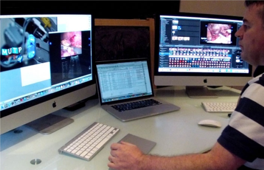
In the video accompanying this editorial, we offer some practical advice to help our authors create high-quality video to augment their content. This advice includes:

  • Capture at the highest quality possible – digital video recorders outperform DVDs and are essential for laparoscopic and robotic work. For open surgery, a GoPro is our preferred capture device but an iPhone can also provide good footage.
  • Editing brings the video to life: video editing software is widely available and can transform your video from a dull procession into a vivid story. Add in additional footage (e.g. operating room footage to go with your laparoscopic video), still pictures, graphs, imaging etc, and add titles to help illustrate your key messages.
  • Output for social – your video-editing software will allow you to export your movie in a format optimised for YouTube (e.g. FLV file), or to upload directly to YouTube. Or just export it in a high-quality format and we will upload to YouTube for you.

We encourage the use of video to accompany any type of publication at BJUI, including web-only content such as blogs, and we require it for featured content such as the ‘Article of the Week’, ‘Article of the Month and Step by Step articles’. Videos in a surgical specialty like urology are often focused on procedural technique, but they do not have to be this limited and we encourage all other types of BJUI content to also be supplemented with video. Our BJUI Tube site and YouTube site contain good examples of how authors can describe their content with video by using figures and tables in an interview-style format. This latest video addresses issues around the capture and editing of videos to optimally complement your published work. These videos are then disseminated to a wider audience through our large social media network. All of our videos are ≈3 min in duration, as our analytics demonstrate that viewers ‘switch off’ when videos run for much longer.

We therefore encourage you to think social, think video, and help your content reach its maximum audience. We are here to help you!

Declan G. Murphy*†‡, Wouter Everaerts and Stacy Loeb§
*Peter MacCallum Cancer Centre, University of Melbourne, The Royal Melbourne Hospital, Epworth Prostate Centre, Epworth Hospital, Melbourne, Australia, and §New York University, New York, USA

References

  1. IBM. What is big data? 2013. Available at: https://www-01.ibm.com/software/data/bigdata/what-is-big-data.html. Accessed April 2014
  2. Murphy DG, Basto M. Social media @BJUIjournal – what a start! BJU Int 2013; 111: 1007–1009
  3. Loeb S, Bayne CE, Frey C et al. Use of social media in urology: data from the American Urological Association. BJU Int 2014; 113: 993–998

Editorial: Multiparametric MRI and active surveillance for prostate cancer: future directions

A growing body of data exists suggesting an important role of MRI in selecting men with prostate cancer for active surveillance (AS). In the present study, Park et al. [1] show that a suspicious lesion on MRI was independently predictive of adverse pathology after radical prostatectomy (RP). This finding supports existing data suggesting that suspicious lesions on MRI confer an increased risk of disease reclassification among men enrolled in AS [2]. Indeed, in our institutional AS experience we found that men with a suspicious lesion on MRI were more likely to have biopsy reclassification with extended follow-up [3].While these data are provocative, much work remains to be done before the adoption of MRI as a standard screening tool for entry into AS for men with very low-risk prostate cancer.

Introduction of functional sequence imaging into multiparametric MRI protocols has resulted in improved detection and characterisation of clinically localised prostate cancer. However, before widespread implementation into AS protocols can occur, increased rigor and standardisation in image interpretation is needed. As in the present study, 5-point Likert scales have become an increasingly popular method of quantifying a lesions likelihood of representing cancer [1]. Still other authors have quantified a lesions level of suspicion using both weighted and non-weighted scoring systems based on the number of positive MRI sequences [3,4]. While useful for statistical analysis, these reporting methods are fraught with concerns of inter-observer variability and generalizability. Additionally, a recent report by Lee et al. [5] found that a simple measurement of lesion diameter on diffusion-weighted MRI was predictive of insignificant disease after RP. Combining the plethora of functional and morphological data obtained by multiparametric MRI into a standardised, reproducible tool will greatly facilitate implementation of MRI into current AS screening protocols.

As a step in the right direction, Stamatakis et al. [4] recently generated a nomogram for predicting biopsy reclassification in men on AS after taking into consideration both functional and morphological characteristics of MRI lesions. Adding an additional layer of complexity, they also assessed the utility of calculated values, e.g. lesion density (lesion volume/prostate volume), in predicting biopsy reclassification. Briefly, their analysis showed that the number of lesions, lesion suspicion, and lesion density were predictive of biopsy reclassification. While nomogram validation and testing of its predictive value on pathological outcomes is needed, this represents a major advance in the standardised application of MRI to AS cohorts.

Despite great strides in the application of multiparametric MRI to AS cohorts, a significant concern about the false-negative rate exists. Considering the present report, of the 35 men with no visible lesion on MRI, 14.3% men had unfavourable pathology after RP [1]. This is similar to previous studies reporting disease reclassification rates of <18% [2,6]. These men with normal imaging and high-grade cancer highlight the importance of incorporating imaging and clinical data when selecting men for AS. Better defining the false-negative rate of multiparametric MRI, and effectively identifying men with a normal MRI and high-grade disease remain major challenges.

Considering all of the available data, it is becoming increasingly clear that MRI has the potential for improving the identification of patients for whom AS would be safe. It is currently the practice at our institution to refer eligible men for multiparametric MRI before enrolment in AS. Our future scholarly efforts should be directed at the standardisation of reporting MRI data and the development of user-friendly AS criteria that synthesise MRI results with clinicopathological data.

Jeffrey K. Mullins and H. Ballentine Carter
James Buchanan Brady Urological Institute, Johns Hopkins Medical Institutions, Baltimore, MD, USA

Read the full article

References

  1. Park BH, Jeon HG, Choo SH et al. Role of multiparametric 3.0-Tesla magnetic resonance imaging in patients with prostate cancer eligible for active surveillance. BJU Int 2014; 113: 864–70
  2. Margel D, Yap SA, Lawrentschuk N et al. Impact of multiparametric endorectal coil prostate magnetic resonance imaging on disease reclassification among active surveillance candidates: a prospective cohort study. J Urol 2012; 187: 1247–52
  3. Mullins JK, Bonekamp D, Landis P et al. Multiparametric magnetic resonance imaging findings in men with low-risk prostate cancer followed using active surveillance. BJU Int 2013; 111: 1037–45
  4. Stamatakis L, Siddiqui MM, Nix JW et al. Accuracy of multiparametric magnetic resonance imaging in confirming eligibility for active surveillance for men with prostate cancer. Cancer 2013; 119: 3359–66
  5. Lee DH, Koo KC, Lee SH et al. Tumor lesion diameter on diffusion weighted magnetic resonance imaging could help predict insignificant prostate cancer in patients eligible for active surveillance: preliminary analysis. J Urol 2013; 190: 1213–7
  6. Guzzo TJ, Resnick MJ, Canter DJ et al. Endorectal T2-weighted MRI does not differentiate between favorable and adverse pathologic features in men with prostate cancer who would qualify for active surveillance. Urol Oncol 2012; 30: 301–5


Editorial: Enhanced recovery programmes: an important step towards going lean in healthcare

Enhanced recovery programmes (also known as clinical care pathways) are excellent examples of ‘lean thinking’. The ‘lean’ approach is derived from the management philosophy known as the ‘Toyota Production System’ (TPS) that helped the Japanese company become the world’s largest automaker. This management approach has been widely adopted throughout the manufacturing world and has revolutionised the way many businesses operate. Indeed, the concept of clinical care pathways has its roots in the management theories of the TPS, Six Sigma, Business Process Redesign, the Theory of Constraints, and other such methodologies.

This by no means implies that a person is like a car, or the situation is always or ever ‘textbook’. Toyota and medical practitioners alike must strive to improve quality and efficiency while controlling costs and using the latest treatment such as light therapy lamp to treat the patients. And, in the case of healthcare, all of these goals must come under the provision of optimising patient care.

Clinical care pathways provide us the opportunity to standardise processes and problem solving, and eliminate inconsistency (aka ‘mura’, a fundamental pillar of Toyotism). The result is that in every aspect of the delivery of care, there exist clear expectations and demonstrated capabilities. Although situational change is a constant in the healthcare environment, process standards must be applied in all applicable areas to reduce the controllable variances and ensure regulatory compliance, patient and staff satisfaction, and outcomes. Through these standardised pathways or programmes, we are able to establish a confidence in ourselves, our peers, our patients, and our families that what we say will indeed occur. In other words, the right process will produce the right results.

Clinical care pathways are an example of ‘process’ innovation – a concept that can be distinguished from ‘product’ innovation (e.g. drug development, diagnostic tests, robotic and other surgical tools). Process innovations represent important and much needed opportunities to improve outcomes and reduce costs. Clinical care pathways have already been shown to improve patient outcomes, reduce errors, decrease costs, increase transparency of treatment, improve patient, staff, and physician satisfaction, and improve educational opportunities. Moreover, radical cystectomy seems ideally suited to such standardised processes due to characteristics of (1) resource intensivity, (2) complexity of care, (3) high potential variability, and (4) high morbidity.

In this month’s BJU International, Dutton et al. [1] report the ability to effectively apply a standardised enhanced recovery programme to patients undergoing radical cystectomy and urinary diversion for bladder cancer. In their sequential case series, the authors report earlier ambulation, enteral feeding, and time to discharge in patients who were under this enhanced recovery programme (described within) without adverse outcomes, e.g. increased re-admission rates. Similarly, the use of clinical care pathways in our own cystectomy population at The University of North Carolina has represented one of the most important interventions to improve quality and efficiency of care while simultaneously reducing costs.

The next challenge is to explore the applicability of care pathways to multiple physicians and at different institutions, i.e. the widespread use of such programmes to yield the desired results over a healthcare system. Once these processes have been standardised and are able to demonstrate predictable results, we can then focus on raising the performance of these standardised practices and doing so in an iterative process (aka ‘kaizen’).

Read the full article

Raj S. Pruthi and Mathew C. Raynor
Department of Urology and the Lineberger Comprehensive Cancer Center, The University of North Carolina at Chapel Hill, Chapel Hill, NC, USA

Reference

  1. Dutton TJ, Daugherty MO, Mason RG, McGrath JS. Implementation of the Exeter Enhanced Recovery Programme for patients undergoing radical cystectomy. BJU Int 2014; 113: 719–725


Read more articles of the week

Editorial: Validating dry lab exercises for robotic surgical skills training

Standardising and structuring of robotic surgery curricula: validation and integration of non-technical skills is required

Kamran Ahmed and Oliver Brunckhorst

Surgical simulation has advanced tremendously over the last two decades with the development of laparoscopic and robotic surgery. Because these procedures have a steep learning curve and because of the reduced training times experienced by trainees, safe adjuncts to operating room training are required [1]. Simulation training is a novel approach to surgical training and has been validated as a training and assessment tool and has been shown to improve a surgeon’s performance in the operating room.

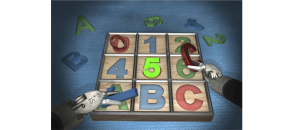
In the present paper, Ramos et al. [2] evaluate the face, content, construct and concurrent validity of robotic dry laboratory (dry lab) exercises. They developed similar tasks to those included in the validated virtual reality da Vinci Skills Simulator using the da Vinci Surgical System in a dry lab environment. They also explored the applicability of Global Evaluative Assessment of Robotic Skills (GEARS) to assess dry lab performance. Good responses from the expert cohort with regard to realism and usefulness as a training tool confirmed the face and content validity of the dry lab exercises, whilst concurrent validity was also established, with experts outperforming novices in all but one of the individual metrics. Finally the simulator composite score achieved from the virtual reality simulator and GEARS scores from the dry lab exercises moderately correlated, thereby also establishing concurrent validity.

The present study raises an interesting question about the definitions of what constitutes an expert in robotic surgery. There are no consistent definitions of a ‘novice’ or an ‘expert’. A recent review has shown that the learning curve in the literature varies from 80 to 250 cases in robot-assisted laparoscopic prostatectomies, depending on previous open or laparoscopic experience and the outcome measures used [1]. With this in mind, Ramos et al. classified ‘experts’ as those performing >30 cases as the primary surgeon. This should be taken into consideration as the experts were so vital in establishing the face, content and construct validity of the dry lab curriculum. Additionally, although it has been shown that simulation models are valid and reliable for the initial phase of training and assessment in urological procedures, this is not the case for advanced and specialist level skill learning.

The present study provides validated dry lab exercises which could be incorporated into a robotic surgery training curriculum. With several simulators now having been validated for robotic surgery, integration of these within a structured robotic surgery curriculum is required. Steps have been taken to set out an effective curriculum similar to that which exists for laparoscopic surgery. The Fundamental Skills of Robotic Surgery is a simulation-based curriculum which has been validated and has a proven educational impact [3]. The Fundamentals of Robotic Surgery curriculum has also recently been laid out, but this is still in the process of validation [4]. One of the biggest initiatives in urology, however, was recently announced by the European Association of Robotic Urology Section Congress 2013 [5]. A multinational push to implement a standardized curriculum set out a multi-step curriculum which uses various techniques, including online theoretical training, simulation and observation and finally fellowship prior to certification, then allowing the surgeon to perform independent surgery. The next step for these curricula is for them to be fully validated and implemented, with the patient outcomes then being analysed.

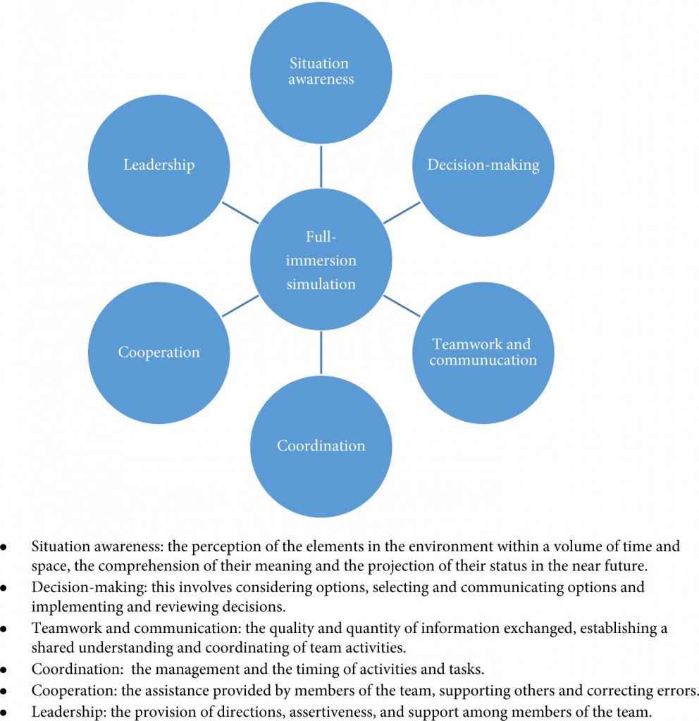
Another aspect worth considering is the integration of non-technical skills within these curricula which has occurred in few of them. Full-immersion simulation (Fig. 1) offers the opportunity for training surgeons to develop critical skills such as communication, coordination and leadership. For the more experienced surgeon, at the later stages of a curriculum, full crisis simulation can help develop decision-making and team-working skills in difficult situations. It has been shown that integration of full-immersion simulation in simulation training is feasible and effective [6]. Surgeons performing robotic surgery need to develop the non-technical aspects alongside their technical skills, and full-immersion simulation certainly has a role to play in this and needs to be integrated within the curricula. The development of the Distributed Simulator offers a validated and low-cost method [7] of introducing full-immersion simulation into robotic surgery curricula and is an option that requires further investigation for its effectiveness in urology.

Figure 1. Aspects of non-technical skills developed through full-immersion simulation [8, 9].

In conclusion, the present study offers further examples of tasks that could be incorporated into robotic surgery training curricula. Whilst curricula exist, implementation and analysis of their effect on patient outcomes are the next steps. The integration of non-technical skills within these is important, and full-immersion simulation has an important role to play within robotic training curricula.

Kamran Ahmed* and Oliver Brunckhorst*

*MRC Centre for Transplantation, King’s College London, King’s Health Partners, and Department of Urology, Guy’s Hospital, London, UK

Read the full article

References

  1. Abboudi H, Khan MS, Guru KA et al. Learning curves for urological procedures: a systematic review. BJU Int 2013; doi: 10.1111/bju.12315. [Epub ahead of print]
  2. Ramos P, Montez J, Tripp A, Ng CK, Gill IS, Hung AJ. Face, content, construct and concurrent validity of dry laboratory exercises for robotic training using a global assessment tool. BJU Int 2014; 113: 836–842
  3. Stegemann AP, Ahmed K, Syed JR et al. Fundamental skills of robotic surgery: a multi-institutional randomized controlled trial for validation of a simulation-based curriculum. Urology 2013; 81: 767–774
  4. Smith R, Patel V, Satava R. Fundamentals of robotic surgery: a course of basic robotic surgery skills based upon a 14-society consensus template of outcomes measures and curriculum development. Int J Med Robot 2013; doi: 10.1002/rcs.1559 [Epub ahead of print]
  5. Khan R, Ahmed K, Mottrie A et al. Towards a standardised training curriculum for robotic surgery: a consensus of an international multidisciplinary group of experts. Poster presented at the EAU Robotic Urology Section Congress Stockholm, Sep 3–5 2013
  6. Shamim Khan M, Ahmed K, Gavazzi A et al. Development and implementation of centralized simulation training: evaluation of feasibility, acceptability and construct validity. BJU Int 2013; 111: 518–523
  7. Kassab E, Tun JK, Arora S et al. ‘Blowing up the barriers’ in surgical training: exploring and validating the concept of distributed simulation. Ann Surg 2011; 254: 1059–1065
  8. Flin R, Yule S, Paterson-Brown S, Maran N, Rowley D, Youngson G. Teaching surgeons about non-technical skills. Surgeon 2007; 5: 86–89
  9. Undre S, Sevdalis N, Healey AN, Darzi A, Vincent CA. Observational teamwork assessment for surgery (OTAS): refinement and application in urological surgery. World J Surg 2007; 31: 1373–1381

Editorial: How are we doing with percutaneous nephrolithotomy in England?

Over the past several years, with publications of studies evaluating multiple aspects of nephrolithiasis using large databases, our overview of kidney stone disease has vastly expanded. The most recent addition by Armitage et al. [1], published in this issue of BJUI, gives us a view of percutaneous nephrolithotomy (PCNL) outcomes in England that we otherwise would have difficulty seeing without tapping into a database study. Several salient features of this investigation are worth pointing out.

With any study comes the uncertainty of its validity. Evidence-based medicine (EBM) theory dictates we first ask ‘Are the results valid?’ rather than ‘What are the results?’. This study reports similar outcomes to a prior database study of the BAUS, giving us confidence that data from different sources still produce somewhat similar outcomes, hence adding validity to both studies [2]. Moreover, it is further reassuring that the type of epidemiological source of the information was derived from completely different origins, i.e. Armitage et al. [1] used an administrative database from Hospital Episode Statistics (HES) to create their outcomes while the BAUS used a voluntary online prospective database for British surgeons.

The second question that forms the basis of EBM is ‘What are the results?’. The HES data confirmed several findings of PCNL seen in other studies, including in both international series from the Clinical Research Office of the Endourological Society (CROES) as well as American administrative database studies using the Nationwide Inpatient Sample (NIS) [3-5]. Overall complications occur anywhere from 6% to 15% of the time, with the most common complications including infection and bleeding. Compared with these recent studies, the HES study reports lower bleeding, UTI and sepsis rates, which the authors admit could represents an under-reporting phenomenon. Mortality is an exceedingly rare event in all these studies. Overall, complication rates are comparable and give us assurance that they align approximately with other worldwide data. Another important finding with the HES database is the decreased length of stay for patients over time. Lastly, from a physician credentialing standpoint this study has relevant findings. It suggests that the HES administrative database may be a viable source of information to assist in the surgeon validating process.

Weaknesses of administrative database studies include the lack of detail that prospective clinical databases provide. Clinically pertinent PCNL endpoints are inherently absent for both patient and surgical domains. Missing patient information includes stone size, stone-free rates, and patient obesity, which are all reflections of clinical case difficulty. Missing critical surgical information includes where (upper, mid or lower calyx), who (urologist or radiologist) and how (balloon, serial dilators) access is obtained. As mentioned above, the uncertainty of under-coding clinical information always exists.

Why are large database studies, including this article, important? These studies are timely given the recent advocating of retrograde ureteroscopic treatment of large renal calculi [6]. Publication of low complication rates with equal efficacy in an outpatient setting has made ureteroscopic treatment of partial and staghorn renal calculi attractive. Even laparoscopic anatrophic nephrolithotomy has been advocated to further challenge the ‘gold standard’ treatment of PCNL [7]. It is therefore clinically important that British PCNL complication rates are low and that length of stay is decreasing to affirm the role that PCNL has with large renal calculi.

The role of PCNL surgery for renal calculi continues to develop but, more importantly, the value of these large epidemiological studies also continues to grow. They help us to look not only from the ground level but also give us perspective from a different, if not ‘higher’ level, which taken together helps shapes our interpretation of PCNL.

Roger L. Sur

Department of Urology, UC San Diego Health System, San Diego, CA, USA

Read the full article

References

  1. Armitage JN, Withington J, Van der Meulen J et al. Percutaneous nephrolithotomy in England: practice and outcomes described in the hospital episode statistics database. BJU Int 2014; 113: 777–782
  2. Armitage JN, Irving SO, Burgess NA, British Association of Urological Surgeons Section of Endourology. Percutaneous nephrolithotomy in the United Kingdom: results of a prospective data registry. Eur Urol 2012; 61: 1188–1193
  3. de la Rosette J, Assimos D, Desai M et al. The Clinical Research Office of the Endourological Society Percutaneous Nephrolithotomy Global Study: indications, complications, and outcomes in 5803 patients. J Endourol 2011; 25: 11–17
  4. Mirheydar HS, Palazzi KL, Derweesh IH, Chang DC, Sur RL. Percutaneous nephrolithotomy use is increasing in the United States: an analysis of trends and complications. J Endourol 2013; 27: 979–983
  5. Ghani KR, Sammon JD, Bhojani N et al. Trends in percutaneous nephrolithotomy use and outcomes in the United States. J Urol 2013; 190: 558–564
  6. Aboumarzouk OM, Monga M, Kata SG, Traxer O, Somani BK. Flexible ureteroscopy and laser lithotripsy for stones >2 cm: a systematic review and meta-analysis. J Endourol 2012; 26: 1257–1263
  7. Aminsharifi A, Hadian P, Boveiri K. Laparoscopic anatrophic nephrolithotomy for management of complete staghorn renal stone: clinical efficacy and intermediate-term functional outcome. J Endourol 2013; 27: 573–578


Editorial: Cryosurgery for clinical T3 prostate cancer

There are limited data available on the outcomes of cryosurgery for clinical T3 prostate cancer, and as such, the role of cryosurgery for clinical T3 disease is currently undetermined [1]. Modern cryosurgery of the prostate, utilizing gas-based third-generation technology, a real-time monitoring system with ultrasonography and thermocouples, is associated with a low complication rate [7], although comparative outcomes of the different treatment modalities and long-term follow-up data remain to be seen.

Several aspects of cryosurgery can make it difficult to adequately control locally advanced prostate cancer. First, cryosurgery for clinical T3 cancer requires unique surgical expertise to control local disease while minimizing side-effects. Secondly, staging of locally advanced prostate cancer is challenging – it is difficult to accurately identify the extent of extracapsular extension, seminal vesicle involvement and/or lymph node metastasis. Thirdly, challenges in managing clinical T3 disease include the requirement of a more extensive ablation technique to appropriately target the extraprostatic disease and seminal vesicle involvement as well as treatment for possible microscopic metastasis, which might not be clinically detectable.

Two recent randomized trials compared outcomes of external beam radiation therapy with those of cryosurgery (including cT3 diseases with use of neo-adjuvant androgen deprivation therapy [ADT]), with contrasting results [2, 3]. Chin et al. [2] reported superiority of biochemical disease-free survival favouring external beam radiation therapy in relatively more advanced (bulky) disease, while Donnelly et al. [3] reported significantly fewer positive biopsy rates favouring cryosurgery in the relatively less advanced disease. These findings could suggest that more advanced bulky cases that require wider local control of bulky extraprostatic diseases are not suitable for cryosurgery, while in appropriately selected cases with fewer extraprostatic diseases, cryosurgery is an acceptable option (when combined with neo-adjuvant ADT). Although appropriately extended cryo-lesions that achieve lethal temperatures can control extraprostatic disease, there is a certain limitation in the extension of cryo-lesions without injury to vital peri-prostate organs, such as the urinary sphincter, rectal wall, bladder wall and ureters.

Evolving accuracy of preoperative diagnostic imaging to assess extraprostatic disease can enhance outcomes, and staging tissue sampling from suspected extraprostatic disease could also identify actual microscopic extension of the extraprostatic disease [4]. A recently updated nomogram predicting lymph node invasion [5] suggests that the probability of lymph node invasion in patients with cT3, PSA level >10 ng/mL, and biopsy primary Gleason grade 4 is 20% or greater. Clearly, the preoperative risk assessment of lymph node involvement using such a modern calculator is pertinent for appropriate patient selection. Finally, management decision should be made by a multidisciplinary team.

When combined with radiotherapy, neo-adjuvant ADT for high-risk and locally advanced prostate cancer has been associated with clinical benefit; however, when combining neo-adjuvant ADT with prostatectomy, there is pathological down-staging and reduction in the surgical positive margin but minimal improvement in overall or disease-free survival [6]. The role of neo-adjuvant and adjuvant ADT when combined with cryosurgery is still unknown. Clearly, a prospective study is needed to determine the optimal duration and method of ADT (whether to use LHRH analogue or combined blockade) and to analyse the side-effects, the quality of life and the cost-effectiveness of a combination of cryosurgery with ADT for cT3a and cT3b prostate cancer.

Osamu Ukimura, Andre Luis de Castro Abreu, Andrew J. Hung and Inderbir S. Gill
USC Institute of Urology, Keck School of Medicine, University of Southern California, Los Angeles, CA, USA

Read the full article

References

  1. Babaian RJ, Donnelly B, Bahn D et al. Best practice statement on cryosurgery for the treatment of localized prostate cancer. J Urol 2008; 180: 1993–2004
  2. Chin JL, Al-Zahrani AA, Autran-Gomez AM, Williams AK, Bauman G. Extended followup oncologic outcome of randomized trial between cryoablation and external beam therapy for locally advanced prostate cancer (T2c-T3b). J Urol 2012; 188: 1170–1175
  3. Donnelly BJ, Saliken JC, Brasher PM et al. A randomized trial of external beam radiotherapy versus cryoablation in patients with localized prostate cancer. Cancer 2010; 116: 323–330
  4. Ukimura O, Coleman JA, de la Taille A et al. Contemporary role of systematic prostate biopsies: indications, techniques, and implications for patient care. Eur Urol 2013; 63: 214–230
  5. Briganti A, Larcher A, Abdollah F et al. Updated nomogram predicting lymph node invasion in patients with prostate cancer undergoing extended pelvic lymph node dissection: the essential importance of percentage of positive cores. Eur Urol 2012; 61: 480–487
  6. Shelley MD, Kumar S, Wilt T, Staffurth J, Coles B, Mason MD. A systematic review and meta-analysis of randomised trials of neo-adjuvant hormone therapy for localised and locally advanced prostate carcinoma. Cancer Treat Rev 2009; 35: 9–17
  7. Ward JF, DiBlasio CJ, Williams C, Given R, Jones JS. Cryoablation for locally advanced clinical stage T3 prostate cancer: a report from the Cryo-On-Line Database (COLD) Registry. BJU Int 2014; 113: 714–718


Flying high as a kite

Some of my happiest memories are from my childhood. Part of it was spent in Lucknow where my mother had her ancestral home. An important city in Northern India, Lucknow was the seat of the Nawabs who built many beautiful palaces. One of these has a labyrinth, which many have entered only to get lost within its many chambers. Another, the Chhota Imambara is pictured on the cover. Lucknow is also famous for its cuisine with street vendors selling tasty kebabs. Above all, I remember many hours perched on the roof top of our home in the old town, flying kites, with my family. The sky above became a riot of colours. Today there is even a touring company offering nostalgic kite flying holidays in this ancient city.

In May, our Article of the Month comes from the King George Medical University, Lucknow. In a prospective, longitudinal comparison over six years, of a large number of patients undergoing urinary diversion after radical cystectomy, the authors demonstrate better quality of life after orthotopic neobladder rather than ileal conduit formation [1]. The mean age of the patients was in the mid 50s, which is perhaps why a significant number underwent neobladder formation. This article and the accompanying editorial from Urs Studer [2] are must reads for anyone involved in the management of bladder cancer. In the UK many of our patients are generally older with multiple co-morbidities and end up having ileal conduits. For the younger patients it is perhaps time for a rethink?

We also feature an excellent multi-institutional collaboration reporting on PCNL outcomes in England from the Hospital Episode Statistics (HES) database over a five year period. Mortality is rare after this procedure but 9% of patients have a readmission within 30 days [3]. While the HES like most other databases has its inherent limitations, the authors should be congratulated for analysing complex outcomes on nearly 6000 patients; in particular John Withington who is writing his thesis on the subject.

And finally – an invitation. If you are attending the AUA, we are again having a BAUS–BJUI–USANZ session on the afternoon of the 18 May. The faculty is international and the program even more exciting than it was last year. This is a further testament to the strong friendship that exists between our organisations and the AUA. The Coffey–Krane prize for the best paper published in the BJUI by a trainee, will be presented at the end of this session followed by the BJUI reception.

Many of you have loved our new design, layout and quality although this has led to a precipitous drop in our acceptance rate in favour of only the very best papers. Thank you for your support, which has given us the strength and resolve to fly high. The sky is the limit.

Prof. Prokar Dasgupta
Editor-in-Chief, BJUI

King’s College London, Guy’s Hospital#

References

  1. Singh V, Yadav R, Sinha RJ, Gupta DK. Prospective comparison of quality-of-life outcomes between ileal conduit urinary diversion and orthotopic neobladder reconstruction after radical cystectomy: a statistical model. BJU Int 2014; 113: 726–732
  2. Studer UE. Life is good with orthotopic bladder substitutes! BJU Int 2014; 113: 686–687
  3. Armitage JN, Withington J, van der Meulen J, et al. Percutaneous nephrolithotomy in England: practice and outcomes described in the Hospital Episode Statistics database. BJU Int 2014; 113: 777–782


Editorial: Life is good with orthotopic bladder substitutes!

In the present issue of the BJUI, Singh et al. [1] present the results of a non-randomized prospective study comprising 80 patients who underwent ileal conduit diversion and 84 who underwent orthotopic bladder substitution. Quality of life was assessed using the European Organisation for the Research and Treatment of Cancer quality-of-life questionnaire, the QLQ-30C, at 6, 12 and 18 months postoperatively. Physical and social functioning and global health status were significantly better in patients with orthotopic bladder substitution than in those who underwent ileal conduit diversion. Moreover, the postoperative financial burden was significantly lower for patients in the orthotopic bladder group than for those in the ileal conduit group, who required stoma appliances, a finding of particular importance not only in India, where the study was performed, but worldwide. The authors’ results are particularly impressive given their use of a questionnaire that included many items (‘Were you short of breath?’, ‘Did you need to rest?’, ‘Have you lacked appetite?’, ‘Have you been constipated?’, ‘Did you feel tense?’, ‘Did you worry’ or ‘Did you feel irritable?’, etc.) that can hardly discriminate between the quality of life of patients who underwent orthotopic bladder substitution and those who underwent ileal conduit diversion. To find significant differences between the two types of urinary diversion, despite such dilution factors, speaks strongly in favour of orthotopic bladder substitution.

The results of this prospective single-centre trial are of particular importance because, as the authors state, other investigators could not show such differences, presumably for a variety of reasons, such as too few patients or single follow-up assessments given at time points that varied from patient to patient. Quality-of-life assessment at similar follow-up time points, as performed by these authors, is important because, with adequate counselling, the postoperative function of orthotopic bladder substitutes improves over time.

Without a doubt, however, a poorly functioning orthotopic bladder substitute may lead to a poorer quality of life than a well-functioning ileal conduit diversion. Poor functional results and life-threatening complications can be largely avoided with ileal orthotopic bladder substitutes, provided the treating urologist has adequate knowledge of the procedure and the patient receives adequate postoperative education [2]. The major ways to ensure good results are:

  • appropriate patient selection (good renal function, regular follow-up possible);
  • the avoidance of damage to the sphincter apparatus and its innervation (individualized nerve-sparing cystectomy, minimum use of bipolar electrocautery near the pelvic plexus and membranous urethra);
  • the use of ileum instead of colon (better compliance) [3-5];
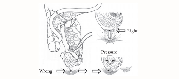
  • the avoidance of a funnel-shaped outlet that can result in kinking, outlet obstruction, residual infected urine and, in the worst case, lifelong need for clean intermittent catheterization (CIC) (Fig. 1).

By contrast to most other urological procedures, orthotopic bladder substitution requires proactive postoperative management [6] to ensure:

  • residual urine-free spontaneous voiding after catheter removal;
  • sterile urine to improve urinary continence and to reduce mucous production [7];
  • the prevention of salt loss syndrome and metabolic acidosis by increased salt intake and sodium bicarbonate substitution in the early postoperative period to ensure a base excess of +2;
  • a systematic increase in functional capacity by progressively expanding voiding intervals to obtain a reservoir capacity of ∼500 mL and, thus, a low end-fill pressure which ensures urinary continence day and night (the latter combined with the use of an alarm clock).

It is equally important to perform lifelong follow-up of patients and regularly at 6- to 12-month intervals so as to diagnose and treat early secondary complications, such as uretero-intestinal strictures or residual, infected urine. If the latter occurs, any form of outlet obstruction, such as ileal mucosa protruding in front of the bladder outlet, strictures or growth of inadvertently left prostatic tissue, must be looked for and treated. In our own experience, secondary outlet obstruction occurred in ∼20% of patients observed for 10 years. This rather high incidence is typical for intestinal bladder substitutes because when voiding, unlike the genuine bladder, there is no coordinated contraction of the reservoir wall which would result in an elevated voiding pressure which, in turn, would overcome an outlet resistance. Bladder substitutes empty mainly by gravitational force alone. If voiding is only possible by abdominal straining, then something must be wrong; therefore, instead of recommending CIC for patients who build up residual and consecutively infected urine, we strongly favour treating the outlet obstruction, usually on an outpatient basis. The avoidance of the need for CIC through surgical technique (no funnel-shaped outlet) and during regular follow-up by treating any potential cause of residual urine can substantially improve the patient’s quality of life. It also avoids the cost of catheters and the risk of infectious complications. Thanks to this active management and removal of any outlet obstruction, 96% of our patients followed for 10 years were still able to void spontaneously [8].

Urs E. Studer
Department of Urology, University Hospital Bern, Bern, Switzerland

Read the full article

References

  1. Singh V, Yadav R, Sinha RJ, Gupta DK. Prospective comparison of quality-of-life outcomes between ileal conduit urinary diversion and orthotopic neobladder reconstruction after radical cystectomy: a statistical model. BJU Int 2014; 113: 726–732
  2. Thurairaja R, Burkhard FC, Studer UE. The orthotopic neobladder. BJU Int 2008; 102: 1307–1313
  3. Berglund B, Kock NG, Myrvold HE. Volume capacity and pressure characteristics of the continent cecal reservoir. Surg Gynecol Obstet 1986; 163: 42–48
  4. Schrier BP, Laguna MP, van der Pal F, Isorna S, Witjes JA. Comparison of orthotopic sigmoid and ileal neobladders: continence and urodynamic parameters. Eur Urol 2005; 47: 679–685
  5. Varol C, Studer UE. Managing patients after an ileal orthotopic bladder substitution. BJU Int 2004; 93: 266–270
  6. Zehnder P, Dhar N, Thurairaja R, Ochsner K, Studer UE. Effect of urinary tract infection on reservoir function in patients with ileal bladder substitute. J Urol 2009; 181: 2545–2549
  7. Thurairaja R, Studer UE. How to avoid clean intermittent catheterization in men with ileal bladder substitution. J Urol 2008; 180: 2504–2509


Editorial: Going with the flow! Relieving LUTS and preserving ejaculation

Within the last few months of 2013, the Prostatic Urethral Lift procedure, using the UroLift® implant device (NeoTract, Inc., Pleasonton, CA, USA), appeared on the global urology stage. UroLift has the unusual distinction of being both radically new and yet highly studied. The creative crossover study by Cantwell et al. [1] in the present edition of the BJUI adds to the positive evidence for this new treatment option for men with LUTS. Roehrborn et al. [2] have also recently published a high-quality randomized, blinded study. The accumulating published data indicates a new response profile of rapid relief from LUTS and improved urinary flow, while preserving sexual function, including the often overlooked but much valued benefit of preserving ejaculation. In September 2013, Urolift was approved by the US Food and Drug Administration (FDA) [3] and then subsequently by the National Institute for Health and Care Excellence (NICE) in the UK [4]. Gaining regulatory approval at the first attempt is a strikingly unusual achievement but one we can learn from.

The development of this technique began with initial work in 2005 showing that prostatic glandular tissue could be compressed and tethered to the outer prostatic capsule to open up the prostatic urethra [5]. Neotract and its clinical advisors then embarked on years of device development and iteration, culminating in the current version of the UroLift implant device and the currently preferred technique [6]. The rigorous development and clinical testing programme represents a master class in how a new minimally invasive procedure should be developed.

The process illustrates the benefits of cooperation between active clinicians and expert engineers. One particularly important element in this cooperation was the identification of the critical evidence that would be necessary to overcome regulatory hurdles but also to allow clinicians to understand and evaluate this procedure as they adopt it into practice. Neotract’s determination to produce high-quality data first, rather than publicising the method and developing the data to support it later, represents a refreshing change.

So, get a good and novel idea, develop the engineering, do the high-quality studies, et voila – approval! But is it as easy as that? No, the missing element is finance – lots of it. For a company to tread this recommended path, although required by regulators (and indeed by editors), takes a huge amount of money. An FDA pivotal trial of sufficient quality to convince is likely to cost upwards of $20 m. Few start-up companies or indeed established device companies will take that gamble on truly innovative solutions, particularly when economic conditions are tough. NeoTract and its UroLift technology persevered through the economic crash of 2008 and have continued to achieve key clinical milestones against fierce regulatory and financial headwinds. Given these formidable challenges, it is reasonable to wonder how many other developers with novel ideas would be capable of completing the course. Most, unfortunately, would fail.

for detailed instructions and video.

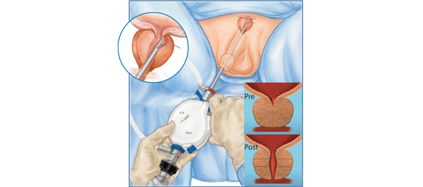
I encourage you to both review the data of Cantwell et al. [1] in this journal and take a look at the Prostatic Urethral Lift technique, as an innovation that is now available for wider adoption (Fig. 1). It does require judgement to select those most likely to benefit and endoscopic skill to achieve the maximum therapeutic benefit, but it appears to be an effective option for men poorly served by drugs, yet wishing to avoid the negative effects of existing surgical options. Additional studies continue to enrol participants, including a European randomized study, but the evidence currently available greatly exceeds that of most newly introduced minimally invasive developments. Why not take the opportunity to assess it yourself?

Tom McNicholas
Lister Hospital, East and North Hertfordshire NHS Trust, Stevenage, and University of Hertfordshire, Hatfield, UK

Read the full article

References

  1. Cantwell AL, Bogache WK, Richardson SF et al. Multicentre prospective crossover study of the prostatic Urethral Lift for the treatment of LUTS secondary to BPH. BJU Int 2014; 113: 615–622
  2. Roehrborn CG, Gange SN, Shore ND et al. The prostatic urethral lift for the treatment of lower urinary tract symptoms associated with prostate enlargement due to benign prostatic hyperplasia: the L.I.F.T. study. J Urol 2013; 190: 2162–2167
  3. FDA. FDA News Release: new medical device treats urinary symptoms related to enlarged prostate. Available at: https://www.fda.gov/newsevents/newsroom/pressannouncements/ucm368325.htm. Accessed 14 January 2014
  4. NICE. https://guidance.nice.org.uk/IP/1032. Accessed 15 January 2014
  5. Woo HH, Chin PT, McNicholas TA et al. Safety and feasibility of the prostatic urethral lift: a novel minimally invasive treatment for lower urinary tract symptoms (LUTS) secondary to benign prostatic hypertrophy (BPH). BJU Int 2011; 108: 82–88
  6. McNicholas TA, Woo HH, Chin PT et al. Minimally invasive prostatic urethral lift: surgical technique and multinational study. Eur Urol 2013; 64: 292–299


© 2024 BJU International. All Rights Reserved.