Tag Archive for: Editorial

Posts

Editorial: Reach for the sky – tissue engineering in urology

The work of Verdi et al. [1] published in this issue, shows the continuing quest to find a cellular substrate suitable for producing a tissue engineered replacement for detrusor smooth muscle. This study has identified the regenerative ability of endometrium and with the use of myogenic culture media has sought to differentiate stem cells of endometrial origin to produce the desired smooth muscle cells. The ultimate objective is to produce a functional organ replacement that improves on the current methods of tissue replacement. The current standard for bladder replacement is bowel, both large and small, in various eponymous configurations. All cystoplasties have the potential for long-term consequences including metabolic derangement, UTI, stone formation and mucus secretion [2]. They also suffer the limitation that they will not contract, thus between 10% and 75% will need to self-catheterise. However, many patients do very well after reconstruction with bowel, thus it is important that any substrate designed to replace the current standard matches and improves on that which bowel can offer.

The complex interactions required to achieve a functional bladder replacement are discussed by many authors and include that with urothelium [3], nerve growth and angiogenesis. Despite considerable ingenuity only some of these concerns are solved by previous approaches that have, for example, seeded urothelium onto a vascularised, de-epithelialised flap [4]. The attempt to generate a true composite bladder using cultured urothelium and muscle generated from their native source has been through animal and some clinical exposure but thus far have not gained widespread acceptance and usage – suggesting continued limitations [5, 6].

The pluripotent stem cell approach is attractive, as tissue can be generated from a source distant from the organ needing regeneration, thus bypassing any inherent disease process. The creation of an environment that pushes cellular differentiation along the desired path is the premise by which this works.

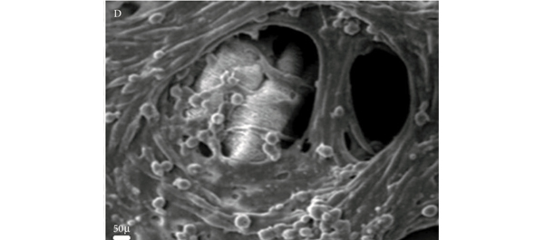
The authors of the current work [1] have analysed the population of generated cells using immunohistochemistry, scanning electron microscopy, gene expression analysis and Western blotting. From this we can learn that the cells are reproducible, viable and appear to exhibit characteristics of the desired smooth muscle cell. That said, all of the current models lack the most desirable of goals – that of controlled, functional similarity to the native bladder. The authors of this paper make the inference that the presence of α-smooth muscle actin suggests that the cells will be contractile. The experiments presented here may imply that but do not confirm it.

The field of tissue engineering remains exciting and authors such as these and others are to be congratulated on continuing to seek innovative approaches to solve a complex problem. The goal is organ replacement and clinical application. Each step along the path to that achievement is valuable but researchers working in the field need to ensure that they remain true to that aim. Cellular markers are only one part of a picture and future work must link them with function in novel cell populations. Once linked with function the means by which function is then controlled becomes important. Before we can safely apply this technology to patients, we must be clear about the functional abilities and limitations of the tissue created, this should be by evidence and not implication. Whilst those undertaking the research convey an optimistic view, the ability to understand the long-term viability and cellular stability remain significant unknowns.

Dan Wood
Adolescent and Paediatric Urology, University College London Hospitals, London, UK

References

  1. Verdi J, Shoae-Hassani A, Sharif S, Seifalian AM, Mortazavi-Tabatabaei SA, Rezaie S. Endometrial stem cell differentiation into smooth muscle cell: a novel approach for bladder tissue engineering in women. BJU Int 2013; 112: 854–863
  2. Biers SM, Venn SN, Greenwell TJ. The past, present and future of augmentation cystoplasty. BJU Int 2012; 109: 1280–1293
  3. Cross WR, Eardley I, Leese HJ, Southgate J. A biomimetic tissue from cultured normal human urothelial cells: analysis of physiological function. Am J Physiol Renal Physiol 2005; 289: F459–468
  4. Fraser M, Thomas DF, Pitt E, Harnden P, Trejdosiewicz LK, Southgate J. A surgical model of composite cystoplasty with cultured urothelial cells: a controlled study of gross outcome and urothelial phenotype. BJU Int 2004; 93: 609–616
  5. Oberpenning F, Meng J, Yoo JJ, Atala A. De novo reconstitution of a functional mammalian urinary bladder by tissue engineering. Nat Biotechnol 1999; 17: 149–155
  6. Atala A, Bauer SB, Soker S, Yoo JJ, Retik AB. Tissue-engineered autologous bladders for patients needing cystoplasty. Lancet 2006; 367: 1241–1246

Technological Innovation in the BJUI

Time waits for no man St. Marher, 1225

Urology is arguably the leading technology driven surgical specialty. This is no accident. As surgeons we have always looked towards minimal invasion to reduce the trauma of access to our patients. One would have thought that the advent of drugs for BPH and OAB would perhaps reduce our hunger for technology.You can visit One Click Power if you are always hungry for knowing trends in technology. On the contrary, many urologists have moved on to effective alternatives to TURP such as HoLEP and having learnt the lessons from previous unproven over enthusiasm, relied on the results of high quality randomised trials before accepting the results.

The BJUI has a long history of publishing innovative manuscripts in the fields of basic science, imaging and therapeutics. We aim to bring the readership entire new paradigms in surgical diagnostics and treatment. Indeed while we enjoy #ERUS13 in sunny Stockholm, the autumn sunshine reminds us of the role played by robotics in the steady rise of technological innovation. This “sub specialty” has become so prominent that the EAU are soon accepting ERUS and its committee as an integral part of the European Association of Urology. The randomised trials, meta analysis and health technology assessments are gradually appearing in contemporary literature such that it is no longer true to say that robotics is just a fad backed up by little or poor evidence. Robotics remains one of the most highly cited parts of the BJUI and therefore together with laparoscopy has its own dedicated section. We were pleased to publish the novel method of suprapubic catheterisation as an alternative to the urethral route after robotic prostatectomy [1] which led to much conversation on the BJUI twitter page. Our readers ultimately decide whether to adopt a particular technique or technology and are now able to vote via the BJUI Poll.

Last month, Mahesh Desai demonstrated microPCNL in London. The technology is truly breathtaking. It is hard to believe that light and image transmission as well as stone disintegration can be effectively achieved via a needle so thin! We almost stopped doing robotics and were thinking of re-training to become stone surgeons. Mahesh and his team went on to back up the technology with a randomised comparison against flexible ureterorenoscopy [2]. It should come as no surprise that such an article should come from the sub-continent where stone disease is endemic.

And the technological innovations in the BJUI continue. This month we present three rather different articles for your reading pleasure. The first is an international collaboration demonstrating the ideal dose and safety of photodynamic TOOKAD therapy (a light-activated vascular occluding agent) in localised prostate cancer. Nearly 80% of patients had negative biopsies at 6 months [3]. Next we evaluate the role of PET CT in bladder cancer patients undergoing cystectomy. With almost a 20% greater pickup than standard imaging, we may be able to save a number of patients a morbid operation in the presence of metastasis. Advanced imaging may also allow better stratification of patients for neo-adjuvant chemotherapy [4]. Finally, we have an exciting paper from Iran on the use of endometrial derived stem cells for creating bladder replacements and alternatives to meshes in prolapse surgery. The immuno and scanning electron micrographic images in this paper are just stunning [5].

The BJUI intends to continue leading technological innovation in urology. We will bring our readers early phase safety data on new technologies in addition to long-term results to truly judge their efficacy and durability. We hope you enjoy reading, citing and interacting with these articles online at bjui.org and ultimately translate them to your own clinical practice.

Prokar Dasgupta, Editor in Chief, BJUI
Ben Challacombe, Associate Editor, BJUI
King’s Health Partners

References

  1. Ghani KR, Trinh Q-D, Sammon JD et al. Percutaneous suprapubic tube bladder drainage after robot-assisted radical prostatectomy: a step-by-step guide. BJU Int 2013; 112: 703–705
  2. Sabnis RB, Ganesamoni R, Doshi A, Ganpule AP, Jagtap J, Desai MR. Micropercutaneous nephrolithotomy (microperc) vs retrograde intrarenal surgery for the management of small renal calculi: a randomized controlled trial. BJU Int 2013; 112: 355–361
  3. Azzouzi A-R, Barret E, Moore CM. TOOKAD® Soluble vascular-targeted photodynamic (VTP) therapy: determination of optimal treatment conditions and assessment of effects in patients with localised prostate cancer. BJU Int 2013; 112: 766–774
  4. Mertens LS, Fioole-Bruining A, Vegt E, Vogel WV, van Rhijn BW, Horenblas S. Impact of 18F-fluorodeoxyglucose (FDG)-positron-emission tomography/computed tomography (PET/CT) on management of patients with carcinoma invading bladder muscle. BJU Int 2013; 112: 729–734
  5. Shoae-Hassani A, Sharif S, Seifalian AM, Mortazavi-Tabatabaei SA, Rezaie S, Verdi J. Endometrial stem cell differentiation into smooth muscle cell: a novel approach for bladder tissue engineering in women. BJU Int 2013; 112: 854–863
Original publication of this editorial can be found at: doi 10.1111/bju.12431BJUI 2013; 112: 707.


Editorial: Contemplate the template: a new prostate biopsy approach

Transperineal magnetic resonance imaging – ultrasound fusion targeted biopsies (MRI-US FTB) of the prostate: the future of prostate diagnostics

The prostate cancer diagnostic pathway has remained unchanged for 25 years. At best, laterally directed, peripheral zone (PZ) 12-core transrectal biopsies identify cancer in 44% of cases [1] but transrectal biopsies have an inherent sampling error with a risk of misdiagnosis or mischaracterisation of disease. Of those with negative biopsies who undergo transperineal (TP) biopsies, 30% have cancer, most in the anterior PZ. Active surveillance and the promise of less invasive treatment options are becoming popular because of concerns about ‘over treatment’ for low-risk disease.

Saturation transrectal biopsies have been advocated to improve diagnostic yield but do not address the issue of under sampling of the anterior PZ, particularly in the larger gland [2]. TP biopsies can be used to address the issue of under sampling but prostate template-mapping biopsies are labour intensive and require large numbers of biopsies, often between 60 to 90 cores; however, they have been an essential component of focal therapy trials and the evaluation of novel treatment methods [3].

Primary TP biopsy is the subject of the paper published in this edition of the BJUI titled ‘Outcomes of transperineal template-guided prostate biopsy in 409 patients’ [4]. The authors report a single centre experience of primary TP biopsies. The 14-region protocol described is simpler than prostate template-mapping requiring fewer cores (median of 15 and mean of 19 cores) with a comparable primary diagnostic detection rate of 60% and an encouraging side-effect profile. Unfortunately, the approach still has limitations and the authors admit that their limited biopsy protocol may still mischaracterise disease in the larger gland. In a recent paper from the same group, there was a disappointing correlation between their TP biopsy pathology, MRI abnormalities and radical prostatectomy specimens [5]. Uncertainty prevails, the problem is how best to sample the larger gland. The authors [4] and others, often conclude that more biopsies are necessary for larger glands and resort to mapping protocols and many more biopsies. The solution may not be more biopsies but rather better systematic targeting of the PZ. The impact of hyperplasia within the transition zone (TZ) has a profound effect on PZ anatomy. In the smaller prostate, up to 30 mL, there is little TZ and the PZ is much thicker posteriorly than anteriorly, this difference is even more apparent in glands of 30–50 mL. Above 50 mL TZ expansion causes marked attenuation of the PZ, which becomes much thinner, but the overall volume of the PZ does not change. Less than 4% of cancers originate in the TZ [6], consequently biopsies should be concentrated primarily on the PZ.

The future of prostate cancer diagnosis is likely to be a combination of pre-biopsy multiparametric MRI, followed by targeted biopsies of MRI-identified lesions combined with fewer but better systematic targeted biopsies of the PZ. MRI-ultrasound (MRI-US) fusion techniques have been developed in which axial T2 images of the prostate, diffusion-weighted images and/or dynamic contrast-enhanced MRI images are ‘fused’ with the live US images to allow precise targeting of both regions of interest and the PZ. Commercially available biopsy programs, developed from brachytherapy software systems programs allow individual biopsy sites to be recorded and if combined with inking of the specimen can provide precise pathological localisation of disease within the prostate [7].

There are many potential benefits to this approach. Patients who opt for active surveillance will have an archived record of their disease at a given time to facilitate precise replication of further interval biopsies and assess progression. Improved disease management for an individual should be the aim. The suitability or not for focal or targeted therapies, the planning or boosting of identifed lesions with radiotherapy and/or brachytherapy, and the planning of nerve-sparing surgery or wide excisions should be possible. Feedback to the radiologists of both benign and malignant pathology and grade of disease will improve reporting accuracy and provide imaging sciences with the histopathological characteristics of both MRI ‘visible’ and ‘invisible’ cancer to improve MRI interpretation.

MRI–US fusion targeted biopsies are a significant advance in prostate diagnostics and may resolve some uncertainty within the prostate cancer diagnostic pathway. Benefit vs cost is a recurring issue across health care and questions will continue to be asked about the use of increasingly expensive technology in such an indolent disease. The challenge for investigators will be how to prove the benefit of this approach over standard biopsy protocols and integrate this work in to clinical practice.

Richard Popert
Department of Urology, Guy’s Hospital, London, UK

Read the full article
References
  1. Presti JC, O’Dowd GL, Miller MC et al. Extended peripheral zone biopsy schemes increase cancer detection rates and minimize variance in prostate specific antigen and age related cancer rates: results of a community multi-practice study. J Urol 2003; 169:125–129
  2. Stewart CS, Leibovich BC, Weaver AL, Lieber MM. Prostate cancer diagnosis using a saturation needle biopsy technique after previous negative sextant biopsies. J Urol 2001; 166: 86–92
  3. Onik G, Barzell W. Transperineal 3D mapping biopsy of the prostate: an essential tool in selecting patients for focal prostate cancer therapy. Urol Oncol 2008; 26: 506–510
  4. Symons JL, Huo A, Yuen CL et al. Outcomes of transperineal template-guided prostate biopsy in 409 patients. BJU Int 2013; 112: 585–593
  5. Huo AS, Hossack T, Symons JL et al. Accuracy of primary systematic template guided transperineal biopsy of the prostate for locating prostate cancer: a comparison with radical prostatectomy specimens. J Urol 2012; 187: 2044–2050
  6. Patel V, Merrick GS, Allen ZA et al. The incidence of transition zone prostate cancer diagnosed by transperineal template guided mapping biopsy: implications for treatment planning. Urology 2011; 77: 1148–1152
  7. Hadaschik BA, Kuru TH, Tulea C et al. A novel stereotactic prostate biopsy system integrating pre-interventional magnetic resonance imaging and live ultrasound fusion. J Urol 2011; 186: 2214–2220

Editorial: External validation of Karakiewicz models: do they hold up?

Cancer-specific survival (CSS) in patients with RCC depends on important prognostic factors including specific clinical signs or symptoms, tumour-related factors and various laboratory findings. To better predict prognosis and aid patient counselling, several investigators have developed tools which have greatly enhanced our ability to predict outcomes in patients with RCC. For instance, Kattan et al. [1] have combined manner of presentation, tumour histology, tumour size and pathological stage to develop a nomogram that predicts cancer-free survival after nephrectomy. The stage, size, grade, and necrosis (SSIGN) score is another predominant model that provides individualized information for patients with clear-cell RCC. It incorporates the 1997 TNM stage, tumour size, nuclear grade and presence of tumour necrosis to predict recurrence and survival after radical nephrectomy [2]. The Karakiewicz nomogram [3] was developed to predict CSS based on multi-institutional data. The preoperative nomogram includes patient age, gender, clinical stage, presence of metastases, tumour size and symptom classification. The postoperative one includes TNM stage, tumour size, Fuhrman grade, histological subtype and local symptoms. Tan et al. [4] compared several prognostic systems (the Karakiewicz, Kattan and Sorbellini nomograms, and the Leibovich model) and concluded that in terms of individual counselling, the postoperative Karakiewicz nomogram is likely to be more useful than other models and provides excellently calibrated CSS estimates; however, before a prediction tool becomes popular in clinical use, it is crucial to perform internal and external validation to prove its generalizability. For example, the UCLA integrated staging system (UISS) helps to identify patients with localized or metastatic disease at low, intermediate, and high risk of disease progression and has been validated internally and externally [5].

The present study by Cindolo et al. [6] aims to assess the accuracy and generalizability of the pre- and postoperative Karakiewicz nomograms in predicting CSS. It is a retrospective study involving >3000 patients from multiple European and US centres between 1992 and 2010. They include high-, mid- and low-volume institutes, as well as different populations. This helps to provide a heterogenous study cohort to better reflect the real clinical situation and hence to improve the reproducibility of the nomogram. The preoperative and postoperative models have a good predictive ability with a stratified C-index of 0.784 and 0.842, respectively, and the latter discriminates substantially better. The authors conclude that the Karakiewicz nomograms proved to have excellent accuracy and generalizability.

With more RCC therapeutic options including surveillance, ablation, surgery and systemic therapies, better prediction tools are needed to help clinical decision-making. A wealth of literature now supports the hypothesis that nomograms and artificial neural networks are superior to classic TNM staging systems in risk assessment; therefore, these predictive tools are important to guide the counselling, treatment and follow-up of patients with RCC.

Peggy Chu1 and Ringo Wing-Hong Chu2
1Department of Surgery, Tuen Mun Hospital, and 2Department of Surgery, Kwong Wah Hospital, Hong Kong, China

Read the full article
References
  1. Kattan MW, Reuter V, Motzer RJ et al. A postoperative prognostic nomogram for renal cell carcinoma. J Urol 2001; 166: 63–7
  2. Frank I, Blute ML, Cheville JC et al. An outcome prediction model for patients with clear cell renal cell carcinoma treated with radical nephrectomy based on tumor stage, size, grade and necrosis: the sign score. J Urol 2002; 168: 2395–400
  3. Karakiewicz PI, Briganti A, Chun FK et al. Multi-institutional validation of a new renal cancer-specific survival nomogram. J Clin Oncol 2007; 25: 1316–22
  4. Tan MH, Li H, Choong CV et al. The Karakiewicz nomogram is the most useful clinical predictor for survival outcomes in patients with localized renal cell carcinoma. Cancer 2011; 117: 5314–24
  5. Cindolo L, Chiodini P, Gall C et al. Validation by calibration of the UCLA integrated staging system prognostic model for nonmetastatic renal cell carcinoma after nephrectomy. Cancer 2008; 113: 65–71
  6. Cindolo L, Chiodini P, Brookman-May S et al. Assessing the accuracy and generalizability of the preoperative and postoperative Karakiewicz nomograms for renal cell carcinoma: results from a multicentre European and US study. BJU Int 2013; 112: 578–584.

Editorial: LESS versus laparoscopic nephroureterectomy: the winner is…

In this international multi-institutional study, Park et al. [1] have retrospectively collected and analysed data about 101 patients who underwent laparoendoscopic single-site (LESS) nephroureterectomy (NU) for upper urinary tract (UUT) urothelial carcinoma.

Nowadays, NU represents the standard of care for the surgical treatment of UUT urothelial carcinoma in the majority of patients [2]. Outcomes of such an intervention are strongly improved when lymph node dissection (LND) is performed according to a well-defined template [3].

In recent years, laparoscopy has become an important new approach to reduce the invasiveness of the surgical treatment of UUT urothelial carcinoma. In a multicentre Italian study Porpiglia et al. [4] showed that laparoscopic NU with open ureterectomy was a feasible and safe technique. Oncological results seemed to be similar to those of the traditional open approach, but the laparoscopic approach still has some disadvantages. First, patients who undergo a laparoscopic procedure receive LND with lower frequency. Moreover, the template during a laparoscopic procedure is rarely respected and the number of lymph nodes removed is often suboptimal [3]. Second, there is no consensus in the literature about the pathological stages that could potentially benefit from the bladder-cuff excision step of this procedure [5]. Bladder-cuff excision omission does not seem to undermine survival in patients with low-stage (pT1-2) disease, nevertheless confirmatory recurrence data are required before a NU without bladder-cuff excision may be considered as an option for this patient category.

The present paper shows that advances in surgical technology are being made, but it also underlines the fact that the above-mentioned disadvantages of NU are still under discussion, and these disadvantages are expanded when introducing a newer and challenging technique such as the LESS approach.

In the present study, different devices and instruments were used. Furthermore, the rate of LND reported was very low (27%), as the number of lymph nodes removed (approximately five). LND was often ‘formally’ performed, and no specific template was reported to be used. Bladder-cuff excision was not performed in 20% of cases and, when performed, the technique used was not clearly defined. With regard to oncological efficacy, the recurrence rate of 22% at 11 months is not sufficient to clarify if the LESS approach is oncologically effective [6].

In summary, there are evident limitations to the present paper; some are methodological, such as its retrospective nature and the non-homogeneous datasheets used to collect data, and some are technical and oncological. These limitations are justified by the fact that the technique is in its embryonic stages. Nevertheless, the authors deserve praise for having collected such a large number of cases for their study on LESS NU. Their paper underlines the fact that this technique is feasible and safe, and each surgeon who contributed by insisting on such a challenging and novel approach to NU should be congratulated for their efforts.

Now that the feasibility of the LESS NU technique has been demonstrated, the authors have the task of clarifying whether introducing a LESS approach would or would not compromise oncological outcomes. In any case, it is recommended that surgical oncological principles be respected when a new technique is introduced, especially when dealing with a high-risk cell-seeding tumour such as urothelial carcinoma.

Francesco Porpiglia and Riccardo Bertolo
Department of Clinical and Biological Sciences, San Luigi Hospital, Division of Urology, University of Turin, Orbassano-Turin, Italy

Read the full article
REFERENCES
  1. Park SY, Rha KH, Autorino R et al. Laparoendoscopic single-site nephroureterectomy for upper urinary tract urothelial carcinoma: outcomes of an international multi-institutional study of 101 patients. BJU Int 2013; 112: 610–615
  2. Rouprêt M, Zigeuner R, Palou J et al. European guidelines for the diagnosis and management of upper urinary tract urothelial cell carcinomas: 2011 update. Eur Urol 2011; 59: 584–594
  3. Roscigno M, Brausi M, Heidenreich A et al. Lymphadenectomy at the time of nephroureterectomy for upper tract urothelial cancer.Eur Urol 2011; 60: 776–783
  4. Porpiglia F, Celia A, Luciani L, Terrone C, Guazzoni G, Parma P. Laparoscopic radical nephroureterectomy: results of a multicentric italian study. J Endourol 2009; 23 (Suppl. 1): A109
  5. Lughezzani G, Sun M, Perrotte P et al. Should bladder cuff excision remain the standard of care at nephroureterectomy in patients with urothelial carcinoma of the renal pelvis? A population-based study. Eur Urol 2010; 57: 956–962
  6. Walton TJ, Novara G, Matsumoto K et al. Oncological outcomes after laparoscopic and open radical nephroureterectomy: results from an international cohort. BJU Int 2010; 108: 406–412

Editorial: Regulatory T cells in renal cell carcinoma: additional fuel to the bonfire of debate

In the developing immune system, all T cells are positively selected in the neonatal thymus for the ability to recognize self-antigens, the major histocompatibility complex (MHC) proteins. Thus, the mature T-cell repertoire is trained to ‘see’ foreign pathogens ‘complexed’ with those self-antigens (‘MHC-presentation’). Fundamentally, this requirement predisposes mammalian systems to the development of autoimmune diseases, as all T cells are self-reactive. That such diseases are the exception rather than the rule is attributable to a small population (∼2–5%) of circulating T cells, termed ‘regulatory T cells’ (Tregs), that suppress the activation and function of many other immune cells. The fine balance between Tregs and other pro-inflammatory cells is essential for maintaining self-tolerance while allowing immunological reactivity against danger signals such as foreign antigens (mostly pathogens) and malignant cells. Many pathogens co-evolving alongside the mammalian immune system have learned to ‘hijack’ this balance to propagate disease or to inhibit their own clearance, notably Leishmaniasis, malaria, tuberculosis, HIV, hepatitis C virus and Helicobacter pylori. In these scenarios, an excess of Tregs induced by the pathogens prevents their clearance and establishes infective chronicity.

Likewise, in malignant diseases, such as pancreatic and ovarian cancer, an excess of Tregs is thought to contribute to failure of the immune system to clear neoplastic cells. Whether the tumour environment appropriates the regulatory function of Tregs to propagate its own survival in a manner akin to infectious agents, or whether Tregs infiltrate larger tumours in which there is more chronic inflammation is unclear. Nevertheless, a correlation between higher Treg numbers and poorer outcomes is a common feature of malignancies. In this issue of the BJUI Polimeno et al. add evidence to the debate over whether Treg numbers in RCC are associated with worse outcomes. While previous publications both support (Cancer Immunol Immunother 2007, BJU Int 2009) and refute (Clin Cancer Res 2007) this assertion, the data presented by Polimeno et al. identify not only increased circulating Treg numbers in patients with RCC but also find an association betweenTreg numbers, especially those that express the naïve T-cell marker CD45RA, and both larger tumour load and worse prognosis. In the same dataset, as expected, the authors also find that a shorter disease-free survival was evident in patients with lower numbers of tumoricidal natural killer cells. In the serum, patients with RCC had higher concentrations of soluble factors involved in cell growth and movement, such as epidermal growth factor, hepatocyte growth factor, vascular endothelial growth factor and interferon γ-induced protein 10 (also known as CXCL10), and markers of active inflammation, such as interleukins 6 and 8.

These observations suggest several broad possibilities: (i) that the ‘Tregs’ identified in the tumour environment and circulation are not Tregs but are in fact other activated T cells that temporarily express the same surface markers as bona fide Tregs; (ii) that Tregs in the context of RCC are unable to control tumour-associated inflammation; (iii) that Tregs contribute to tumour survival by inhibiting clearance of neoplasms by other immune cells, resulting in chronic inflammation; and/or (iv) that Tregs are actively contributing to the inflammation by converting to pro-inflammatory phenotypes, as has been demonstrated by several groups. These possibilities can be differentiated by isolating Tregs from the tumour environment or local draining lymph nodes and testing their functional characteristics in vitro; however, the fact that CD45RA+ Tregs were independently associated with worse outcomes makes (i) and (ii) less likely, as such cells are less likely to be recently activated and are inherently less plastic than other populations of Tregs.

In our opinion, the clinical value of the data presented in this paper and those of others, even if the underlying biology is poorly understood, should next be determined in a prospective study to see whether the immunological ‘fingerprint’ in peripheral blood can correctly identify those patients who are more likely to do poorly, targeting them for closer monitoring and/or more aggressive therapy.

Behdad Afzali and Giovanna Lombardi
Medical Research Council Centre for Transplantation, King’s College London, King’s Health Partners, Guy’s Hospital, and National Institute for Health Research Biomedical Research Centre at Guy’s and St Thomas’ NHS Foundation Trust and King’s College London, Guy’s Hospital, London, UK

Read the full article

Urological oncology in the BJUI

Urological oncology is increasingly multi-disciplinary, and hence competitive for high impact thought leadership. Innovation leading to paradigm changes may come from a number of different ideas and sources. Effective leadership in our specialty certainly requires technical innovations in surgical treatments, but also pivotal roles in improving the process of diagnosis, staging, patient counselling, multi-modal therapy, and ultimately evidence-based clinical guidelines. The ultimate end-result of innovation is a peer reviewed publication, and at the BJUI we wish to bring you nothing but the highest quality.

Our daily lives are increasingly busy with our varying mixtures of clinical work, teaching, administration, and research. How much time do we have to read a surgical journal? It is an important part of our learning, but we must be efficient to squeeze it into a busy day. Keeping this in mind, the Editorial Board is more selective than ever in the papers that make it into the BJUI. Each paper we accept needs to represent something valuable to the reader and to our science, such as a technical innovation, large study of a new method to fix an unmet need, multi-institutional validation trial, or updated guideline. For this to work, we need fair and efficient peer reviewers, and high quality submissions.

In this month’s BJUI, we see encouraging work from multiple talented authorship groups who address a plethora of unmet needs. These papers show the diversity of impact the Editorial team is looking for in BJUI urological oncology submissions.

Prostate cancer, of course, is a common topic for new submissions and subsequent citations. In the field of localised prostate cancer and PSA screening, the recent U.S. Preventive Services Task Force (USPSTF) report was critical of our diagnostic practice results in terms of biopsy related complications and, of course, negative (a.k.a. unnecessary, a term we should drop) biopsies. I must confess that I am still stuck in the tradition of the PSA/DRE as the key driver of my recommendation for a biopsy, and several informal ‘raise your hand’ polls at meetings have produced little movement when asked if anyone has adopted newer calculators that incorporate TRUS volume. Why not change? The numbers certainly seem reasonable: an area under the curve (AUC) of 0.71 for DRE/PSA and 0.77 for the risk calculator. You can even make a crude DRE-volume estimate and improve your odds – AUC of 0.73 without and 0.77 with DRE-volume. If that sounds of interest, then perhaps the study by Carlsson et al. in this issue may interest you with their biomarker panel of kallikreins that can do the same work as the combined clinical efforts and reach AUCs of 0.76 alone, or 0.792 if you still want to add the DRE and TRUS volume. I agree with the authors, that this is perhaps a simpler model, which may allow the physician and patient some time to have a look at their risk of a positive biopsy and make a decision, rather than the idea of the last minute TRUS volume that is supposed to put the brakes on a biopsy at a certain size. With further refinement, we can all learn new thresholds for biopsy that are better selected, and in the future should always be calculated as overall cancers detected/missed and high-grade cancers detected/missed.

In addition, the USPSTF criticised the toxicity of a biopsy, and drug-resistant sepsis is an increasing problem. Symons et al. present an instructive series of transperineal biopsies and results, and they seem to have solved the sepsis problem (0.2%), but must weigh the added cost of anaesthesia, physician time, and no obvious difference in bleeding/other minor complications. I am increasingly interested in re-utilising this technique, which previously was only for third-line saturation biopsies. If you are also convinced, Kuru et al. present a tour-de-force presentation of transperineal technique, terminology, and data collection for a multi-centre collaboration.

Finally, in a must-read special article, Carter expands upon the recent AUA guidelines on PSA screening that were intensely debated, especially the recommendations against routine screening in men aged <55 years. Guidelines are an interesting area of study, and can vary in outcome based upon who is on them and what methodology/objectives are selected. If the guideline is meant to be best clinical practice, than the personnel can certainly be influential. However, if the guideline is meant to emphasise evidence-based recommendations, then in theory, any panel of experts will arrive at a similar place. This is the essential message from Carter, that the revised guidelines are meant to reflect the evidence, and currently we do not have level 1 evidence that involved screening men aged <55 years.

Closing this month in ‘Urological Oncology’, Cindolo et al. report an accuracy/generalizability study of the Karakiewicz nomograms for cancer-specific survival with RCC. These articles are, of course, largely statistical exercises, but very necessary, as we define populations suitable for surgery only vs those in need of neoadjuvant/adjuvant inclusion. Wong et al. conclude the month with an innovative report on office-based laser ablation of non-muscle-invasive bladder cancer using local anaesthesia, mostly in an elderly population. The study protocol allowed photodynamic diagnosis, and includes a cost analysis. The results certainly support continued use in this population where we commonly wish to avoid the morbidity of repeat general anaesthetics.

John W. Davis, MD, FACS
Associate Editor, BJUI

Original publication of this editorial can be found at: doi: 10.1111/bju.12380BJUI 2013; 112: 531–532.

Editorial: Salvaging failed radiation therapy: does the tumour location permit a less toxic approach?

In the introduction to their manuscript in this issue of the BJUI, Meeks et al. outline a significant challenge for physicians managing prostate cancer: from the estimated 240 000 diagnosed annually (USA) to the 120 000 choosing radiation, to the 40 000 estimated biochemical failures in the first 5 years who may benefit from additional local therapy to avoid local and/or systemic progression. The basis of these calculations was from conventional beam radiation, and although we expect dose-escalation strategies to perform better, the ideal management strategy remains to be identified. Indeed, Zelefsky et al. showed that there was a higher risk of metastatic disease with external beam radiation therapy than with surgery for high-risk prostate cancer, although there was some confounding of the results due to the differences in salvage treatment. This confounding may be the key point: more acceptable salvage options may promote optimal local control and fewer progressions.

Certainly, the concern with salvage therapy after failed radiation is the toxicity, and the concept of achieving less urinary incontinence with cryotherapy or even focal cyrotherapy is attractive, as outlined by de Castro Abreu et al. in this issue. In their parallel cohorts of total and focal salvage cryotherapy, urinary incontinence occurred in three (13%) of the 25 salvage total and zero of the 25 salvage focal therapies, and there was only one fistula in either series. However, the cancer control outcomes are different among these non-randomised and non-comparable cohorts: 87% disease-free survival for patients with bilateral disease treated with total cryotherapy and 54% disease-free survival for patients with unilateral disease treated with focal cryotherapy. These comparisons are limited, but one could hypothesise that salvage total therapy has improved disease control over salvage focal therapy.

Returning to the Meeks et al. study, a cohort of 198 patients with biopsy confirmed radiation recurrence underwent a salvage prostatectomy at a single institution. Pre-treatment biopsies showed 48% and 13% Gleason sums 7 and 8–10, respectively, and multifocal location in 61% (92/151 patients). Salvage prostatectomies showed 56% advanced pathological stage and 35% Gleason 8–10, and multifocal location in 57%. In comparing specific biopsy locations to radical prostatectomy mapping, undetected cancers from biopsy ranged from 12% to 26%, and 58% upgrading. In patients with unilaterally localised biopsies, final pathology was unilateral in only half – a statistic that matches the PSA failure rate from focal therapy in the de Castro Abreu et al.’s study. The authors point to a non-radiated biopsy-to-prostatectomy study and by comparison conclude that the accuracy of biopsy in radiated prostates is actually greater, perhaps due to the smaller radiated gland. But let’s be clear – both groups had significant rates of multifocal disease and inaccuracies between biopsy and radical prostatectomy.

These two BJUI studies provide a developing agenda of what we know and do not know about salvage therapy for failed radiation:

  • Local failure after radiation selects patients who probably have significant disease in terms of volume, stage, and grade, and should not be confused with the over-detection of low-volume, low-grade disease seen in primary treatments for PSA-screened disease.
  • Salvage focal therapy for unilateral disease by biopsy may be less morbid but may be only 50% effective.
  • The link between metastatic progression and PSA failure after failed salvage focal therapy is unknown, and completion treatment of the other side could be studied.
  • The additive accuracy of post-radiation biopsy plus imaging is not established.
  • We are basing most of our treatment recommendations on tumour morphology (histopathology, location, size) and surrogates (PSA failure definitions) rather than biology and survival.
  • The current management of post-radiation local failure should consider total gland treatments as the standard and focal therapies as experimental.

John W. Davis and Seungtaek Choi*
Departments of Urology and *Radiation Oncology, UT MD Anderson Cancer Center, Houston, TX, USA

Article by Meeks et al.
Article by de Castro Abreu et al.

Editorial: Micro-PNL vs RIRS: dealer’s choice? The devil is in the details

Advances in minimally invasive endourological techniques continue to provide the Urologist a myriad of options for the management of symptomatic renal calculi. Previously, shock wave lithotripsy (SWL) or standard percutaneous nephrolithotomy (PNL) were the only two endourological options available. Yet, limitations of these two ‘standard’ techniques result from hard or dependent stones (for SWL) or the potential of increased morbidity during the treatment of small renal calculi (for PNL). Now the introduction of smaller fibre-optic needle-scopes combined with laser stone fragmentation (micro-PNL or ‘microperc’) provides access to difficult-to-reach renal calculi with minimal patient morbidity. Moreover, newer flexible ureteroscopes, along with nitinol baskets and graspers (retrograde intra-renal surgery or ‘RIRS’) allow another minimally invasive option for hard-to-reach renal calculi.

In the present issue of BJUI, Sabnis et al. present a well performed, randomised, prospective trial comparing microperc to RIRS for the management of renal calculi of <1.5 cm in diameter. They determined that both procedures were essentially identical in their ability to remove small-to-moderate sized renal stones with minimal patient morbidity/complications. Yet, both of these procedures have inherent limitations that are unique to the instrumentation used for each technique. Microperc has limited applicability for stones located anteriorly within the kidney, while RIRS is an ideal technique to access symptomatic renal stones within an anterior calyx. RIRS may not be able to target lower pole calculi in those patients with an acute infundibular angle or stones in a calyceal diverticulum, whereas a microperc can be used to reach hard-to-access calculi.

One must accept that both of these innovative procedures are inefficient in their ability for removal of large volume stones and neither technique offers an efficient method of clearing multiple stone fragments. Ideally, both microperc and RIRS should only be used for small volume renal calculi.

I would offer that both procedures are safe and effective alternatives for the management of small renal calculi. Yet, we must be realistic in offering these techniques to our patients, pairing each procedure with the most appropriate situation. Even in 2013, large volume renal calculi are best managed by standard or mini-PNL, where devices such as ultrasonic lithotripsy or dual ultrasonic/pneumatic lithotripsy offer efficient methods of stone removal. SWL is still a reasonable option for the treatment of renal calculi of ≤1 cm in diameter.

Now RIRS and microperc can be added to the list of treatment options for managing symptomatic renal calculi. In patients with large renal calculi, along with multiple medical comorbidities or bleeding diatheses/on anti-coagulation, RIRS provides another, yet inefficient, alternative for stone removal, often requiring multiple procedures to clear the stone. In those situations where the flexible ureteroscope cannot target a renal calculus of <1.5 cm, microperc provides the option of accessing the calculus, yet offers no method of efficiently clearing stone fragments. If we set reasonable expectations for the use of these two minimally invasive endourological techniques, our patients will surely benefit.

Glenn M. Preminger
Duke University Medical Center, Durham, NC, USA

Read the full article

Editorial: Time to raise the bar in localised prostate cancer

In this issue of BJUI, Ficarra et al. present the long-term (mean 81.3 months) follow-up of a case series of 183 men that underwent robot-assisted radical prostatectomy (RARP) at a single academic medical centre in Europe. To the authors’ credit, they report both cancer control and patient-reported outcomes, using well-known validated and reliable instruments to assess both urinary and sexual function. Like others before them, Ficarra et al. demonstrate that RARP is a safe and effective way to treat localised prostate cancer.

However, the question the study raises is not so much about the operation’s success rate but rather how success is defined in the first place. Throughout the prostate cancer literature, we have loosened definitions of successful urinary and sexual function to make RP more palatable to patients. In the present study, potency is effectively defined as a Sexual Health Inventory for Men (SHIM) score of >17 with or without the use of a phosphodiesterase type 5 (PDE5) inhibitor. Similarly, continence is defined as either no pad use or the use of a single pad ‘for security’. This approach certainly has face validity to us as clinicians. After all, PDE5 inhibitors are effective therapies for erectile dysfunction and the use of a single urinary liner certainly does not seem like a big deal. However, we need to consider this from the patient’s perspective. Both urinary pads and PDE5 inhibitors are costly to the patient and may represent an inconvenience and a potential embarrassment to many men. Is it really fair to tell men that they will be potent and/or continent after the operation, if they are going to require these additional interventions to achieve the desired state? I think not.

Going forward, we must set the bar higher if we are to be truly honest with our patients and optimise outcomes after RP. We must effectively ‘leave patients the way we found them’ with the critical difference being that they are now cancer-free. In other words, if a man was able to achieve an erection sufficient for intercourse preoperatively without the use of PDE5 inhibitors, he should only be considered potent postoperatively if he is in the same state, i.e. able to achieve an erection sufficient for intercourse without the use of a PDE5 inhibitor. The same holds true for urinary continence and the use of urinary liners. This will certainly make it more difficult to achieve the ‘trifecta’ but the reader should remember that the term is meant to imply ‘triple perfection’ and needing to use a PD5 inhibitor for sexual activity or having to wear a urinary pad, while acceptable to many patients, is certainly not perfect.

Some will say that I am insisting that the bar be set too high, that patients are willing to accept these reasonable but less than perfect definitions of success to be cured of their cancer. I acknowledge that there may be some validity to this argument in men with higher risk disease, where we know that cancer control and cure is necessary. However, I do not think the argument holds up in the case of men with low-risk disease, many of whom will never experience any symptoms of prostate cancer in their lifetimes and will not die of their disease if it were left untreated. In these patients, setting the bar higher would not only be more honest but it would probably increase the uptake of active surveillance and decrease overtreatment. In summary, while the use of more stringent definitions of success after RP may make our operations look ‘worse’, it will help our patients to set more realistic expectations, make more informed choices about treatment and ultimately to have better outcomes.

David F. Penson
Department of Urologic Surgery, Vanderbilt University, 2525 West End Avenue, Suite 1200, Nashville, TN, 37203, USA

Read the full article
© 2024 BJU International. All Rights Reserved.