Tag Archive for: Editorial

Posts

Editorial: Sergeant, do you copy?

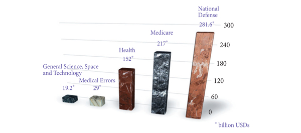
In the Institute of Medicine report published in 1999, it was estimated that 44 000–98 000 patients died annually from preventable medical errors. It was further reported that the annual burden on economy due to preventable medical errors was anywhere between 17–29 billion American dollars. In the USA federal budget 2000–2001, the entire federal resources devoted to general science, space and technology was 19.2 billion American dollars: ≈10 billion less than the cost of medical errors (Fig. 1).

Figure 1. The magnitude of problem caused by medical errors. USDs, American dollars.

On root cause analysis of the errors identified in the Joint Commission on Accreditation and Certification database (2011), it was reported that most of these errors are non-technical, i.e. human factors (72%), leadership (65%), communication breakdown (61%), etc. Furthermore, Greenberg et al. studied the patterns of communication breakdown on the Malpractice Insurers’ Medical Error Prevention Study (MIMEPS) database and concluded that breakdown patterns were similar preoperatively (38%), intraoperatively (30%) and postoperatively (32%). Most errors were due to miscommunication within a single department (78%), as compared with across departments (19%) or institutions (3%). In 49% of the cases, the information was never relayed and in 44% the information relayed was not comprehended appropriately. In all, 29% of these errors involved a surgery attending at transmitting end and 56% at the receiving end of information. In all, 85% of these communications were verbal.

In this issue of BJUI, Ahmed et al. have used the Healthcare Failure Mode and Effect Analysis (HFMEA) model to design a safety checklist specifically for robotic procedures. Checklists have been heavily used in high-risk environments that involve complex technology, e.g. aerospace and nuclear engineering. Robotic surgery is another such high-risk environment, where intraoperative communication is critical. When a surgeon performs a robotic surgery, (s)he is not standing next to the patient (and occasionally not even in the same room!) and relies heavily on his/her assistant. Additionally, the bulky robot takes most of the space around the patient. Small movements of the instruments can cause abrupt and exaggerated movements of the robotic arms, which might injure the bedside assistant, anaesthesiologist, or the patient himself. Last, but not the least, there is a memory clutch on the robotic arms, and its purpose is to ‘remember’ the position of the arms while exchanging the instruments. However, if this clutch is pressed by mistake, all memory is lost and careless insertion of an instrument at this time, making an assumption of memory, can be dangerous and can cause serious injury. The safety checklist described by Ahmed et al. is one of the first checklists specific to robotic surgery. In parallel to this, the Fundamentals of Robotic Surgery (FRS) inter-disciplinary consortium led by Dr Richard Satava has also developed a checklist, specifically for robotic surgery. It will be interesting to study the actual impact of these checklists on prevention of medical errors in robotic surgery. Similar checklists have been validated showing significant clinical correlation using in situ simulation for obstetric emergencies.

Although checklists do help to a certain extent to prevent serious errors, the basics of communications must not be forgotten while communicating to a colleague about patient care. There should be no ambiguity about who is the ‘transmitter’ and who is the ‘receiver’ of information. Both the ‘transmitter’ and ‘receiver’ should have a shared mental model about the purpose of communication (‘transmitter’ is seeking guidance, giving orders, asking for an opinion, referring a case, etc.). Finally, closed-loop communication should be a part of protocol where both the ‘receiver’ and ‘transmitter’ acknowledge the receipt of information, e.g.

Console Surgeon: ‘Please replace the scissors in the right arm with the needle driver’.

Assistant: ‘OK, I am replacing the scissors in your right arm with a needle driver’.

Console Surgeon: ‘Go ahead’.

Assistant: ‘Needle driver coming in’.

Console Surgeon: ‘Perfect. Thank you’.


Sanket Chauhan and Robert M. Sweet
Department of Urology, University of Minnesota Medical School, Minneapolis, MN, USA

Editorial: A promising solution for biofilm inhibition in the bladder, but is the application of wireless capsule cystoscopy practical?

The study by Neheman et al. follows up on an idea first proposed in 2009 by Gettman and Swain to adapt wireless capsule endoscopy (WCE) technology for cystoscopy. Unlike the gastrointestinal tract where the small bowel is not endoscopically accessible making WCE appealing and advantageous, the idea of wireless capsule cystoscopy (WCC) competes with a minor procedure, office cystoscopy, that does not require anaesthesia or sedation and takes only a few minutes to perform. Furthermore, although the authors suggest that WCC would shift the labour associated with bladder cancer monitoring from practising urologists to ancillary health team providers, flexible office cystoscopy is a procedure already routinely performed by physician extenders in many offices. Nevertheless, the concept proposed by Neheman et al. is innovative and intriguing. The potential advantage of a wireless capsule cystoscope placed in the bladder safely for up to a 2-year time period, and thereby reducing the inconvenience and cost of repeated cystoscopies, could be a significant advance.

It should be emphasized that despite the title, no WCE was actually performed. The real value to the present study is the novel anti-biofilm mechanism developed that would be needed for any device implanted in the bladder for the long term. The device was housed in a semi-permeable silicone balloon filled with mineral oil that allowed a continuous slow diffusion of oil across the membrane. Based on the evidence provided in only one animal, it seems this continuous permeation of oil can interfere with surface protein adherence and consequently bacteria adhesion and biofilm creation. Certainly, this concept needs to be tested further in additional animals, aggressively exposed to bacteria, and for longer periods.

Although I am unconvinced that the concept of WCC provides significant value, the development of this biofilm inhibition technique could be pioneering. I read this study and wondered if ureteric stents and Foley catheters could be designed and impregnated with mineral oil to be released gradually. Perhaps the balloon of a Foley catheter could be redesigned and filled with mineral oil that is then released along the catheter’s entire length in a similar fashion. The true Holy Grail is the prevention of encrustation and biofilm formation on these relatively mundane devices whose chronic exchange for many patients is more costly than bladder cancer surveillance. I look forward to additional work from the authors exploring the potential of this technology.

Jeffrey A. Cadeddu
Department of Urology, University of Texas Southwestern Medical Center, Dallas, TX, USA

Read the full article

Social media @BJUIjournal – what a start!

When Prokar Dasgupta assumed the role of new Editor-in-Chief of the BJUI in January 2013, he outlined his vision and some of the major changes that the Journal would make as it transitioned to a new editorial team. After 10 years of progress under John Fitzpatrick, it was clear that we are now working in a much-changed publishing landscape, one which will change even more in the next few years. In particular, the way in which medical professionals receive information and interact with colleagues, patients, journals and other professional groups is unrecognisable from what it was just 2 or 3 years ago.

Social media is the driver of much of this change. It has transformed the way in which the current generation of trainees interact—Facebook, Twitter, YouTube, LinkedIn, Urban Spoon, Expedia, Trip Advisor, Instagram – all of these platforms are key conduits for how Generation Z experiences life. This generation will find the idea of a printed journal arriving in the post every month to be anathema. In a world with an ever-increasing amount of content being produced, and much competition for our limited attention span, Gen Z live their lives through mobile platforms capable of delivering the precise content they want, immediately to their devices. Not just that, this content, whether that be breaking news via Twitter, friend status updates on Facebook, job opportunities via LinkedIn, is delivered through vibrant media that allows them to engage and respond by liking, sharing, favourite-ing, re-tweeting and commenting, even as the content reaches them. All of this activity is done through convenient and increasingly pervasive mobile platforms while on the train to work, while queuing for a coffee, between cases in theatre, during a lecture, first thing in the morning, last thing at night. Gen Z will not seek out this type of content – it will seek them out and be delivered straight to their timeline/twitter-feed.

The BJUI is the first surgical journal to introduce an Associate Editor for Social Media. The aim is to devise and implement a strategy to ensure that the BJUI evolves in this new world; to ensure that the next generation of trainees find us a meaningful organisation to engage with and be informed, educated and entertained by. Our fellow Associate Editor, Matt Bultitude (Web) plays an important role here as do our publishers, our Executive team and Editor-in-Chief.


Our social media platforms

So what have we done? If you are on Twitter or Facebook you will have noticed that BJUI has come to life on these key social media platforms.

Between January and April 2013, our followers on Twitter have grown from by one third to over 1300, and continue to grow at over 100 followers per month. Through Twitter alone, we have generated huge traffic back to our website with over 3500 link clicks from the hundreds of interactions we have had during this period.


Advanced social media metrics allow us to measure all of this activity against other organisations active in urology. For example our Klout score has increased from 46 to 55 with a corresponding increase in our Peerindex rating. We are leading the field across all of the key domains we have targeted to date and continue to make progress as we introduce further changes at www.bjui.org in 2013.

Our Facebook site is now highly engaging and is constantly updated with news and content from our website.


We have recorded over 133 000 page impressions by 23 000 Facebook visitors in the first 3 months of 2013, a huge rise from previously, and all of this traffic gets directed back to content at www.bjui.org, whether that be a Journal article, blog, picture quiz or our new ‘Poll of the Week’.


Our YouTube site is updated with videos from authors and other multimedia content to complement citable articles published in the Journal. You will see a lot more content added here in coming months.


Blogs@BJUI

But perhaps the most talked-about area we have introduced is Blogs@BJUI. And although we are the first mainstream urology journal to introduce a blog site, other journals have done so with great success. In September, we visited the social media team at the BMJ to get some tips on how they had developed their social media strategy into the very successful multi-platform spectacular, which they now oversee. Juliet Dobson, Blogs Editor and Assistant Web Editor at the BMJ offered some excellent advice to help us get up and running and their former Editor, Richard Smith, remains one of the bloggers we most admire. BMJ Blogs is well worth a visit for aspiring bloggers to read some of the best.

We launched our new web journal on the 2 January 2013 to coincide with the new Editor taking the helm, and also published our first blog that day. From then until April 2013, Blogs@BJUI has featured the following:

  • 51 blogs contributed by 25 authors on three continents
  • 193 comments from all over the world, including opinion from some household names in academic urology
  • 16 editorial blogs from our specialty Associate Editors
  • 4 blogs from major urology conferences
  • Multidisciplinary contributions from both authors and comment-leavers

The topics have included everything from urology humour, through the European Working Time Directive, reality TV and an eminent urologist describing his recent personal experience of robotic radical prostatectomy. Our contributors have included many of the key opinion leaders in social media in urology, many of whom are rising stars or already established in academic urology. Also established urology opinion-leaders who are rather new to social media but enjoying the challenge! Other contributors are young trainees who have proved themselves to be talented bloggers already. Blogs@BJUI has been highly successful at driving traffic to the Article of the Week as improving quality remains our main objective.

Also of note is the impact that social media has made at urology conferences in the past few months. As part of a planned strategy, the BJUI social media team has been very active posting updates on Twitter, Facebook and YouTube from major urology conferences, thereby increasing the reach of these meetings to a much larger audience and also allowing those following on social media to engage pro-actively with the conference. This has been a very successful strategy; social media metrics confirm that the BJUI team has been leading the social media revolution at this year’s Annual European Association of Urology (EAU) Congress:


We had set a target that by the end of the first quarter we would have 1000 readers per month visiting Blogs@BJUI. By the end of the February, we had already had over 9000 visits to our blog site! Each reader spent over 3.5 min reading the web journal and many of them left comments or pushed out links using Twitter or Facebook. We have had many comments posted by readers from every corner of the world and have enjoyed some very humorous posts. For us, social media is all about engagement. We want to use these platforms to allow readers to passively engage with us by liking, sharing, tweeting content that they enjoy whether that is a full paper in the BJUI, a blog post, YouTube video, weekly poll or Picture Quiz of the Week. And for those who want to engage more actively, we strongly encourage you to join the conversation and add a comment.

So we have had a great start to our social media push at the BJUI. And there will be a lot more to come in the coming months. For those of you who are new to social media, we encourage you to dip your toes in by reading a blog or two and adding a comment. Before you know it you will have downloaded the Twitter app to your smartphone and you’ll be off and running! For the Twitterati, we thank you for all your enthusiasm in helping us get social media up and running at the BJUI and we look forward to your blogs, mentions, re-tweets and podcasts over the coming months. Social media is all about engagement – join the conversation @BJUIjournal.

Declan G. Murphy and Marnique Basto

Division of Cancer Surgery, University of Melbourne, Peter MacCallum Cancer Centre, Melbourne, Australia


Declan Murphy is Associate Editor for Social Media at the BJUI.
Follow him on Twitter @declangmurphy

Editorial: Robot-assisted partial nephrectomy in patients with recurrent disease: fiction or fact?

In recent decades, the detection of small renal masses (SRMs) has steadily increased with an accompanying shift of treatment towards partial nephrectomy (PN). Indications for PN have successfully expanded to more challenging cases, and robot-assisted PN (RAPN), in particular, has attracted increasing attention (BJUI, Eur Urol); however, despite excellent cure rates for PN, parallel to the increasing number of patients with SRMs undergoing PN, cases of ipsilateral recurrence after PN are also expected to rise. In addition to the incomplete surgical removal of the primary tumour, unknown multifocality or the development of new tumours or metastasis, in a minority of cases recurrence originates at the previous surgical bed and can be considered a proper local recurrence. Retreatment in these patients represents a specific challenge with radical nephrectomy (RN), ablative treatment, repeat PN, and active surveillance in selected cases as therapeutic options. RN should be considered the least attractive option because of the further damage to renal function that it entails, yet it represents one of the most selected options worldwide. Besides ablative techniques, which should be considered under investigational circumstances only, repeat PN is one of the possible options, especially in cases of recurrences attributable to multifocality or bilateral nature.

Repeat open as well as laparoscopic PN (LPN) have been reported sparsely in the literature, but were shown to be associated with good functional and oncological outcomes given adequate laparoscopic experience and patient selection. Such procedures, however, might be challenging and fraught with complications. The role of repeat RAPN (as well as the role of repeat open PN) in this situation is yet to be defined. Nevertheless, given that current data show RAPN to be a more attractive minimally invasive PN technique compared with its standard laparoscopic counterpart, providing equal or better perioperative outcomes, its advantages may even be greater when repeat PN is indicated.

In the current issue of the journal, Autorino et al. report the first study on functional and oncological outcomes and feasibility of repeat RAPN in patients with recurrence after previous PN. Of 490 patients treated with RAPN, nine patients underwent RAPN for recurrent disease at a median time of 39.4 months after previous open or LPN. A total of 12 tumours were removed in these patients, and one third of operations were performed on solitary kidneys. No intra-operative complications were observed, and only two minor complications occurred postoperatively, which were managed conservatively. With regard to functional outcomes, there was a nonsignificant median postoperative decrease in estimated GFR of 7%. More importantly, all patients preserved adequate renal function, which meant that renal replacement therapy was not necessary. With regard to oncological outcomes, all surgical margins were negative and no recurrence was reported.

The results of another study cohort of five patients undergoing repeat RAPN for recurrent disease after open or LPN has recently been published by Jain et al. In their series, surgery was completed in all patients without conversion to RN or an open procedure; furthermore, no complications were reported and the median decrease in GFR was 10%.

Importantly, both series suffer from retrospective evaluation of selective and small sample sizes with a short follow-up, and comparative analysis with other treatment options was not performed; however, the effective comparator for RAPN in this setting has yet to been defined. Bearing in mind current data that demonstrate RAPN to be a preferable minimally invasive PN technique compared with its standard laparoscopic counterparts, the real competitor for RAPN seems to be open surgery. This point was recently also reflected by Mottrie et al. LPN, as a challenging procedure with a long learning curve, limited diffusion and prevalent application in less complex cases, cannot be considered an attractive comparator for RAPN. During the last 8 years, RAPN has become a promising technique which can overcome the technical difficulties of LPN. Three-dimensional vision, ‘endowrist’ technology, and optical magnification allow excellent vision of the operative field and optimum tissue dissection. These technical characteristics help surgeons to minimize ischaemia-time and facilitate accurate tumour excision. Intra-operative ultrasonography, contrast-enhanced sonography, and photodynamic diagnostics can further improve this procedure. It was already shown that the availability of robotic technology is associated with increased use of PN, and, hence, broader diffusion in routine clinical practice may also provide the possibility to outperform results of open PN, even in more complex cases, and will make minimally invasive PN possible and available for more surgeons and patients. Today, the spread of RAPN is only limited by its availability and the associated financial burden. Hence, LPN will currently be considered a cheaper alternative to RAPN in centres with laparoscopic experience and in those which lack the availability of the robot.

Finally, the study from the Cleveland group and the series by Jain et al. provide some valuable support to the feasibility and safety of repeat RAPN and demonstrate that previously performed PN is not a contraindication for RAPN.


Sabine Brookman-May1, Andrea Minervini2, Alessandro Volpe3, Vincenzo Ficarra4, Maciej Salagierski5, Martin Marszalek6,7, Marco Roscigno8, Bülent Akdogan9, Alkuin Vandromme10, Hans Langenhuijsen11, Oscar Rodriguez-Faba12, and Steven Joniau13 for the Renal Cancer Working Group of the Young Academic Urologists (YAU) Working Party of the European Association of Urology (EAU)

1Department of Urology, Ludwig-Maximilians-University, Campus Grosshadern, Munich, Germany, 2Department of Urology, University of Florence, Florence, 3Department of
Urology, University of Eastern Piedmont, Maggiore della Carità Hospital, Novara, 4Department of Oncological and Surgical Sciences, Urologic Unit, University of Padua, Padua,
Italy, 5Department of Urology, Medical University of Łódź, Łódź, Poland, 6Department of Urology and Andrology, Donauspital, Vienna, and 7Department of Urology, Graz Medical University, Graz, Austria, 8Department of Urology, AO Papa Giovanni XXIII, Bergamo, Italy, 9Department of Urology, Hacettepe University, School of Medicine, Ankara, Turkey, 10Klinik für Urologie und Uroonkologie, Klinikum Braunschweig, Germany, 11Laparoscopy, Robotics and Endourology, Radboud University Nijmegen Medical Centre, Nijmegen, The Netherlands, 12Uro-oncology Unit, Fundacio Puigvert, Barcelona, Spain, and 13Department of Urology, University Hospitals Leuven, Leuven, Belgium


Read the full article

Editorial: Androgen deprivation therapy: further confirmation of known harms

Androgen deprivation therapy (ADT) has been an established and effective treatment for men with asymptomatic metastatic prostate cancer for decades. Randomized trials have shown significant survival benefits when ADT is used, coupled with radiotherapy, for patients with locally advanced disease; however it is often used in patients where the benefits are less clear, such as for a rising serum PSA level after radical prostatectomy, and among patients who elect to take a more conservative approach to treatment for low-risk disease. In addition to the absence of data proving benefit, there are a number of adverse consequences attached to androgen deprivation which should be given serious consideration before beginning treatment. Most of the side effects of ADT are linked to its induced hypo-androgenic, and consequently hypo-oestrogenic, state. These include fatigue, vasomotor flushing, loss of muscle mass, weight gain, hyperlipidaemia and insulin resistance.

Osteoporosis and fracture are additional known consequences of ADT with a trend toward greater fracture risk with a higher number of doses of a GnRH agonist and/or longer duration of use. Studies indicate that men with non-metastatic prostate cancer treated with ADT experience an annual loss in bone mineral density of up to nine times that of men in the general population. The use of intermittent ADT, as opposed to continuous use, as a strategy to reduce the negative cardiometabolic and osteoporotic effects is unresolved; however, a report indicating that more recent treatment was associated with a greater risk of fracture, irrespective of cumulative dose, suggests the potential for some reversibility in bone loss post-treatment.

In the present issue of the BJU International, Lu-Yao et al. add to the literature in this area. In a study of nearly 76 000 men with prostate cancer, using data gathered as part of the National Cancer Institute’s Surveillance, Epidemiology and End Results (SEER) cancer registry linked to Medicare claims data, the authors reported that patients at high risk for skeletal complications were, not surprisingly, more likely to experience a fracture associated with ADT use over a 12-year period compared with patients receiving ADT but at low risk for developing such complications. Furthermore, men who experienced a fracture were 40% more likely to die during follow-up than those without fracture. Patients were sorted into risk groups using an index which summed the number of known risk factors for incident fracture identified from Medicare claims in the 12 months before their prostate cancer diagnosis. Unfortunately, owing to the relatively small number of patients with more than one risk factor, the study was limited in its ability to establish a dose – response relationship between the baseline index and fracture risk. SEER-Medicare is an excellent resource to investigate both outcomes and treatment-related expense associated with cancer diagnoses in the USA; however, in this particular study, the reliability of behaviours included in the baseline index (i.e. smoking) is questionable as it would require both patient report of tobacco use as well as physician documentation as a billable claim. Still, one might argue that the heaviest smokers, whose behaviour would most likely be captured as part of a claim, would also be the most important group to capture in an index intended to predict fracture risk.

Interestingly, it was reported that patients at high risk for skeletal complications were significantly more likely to receive ADT than patients at low risk. This was driven in part by the use of primary ADT among elderly men (aged ≥80 years) with prostate cancer and consistent with the notion that when curative treatment is contraindicated (i.e. older patients and those with pre-existing comorbidities) treatment with ADT is more common. Lastly, these findings do not suggest any modification of fracture risk associated with ADT according to baseline risk index, which is consistent with reports of the impact of comorbid conditions and ADT on the risk of incident diabetes and cardiovascular events. This is an important observation in that it says, there is no group that is immune to the adverse effects of ADT – all men are at risk. In absolute terms, however, the men at the greatest risk of an ADT side effect (i.e. a fracture or diabetes) are the men who are at greatest risk of having that side effect even if they were not receiving ADT. The findings of Lu-Yao et al. reinforce the need for careful monitoring of all men receiving ADT. Moreover, when these data are combined with an earlier study that showed that primary ADT was associated with poorer survival than that for men with low-risk prostate cancer who were managed conservatively with observation alone, it should be a wake-up call for us to stop treating non-lethal cancer with lethal and toxic treatments, including ADT.

Jennifer L. Beebe-Dimmer* and Stephen J. Freedland‡§
*Wayne State University School of Medicine, Karmanos Cancer Institute, Detroit, MI, Durham VA Medical Center, and §Duke University School of Medicine, Durham, NC, USA

Read the full article

Editorial: Bipolar plasma enucleation: a new gold standard for BPH?

The history of surgical enucleation for BPH dates back over 100 years and it continues to be the most complete and efficient method of removing adenomata of any size. The popularity and performance of the open approach has declined recently but new enucleation techniques have emerged. In this edition of the journal, Geavlete et al. have studied a recent addition to the endoscopic enucleation armamentarium, namely ‘plasma-button’ bipolar enucleation (BPEP). This procedure is a variation on bipolar endoscopic enucleation using a coiled electrode(or PkEEP) first described in 2006. These authors’ unique contribution to the literature is to compare electrosurgical endoscopic enucleation with open prostatectomy in large prostates (>80 g by TRUS) in a randomized trial and provide Level 1 evidence for this technique. The groups were well-matched preoperatively and were equivalent in terms of operating time, weight of tissue retrieved and postoperative variables up to 12-month follow-up. Significant advantages were noted in perioperative outcomes in favour of the endoscopic technique, particularly those outcomes related to blood loss and subsequent hospital stay. Although not specifically addressed, it is highly likely that substantial cost savings were also achieved and patients returned to normal activities sooner with the endoscopic approach.

Endoscopic enucleation for very large prostates using the Holmium laser as the energy source, was first described over a decade ago. Holmium laser enucleation of the prostate (HoLEP) has been compared with open prostatectomy in two randomized trials (Eur Urol 2006, Eur Urol 2008and similar advantages were noted to those of BPEP in the comparison. The next question is, therefore, which of the endoscopic enucleation techniques is superior? Before this question can be answered, we need to separate those techniques that merely resect large tissue fragments (a ‘mega-resection’), and call themselves ‘enucleation’, from those that truly involve complete enucleation of the anatomical lobes using established surgical planes. HoLEP clearly falls into the latter category but electrosurgical methods may or may not because the actual surgical plane, with both electrosurgery and continuous laser wavelengths such as the Thulium : YAG, 532 nm and Diode lasers, is more difficult to achieve and follow. Exponents of these alternative energy sources perform a variety of different procedures, ranging from resection and vaporization hybrids through to a true enucleation technique, all under the banner of ‘enucleation’. For example, green EP with a side-firing fibre, can be a true enucleation technique if blunt dissection is also employed or a ‘mega-resection’ if the laser energy is merely used to cut off the lobe as a single large fragment. The use of the morcellator is also variable, with some authors instead reverting to the resectoscope to resect the lobes while they remain attached at the bladder neck.

The movement back to enucleation techniques, which also yield tissue for analysis, is partly attributable to the desire to detect transition zone cancers but, more importantly, to address the inadequacy of other endoscopic procedures in treating the growing number of huge glands confronting the urologist as a long-term result of the rise of medical therapy. Traditionally, glands > 80–100 g have been thought to be unsuitable for TURP and morbidity becomes significant although laser techniques such as 532 nm vaporization with high-powered devices have been employed in large glands, albeit with prolonged operating times. Unsurprisingly, the retropubic and suprapubic techniques have also been re-visited by robotic surgeons but with more morbidity than HoLEP, although this will probably improve.

Endoscopic enucleation seems to be here to stay with mounting scientific and popular support. It remains to be seen which variation will gain ascendancy in the coming years, but commercial considerations rather than science will probably be the major determining factor.

Peter J. Gilling
Department of Urology, Tauranga Hospital, Tauranga, New Zealand

Read the full article

Editorial: Incorporating prognostic grade grouping into Gleason grades

The ‘Gleason Grading System’ first proposed by Donald Gleason in 1966 was a revolutionary system for its time. As it advocated the use of a sum score that combined the two most common patterns of prostate cancer seen in a radical prostatectomy specimen to predict the biological outcome of the tumour, rather than the worst pattern that was in common usage with other tumour types, it was truly innovative. Furthermore, although several other classification systems for prostate cancer have been proposed since then, none has stood the test of time as well as the Gleason system and certainly no other system is in widespread use internationally.

Gleason and Mellinger went on to make adjustments and modifications to this classification system in 1974 and 1977, as the series of cases examined was expanded from the original 270 patients to >1000 patients.

Since then, there have been further changes to the Gleason Grading System with the advent of immunocytochemistry and in terms of clarification of the size and spacing of individual acini that are seen in the various patterns originally illustrated by Gleason. A tertiary pattern of prostate cancer, mentioned in passing by Gleason, has also become more clearly identified in a proportion of cases.

Possibly the most important advance regarding the Gleason Grading System was the result of an International Consensus Conference of Urological Pathologists in 2005. This meeting, comprising >80 specialist pathologists from 20 countries, published the updated or ‘Modified Gleason Grading System’. These guidelines were based on the changes in practice that had taken place in the diagnosis and treatment of prostate cancer in the previous 40 years and included evidence for the confirmation that Gleason 1 and 2 patterns should not be assigned on prostatic needle biopsy specimens and that all cribriform areas of tumour were best regarded as Gleason pattern 4 rather than Gleason pattern 3.

Although these modifications have been useful for the surgeon and pathologist, they have not clarified the Gleason grading system for the patient. It is not easy to explain or to understand why a system that in theory could produce a range of Gleason sum scores from 2 to 10, is in practice actually limited on prostatic biopsy to Gleason sum score 6 to 10. Thus, rather confusingly, Gleason 6 is the most favourable category of prostatic carcinoma in terms of prognosis, rather than indicating a ‘middle-of-the-scale’ tumour.

The paper presented in this issue of BJUI, ‘Prognostic Gleason grade grouping: data based on the modified Gleason scoring system’, attempts to compensate for this by allowing the categorisation of prostatic carcinoma not only in terms of Gleason sum score, but also into prognostic groups I to V that correlate with the sum score and may be easier for the patient to appreciate.

This is an important next step in the development of the Gleason Grading System and hopefully one that will be embraced by surgeons and pathologists and more easily accepted by patients.

Alex Freeman
Department of Histopathology, University College London Hospital, London, UK

Read the full article

Editorial: Targeting the pro-survival side-effects of androgen-deprivation therapy in prostate cancer

In this paper, Bennett et al. [1] report the effects of an anti-androgen drug on autophagy and the subsequent impact on response to androgen-deprivation therapy alone or combined with exiting chemotherapeutic treatments.

With an estimated 238,590 newly diagnosed cases and 29,720 deaths for 2013 in the USA, prostate cancer is, after skin cancer, the second most common cancer in men. Although the disease initially responds well to therapy, the cancer recurs in most patients within 1–2 years of the initial response. Few therapeutic options exist for patients with recurring prostate cancer and docetaxel is considered the standard of care. But despite clinical benefits, its effect is mainly palliative and often short-lived, and all patients eventually develop progressive disease with a median survival of 1–12 months. In addition, the decision of when to initiate docetaxel-based chemotherapy is an important one that is not clearly addressed by current treatment guidelines.

Autophagy is a lysosomal catabolic pathway that promotes cell survival in response to starvation or other cellular stresses by degrading and recycling macromolecules and organelles. In recent years, this cellular process has been implicated in the aetiology of cancer; the roles it plays, however, in the development and maintenance of cancer appear conflicting [2–6]. Indeed, tumour cells appear to disable autophagy at an early stage, thereby facilitating the onset of tumorigenesis, whereas in existing malignancies autophagy is activated as a means of stress adaptation, resulting in metastatic dissemination [7–9]. Autophagy is also induced by nearly every anti-cancer treatment as an adaptive pro-survival mechanism against cytotoxic agents and may therefore favour radio- and chemo-resistance [10–12].

Bennett et al. [1] are the first to show the induction of autophagy due to suppression of androgen function in the absence of other cellular stresses in an androgen-sensitive cell line. The authors showed that anti-androgen treatment induced autophagy in LNCaP prostate cancer cells, resulting in a pro-survival effect that was abolished by pharmacological inhibition of autophagy, a response that is similar to that seen in tamoxifen-resistant breast cancer cells. Their study highlights the potential of combining anti-androgen therapy with autophagy inhibition in the treatment of prostate cancer. The mechanism by which anti-androgen therapy activates autophagy is unclear, but this study suggests that modulation of mammalian target of rapamycin (mTOR) signalling, a major cellular metabolism switch, may underlie this effect. Thus, agents that inhibit the pathway combined with inducers of metabolic stress or chemotherapeutic agents could enhance anti-cancer therapy by inhibiting stress adaptation and increasing cell damage. The search for novel inhibitors of the pathway is crucial in the fight against cancer.

Clearly, to this day, there are no simple rules for the outcome of targeting autophagy as a cancer therapy. The apparent conflicting effects of activating or inhibiting autophagy at various stages of the disease are likely to be dictated by the genetic background as well as the environmental cues tumour cells are exposed to. One of the main challenges in prostate cancer therapy is to determine the precise timing of drug application. Therefore, the identification of a ‘fingerprint’, including the aforementioned parameters, in prostate cancer is crucial for the selection of an effective treatment. The present study opens up potential new avenues in the treatment of prostate cancer but further in vitro and in vivo studies will be necessary for efficiently translating this knowledge into the clinic.

Vincent Zecchini and David E. Neal
Department of Uro-Oncology, University of Cambridge, Cambridge, UK

Read the full article

REFERENCES

  1. Bennett HL, Stockley J, Fleming JT et al. Does androgen-ablation therapy (AAT) associated autophagy have a pro-survival effect in LNCaP human prostate cancer cells? BJU Int 2013; 111: 672–82
  2. Oh SH, Lim SC. Endoplasmic reticulum stress-mediated autophagy/apoptosis induced by capsaicin (8-methyl-N-vanillyl-6-nonenamide) and dihydrocapsaicin is regulated by the extent of c-Jun NH2-terminal kinase/extracellular signal-regulated kinase activation in WI38 lung epithelial fibroblast cells. J Pharmacol Exp Ther 2009; 329: 112–22
  3. Qu X, Yu J, Bhagat G, Furuya N et al. Promotion of tumorigenesis by heterozygous disruption of the beclin 1 autophagy gene. J Clin Invest 2003; 112: 1809–20
  4. White E, DiPaola RS. The double-edged sword of autophagy modulation in cancer. Clin Cancer Res 2009; 15: 5308–16
  5. Yue Z, Jin S, Yang C, Levine AJ, Heintz N. Beclin 1, an autophagy gene essential for early embryonic development, is a haploinsufficient tumor suppressor. Proc Natl Acad Sci USA 2003; 100: 15077–82
  6. Liang XH, Jackson S, Seaman M et al. Induction of autophagy and inhibition of tumorigenesis by beclin 1. Nature 1999; 402: 672–6
  7. Chiarugi P, Giannoni E. Anoikis: a necessary death program for anchorage-dependent cells. Biochem Pharmacol 2008; 76: 1352–64
  8. Douma S, Van Laar T, Zevenhoven J et al. Suppression of anoikis and induction of metastasis by the neurotrophic receptor TrkB. Nature 2004; 430: 1034–9
  9. Yap KL, Zhou MM. Keeping it in the family: diverse histone recognition by conserved structural folds. Crit Rev Biochem Mol Biol 2010; 45: 488–505
  10. Chen S, Rehman SK, Zhang W et al. Autophagy is a therapeutic target in anticancer drug resistance. Biochim Biophys Acta 2010; 1806: 220–9
  11. Liu L, Yang M, Kang R et al. HMGB1-induced autophagy promotes chemotherapy resistance in leukemia cells. Leukemia 2011; 25: 23–31
  12. Lomonaco SL, Finniss S, Xiang C et al. The induction of autophagy by gamma-radiation contributes to the radioresistance of glioma stem cells. Int J Cancer 2009; 125: 717–22

Bringing science closer to urologists

The BJUI has always promoted the best in basic science through its ‘Investigative Urology’ section. However, the new editorial team noticed a small problem – these articles were rarely cited, probably because they were rarely read. As we started speaking to our readers, the truth became rapidly obvious. Most urologists, being clinicians, could not understand the scientific content of these articles. Here was a major challenge. How were we going to attract our surgical readership to science?

Whilst maintaining our commitment to quality, we took three bold steps in discussion with our readership:

  1. Rename the section ‘Translational Science’, so as to highlight the potential clinical relevance of the best basic science papers.
  2. Assemble an editorial team of the best clinician-scientists, not just from molecular and cellular biology but other diverse fields, such as immunology, imaging, engineering and computational sciences.
  3. Precede original science papers with ‘Science made Simple’ articles. These were inspired by the highly successful For Dummies series from Wiley.

The idea behind For Dummies is making everything easier. With >250 million books in print and >1800 titles, For Dummies is the most widely recognised and highly regarded reference series in the world. Since 1991, For Dummies has helped millions make everything easier. Now, Dummies.com is bringing the ‘how-to’ brand online, where readers find proven experts presenting even the most complex subjects in plain English. Whether that means directions on how to hook up a home network, carve a turkey, knit your first scarf, or load your new iPod, you can trust Dummies.com to tell it like it is, without all the technical jargon. For Dummies is a simple, yet powerful concept. It relates to the anxiety and frustration that people feel about technology by poking fun at it with books that are insightful and educational and make difficult material interesting and easy.

Thus we originally thought of publishing articles entitled ‘Science for Dummies’.

Thankfully during a Visiting Professorship in Detroit, one of our science colleagues politely pointed out that urologists are anything but dummies. We have to thank her for suggesting a change of name to ‘Science made Simple’. The format is straightforward – simple language, to the point, along with a simple diagram.

This month we feature an original article on gene fusions in prostate cancer in particular TMPRSS2:ERG. This is made simple by a For Dummies style explanation from Deloar Hossain and David Bostwick. You only have to see the vividly simple diagram to understand how a genetic deletion or translocation can make the joining of two genes possible. Important discoveries of the future will occur if top scientists wherever they maybe, work more closely with their clinical counterparts. We are keen to attract the best science to the BJUI by providing an attractive publishing platform to our best scientists. We also hope that you, our readers will enjoy this new format, engage with quality science in the BJUI, cite these important papers and ultimately relate to their clinical relevance for the benefit of your patients.

Dirk De Ridder, Associate Editor BJUI
Jo Wixon, Publisher BJUI
Prokar Dasgupta, Editor-in-Chief BJUI and King’s Health Partners


Editorial: Think irritable bowel syndrome when treating overactive bladder

The bladder and bowel are functionally related organs; they lie in close proximity, have similar innervations and some structural similarities, albeit having different functional characteristics; they are both critical for the storage, collection and expulsion of waste products. Several previous clinical reports have suggested that LUTS, such as overactive bladder syndrome (OAB), can occur concurrently with disorders of the colon, such as irritable bowel syndrome (IBS).

In the study entitled ‘Relationship between overactive bladder and irritable bowel syndrome: a large-scale internet survey in Japan using the overactive bladder symptom score and Rome III criteria’, Matsumoto et al. investigate the prevalence of OAB and IBS in Japan using a large scale internet based survey. In all, 10 000 randomly selected participants completed the surveys with equal numbers of men and women. Subjects were grouped according to age and gender and the prevalence and severity of OAB was assessed using the OAB symptom score (OABSS). The OABSS as an assessment tool combines OAB symptoms into a single score. Four main criteria were examined (daytime frequency, night-time frequency, urgency and urgency incontinence) and disease severity was assessed by overall score value (5, mild; 6–11, moderate; and >12 severe). Similar epidemiological studies have been conducted in the past; however, this is the first study to use the OABSS to assess OAB in a general population. IBS was assessed using the IBS module of the ROME III criteria.

The study found that in the population studied, the overall prevalence of OAB was 9.3% (with 9.7% of men and 8.9% of women affected) and increased with advancing age. Of those affected, 59% reported mild symptoms, 40% reported moderate symptoms and 1% reported sever symptoms. The prevalence of IBS was greater, with 21.2%  of people reporting symptoms (18.6% of men and 23.9% of women); however, conversely the incidence of IBS was reduced with age. Consistent with previous epidemiological studies conducted in Europe and the USA, 33.3% of participants reporting OAB symptoms also had concurrent IBS (32.0% men and 34.8% women), interestingly though, the prevalence of concurrent IBS and OAB was unaffected by age, suggesting that age is not a contributing factor to this relationship.

The exact aetiology of OAB and IBS, by virtue of the non-specific nature of both symptom syndromes, cannot be clearly defined. However, both disorders are characterised by at least increased frequency of visceral emptying due to increased sensation and in many cases motor hyperactivity. In the LUT this takes the form of urgency with associated detrusor overactivity in 40–90% of patients and in the bowel it manifests as pain and discomfort. Experimental studies in rodent models have shown that initiation of bladder overactivity using chemical agents, such as cyclophosphamide, can induce hypersensitivity of the colon and conversely induction of colitis can lead to altered bladder function resembling OAB (Bielefeldt K et al., Brumovsky PR et al., Pezzone MA et al.). The concurrence of these disorders suggests that there may be a common underlying pathology or dysfunction at least in a subset of patients.

One theory put forward to explain the concurrence of OAB and IBS is that of cross-organ sensitisation, whereby sensory innervation of the bladder and bowel interact. These interactions can occur at multiple levels. In the periphery, there is evidence for afferent fibres, which extensively branch and innervate multiple target structures. These dichotomising afferents converge at a single neurone in the dorsal root ganglion (DRG). Studies using retrograde tracers injected into the colon and bladder wall have identified specific DRGs neurones that receive projections from both organs, although the numbers or these neurones are low. Sensitisation of the endings in one organ by local inflammation damage or injury would probably impact on overall sensitivity after upregulation in excitability in all terminal receptive fields.

In addition to peripheral mechanisms, sensitisation of central pathways could also be a contributing factor in cross-organ sensitisation. Spinal neurones receiving afferent input from the bladder have been shown to respond to afferent input from other pelvic structures including the colon. Second-order neurones in the spinal cord therefore receive convergent input from various visceral structures, as well as somatic inputs. This theory provides an explanation for the phenomenon of referred pain, where sensations from the viscera are experienced in the associated somatic sensory fields. Such viscero-somatic convergence has been extensively investigated (the most common example of this is angina), but only recently has viscero-visceral referral received attention. Clearly much research is still required to understand these interactions; however; this study clearly highlights the concurrence of bladder and bowel disorders. Understanding the mechanism(s) involved could have important implications for future therapeutic interventions aimed at treating both OAB and IBS.

Donna Daly and Christopher Chapple*

Department of Biomedical Science, University of Sheffeld and *Department of Urology, The Royal Hallamshire Hospital, Sheffeld Teaching Hospitals NHS Foundation Trust, Sheffield, UK

Read the full article
© 2024 BJU International. All Rights Reserved.