Tag Archive for: frozen section

Posts

Article of the week: The impact on oncological outcomes after RP for PCa of converting soft tissue margins at the apex and bladder neck from tumour‐positive to ‐negative

Every week, the Editor-in-Chief selects an Article of the Week from the current issue of BJUI. The abstract is reproduced below and you can click on the button to read the full article, which is freely available to all readers for at least 30 days from the time of this post.

In addition to the article itself, there is an editorial written by a prominent member of the urological community and the authors have also kindly produced a video describing their work. These are intended to provoke comment and discussion and we invite you to use the comment tools at the bottom of each post to join the conversation. 

If you only have time to read one article this week, it should be this one.

The impact on oncological outcomes after radical prostatectomy for prostate cancer of converting soft tissue margins at the apex and bladder neck from tumour‐positive to ‐negative

Sahyun Pak*, Sejun Park, Myong Kim*, Heounjeong Go, Yong Mee Choand Hanjong Ahn*


*Department of Urology, Asan Medical Center, University of Ulsan College of Medicine, Seoul, Department of Urology, Ulsan University Hospital, University of Ulsan College of Medicine, Ulsan and Department of Pathology, Asan Medical Center, University of Ulsan College of Medicine, Seoul, Korea


Abstract

Objectives

To assess the impact of conversion from histologically positive to negative soft tissue margins at the apex and bladder neck on biochemical recurrence‐free survival (BCRFS) and distant metastasis‐free survival (DMFS) after radical prostatectomy (RP) for prostate cancer.

Materials and Methods

The records of 2 013 patients who underwent RP and intra‐operative frozen section (IFS) analysis between July 2007 and June 2016 were reviewed. IFS analysis of the urethra and bladder neck was performed, and if malignant or atypical cells remained, further resection with the aim of achieving histological negativity was carried out. Patients were divided into three groups according to the findings: those with a negative surgical margin (NSM), a positive surgical margin converted to negative (NCSM) and a persistent positive surgical margin (PSM).

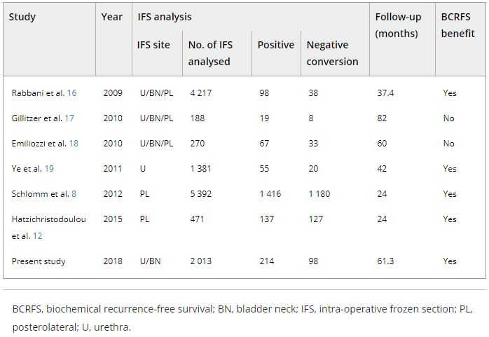
Table 4. Impact of converting margins from tumour‐positive to ‐negative on biochemical recurrence

Results

Among the 2 013 patients, rates of NSMs, NCSMs and PSMs were 75.1%, 4.9%, and 20.0%, respectively. The 5‐year BCRFS rates of patients with NSMs, NCSMs and PSMs were 89.6%, 85.1% and 57.1%, respectively (P < 0.001). In both pathological (p)T2 and pT3 cancers, the 5‐year BCRFS rate for patients with NCSMs was similar to that for patients with NSMs, and higher than for patients with PSMs. The 7‐year DMFS rates of patients with NSMs, NCSMs and PSMs were 97.8%, 99.1% and 89.4%, respectively (P < 0.001). Among patients with pT3 cancers, the 7‐year DMFS rate was significantly higher in the NCSM group than in the PSM group (98.0% vs 86.7%; P = 0.023), but not among those with pT2 cancers (100% vs 96.9%; P = 0.616). The 5‐year BCRFS rate for the NCSM group was not significantly different from that of the NSM group among the patients with low‐ (96.3% vs 95.8%) and intermediate‐risk disease (91.1% vs 82.8%), but was lower than that of the NSM group among patients in the high‐risk group (73.2% vs 54.7%).

Conclusions

Conversion of the soft tissue margin at the prostate apex and bladder neck from histologically positive to negative improved the BCRFS and DMFS after RP for prostate cancer; however, the benefit of conversion was not apparent in patients in the high‐risk group.


Editorial: Conversion to negative surgical margin after intraoperative frozen section – (un)necessary effort and relevance in 2019?

The assessment and impact of positive surgical margins (PSMs) at the time of radical prostatectomy (RP) have been discussed for many decades. The determination and reporting should be performed in a standardised fashion according to the International Society of Urological Pathology [1]. The SM is considered positive if tumour cells touch the inked surface of the RP specimen. However, reasons for difficulty in truly differentiating between negative SMs (NSMs) and PSMs include iatrogenic disruption of the prostatic capsule, penetration of ink into small cracks on the outside, or cases in which prostate cancer cells are very close to, but not definitely touching, the inked margins.

A systematic review by Yossepowitch et al. [2] found a contemporary PSM rate of 15% (range 6.5–32%), which increases with extracapsular extension. In addition, the likelihood of PSM is strongly influenced by surgeon experience, independent of the surgical technique. Although PSM is considered an adverse pathological outcome and associated with an increased risk of biochemical recurrence (BCR), the impact on long‐term survival and actual prognostic value remains debatable. The association with other endpoints, such as prostate‐cancer specific mortality and overall survival, is controversial and may be primarily influenced by other risk factors, such as preoperative PSA level, Gleason score, and pathological T‐stage [2].

The role of intraoperative frozen section analysis in order to reduce the PSM rate continues to evolve. In a study by von Bodman et al. [3], 92.3% of patients with a PSM on frozen‐section analysis could ultimately be converted to a NSM. Similar findings were reported by Schlomm et al. [4] in 5392 patients using the intraoperative neurovascular structure‐adjacent frozen section examination (NeuroSAFE) technique, PSMs were detected in 25%, leading to re‐resection and conversion to definitive NSMs in 86% of these patients. In the setting of increasing experience with intraoperative frozen section analysis, a false‐positive SM status was found in only 48 patients (3.3%).

The study by Pak et al. [5], published in this issue of the BJUI, reported that specimens with initial PSMs were converted to NSMs upon permanent specimen evaluation (NCSM) in 4.9% of 2013 men undergoing RP. In this subgroup, the 5‐year BCR‐free survival (BCRFS) rates did not differ from those observed in National Comprehensive Cancer Network (NCCN) low‐ and intermediate‐risk patients with an initially NSM. However, the benefit of conversion from an initial PSM to final NSM was not apparent in high‐risk patients, as the authors found a significantly lower rate of BCRFS amongst this NCSM group. In multivariate analysis, NCSM status was independently associated (hazard ratio 0.624, P = 0.033) with BCR but not distant metastasis. These findings corroborate the findings of the Schlomm et al. [4] study, in which the BCRFS rates of propensity score‐based matched patients with conversion to NSMs did not differ significantly from patients with primarily NSMs.

What is the current role of intraoperative frozen section analysis during RP? How important is it to achieve NSMs in contemporary practice? In whom and how should the assessment be performed? Although it is clearly desirable to completely remove the entire tumour at the time of surgery, and NSMs are a surrogate marker of adequate local excision, the devil is in the details. First, in this study [5], the authors only assessed SMs at the bladder neck and apex. Although the apex is one of the most frequent locations for PSMs, other and/or multiple sites of PSMs are possible and could have been missed. Alternatively, the NeuroSAFE method is able to assess the entire laterorectal circumference albeit with the trade‐off of more extensive pathological involvement and assessment. Second, intraoperative frozen section analysis, and manoeuvers for NCSM, may ultimately be necessary and beneficial in only a small number of patients currently undergoing RP. An increasing proportion of men harbour more aggressive, higher‐risk disease in whom PSMs may have no impact on oncological outcomes or treatment decisions. In these men, long‐term cancer outcomes are probably more related to risks of unsuspected metastatic disease rather than residual, microscopic cancer within the prostatic fossa. As suggested in this study [5], an initial PSM in high‐risk men, independent of ultimate NCSM, may be a surrogate for non‐localised disease and poorer outcomes; PSMs were found in 53% of men with pT3b. In low‐risk men, the issues are whether active surveillance is a more appropriate initial management strategy and that routine intraoperative frozen section analysis may not be worthwhile with a PSM rate of only 10%. How does this alter the decision for adjuvant therapy? Adjuvant radiotherapy is probably under‐utilised in men with PSMs after RP (~11%), and NCSM may spare men from unnecessary treatment, particularly with lower‐risk disease [6]. However, men with PSMs and additional adverse pathological features, such as extraprostatic extension or seminal vesicle invasion, should probably receive adjuvant therapy, primarily driven by T stage.

The incremental value and potential clinical benefit of intraoperative frozen section analysis to achieve NSMs remain to be determined. Although one would suspect that PSM leading to excision of additional tissue could lead to worse functional outcomes, the study from Mirmilstein et al. [7] is reassuring. Despite higher Gleason score and pT stage in those undergoing the NeuroSAFE approach, the PSM rate was lower in this group (9.2%) compared with those undergoing standard intraoperative nerve‐sparing while leading to greater bilateral nerve preservation, higher potency rates at 12 months, and pad‐free continence.

In the future, other methods may guide surgical decision‐making and may eventually alter PSM rate including preoperative MRI of the prostate to evaluate extracapsular extension, genomic risk scores, or real‐time, near‐infrared fluorescent surgical guidance with prostate‐specific membrane antigen ligands [8]. However, one should not forget that outcomes are not solely based on the SM status. Various pathological and clinical factors and patients’ comorbidities and preference should be taken into consideration in the surgical management and that evaluation of validated oncological and functional outcomes is critical.

by Annika Herlemann and Maxwell Meng

References

  1. Tan, PHCheng, LSrigley, JR et al. International Society of Urological Pathology (ISUP) Consensus Conference on Handling and Staging of Radical Prostatectomy Specimens. Working group 5: surgical margins. Mod Pathol 20112448– 57
  2. Yossepowitch, OBriganti, AEastham, JA et al. Positive surgical margins after radical prostatectomy: a systematic review and contemporary update. Eur Urol 201465303– 13
  3. Bodman, CBrock, MRoghmann, F et al. Intraoperative frozen section of the prostate decreases positive margin rate while ensuring nerve sparing procedure during radical prostatectomy. J Urol 2013190515– 20
  4. Schlomm, TTennstedt, PHuxhold, C et al. Neurovascular structure‐adjacent frozen‐section examination (NeuroSAFE) increases nerve‐sparing frequency and reduces positive surgical margins in open and robot‐assisted laparoscopic radical prostatectomy: experience after 11,069 consecutive patients. Eur Urol 201262333– 40
  5. Pak, SPark, SKim, MGo, HCho, YMAhn, HThe impact on oncological outcomes after radical prostatectomy for prostate cancer of converting soft tissue margins at the apex and bladder neck from tumour‐positive to ‐negative. BJU Int 2019123811– 7
  6. Ghabili, KNguyen, KHsiang, W et al. National trends in the management of patients with positive surgical margins at the time of radical prostatectomy. J Clin Oncol 201836 (Suppl.)111
  7. Mirmilstein, GRai, BPGbolahan, O et al. The neurovascular structure‐adjacent frozen‐section examination (NeuroSAFE) approach to nerve sparing in robot‐assisted laparoscopic radical prostatectomy in a British setting – a prospective observational comparative study. BJU Int 2018;121854– 62
  8. Neuman, BPEifler, JBCastanares, M et al. Real‐time, near‐infrared fluorescence imaging with an optimized dye/light source/camera combination for surgical guidance of prostate cancer. Clin Cancer Res 201521771– 80


Video: The impact on oncological outcomes after RP for PCa of converting soft tissue margins at the apex and bladder neck from tumour‐positive to ‐negative

The impact on oncological outcomes after radical prostatectomy for prostate cancer of converting soft tissue margins at the apex and bladder neck from tumour‐positive to ‐negative

Abstract

Objectives

To assess the impact of conversion from histologically positive to negative soft tissue margins at the apex and bladder neck on biochemical recurrence‐free survival (BCRFS) and distant metastasis‐free survival (DMFS) after radical prostatectomy (RP) for prostate cancer.

Materials and Methods

The records of 2 013 patients who underwent RP and intra‐operative frozen section (IFS) analysis between July 2007 and June 2016 were reviewed. IFS analysis of the urethra and bladder neck was performed, and if malignant or atypical cells remained, further resection with the aim of achieving histological negativity was carried out. Patients were divided into three groups according to the findings: those with a negative surgical margin (NSM), a positive surgical margin converted to negative (NCSM) and a persistent positive surgical margin (PSM).

Results

Among the 2 013 patients, rates of NSMs, NCSMs and PSMs were 75.1%, 4.9%, and 20.0%, respectively. The 5‐year BCRFS rates of patients with NSMs, NCSMs and PSMs were 89.6%, 85.1% and 57.1%, respectively (P < 0.001). In both pathological (p)T2 and pT3 cancers, the 5‐year BCRFS rate for patients with NCSMs was similar to that for patients with NSMs, and higher than for patients with PSMs. The 7‐year DMFS rates of patients with NSMs, NCSMs and PSMs were 97.8%, 99.1% and 89.4%, respectively (P < 0.001). Among patients with pT3 cancers, the 7‐year DMFS rate was significantly higher in the NCSM group than in the PSM group (98.0% vs 86.7%; P = 0.023), but not among those with pT2 cancers (100% vs 96.9%; P = 0.616). The 5‐year BCRFS rate for the NCSM group was not significantly different from that of the NSM group among the patients with low‐ (96.3% vs 95.8%) and intermediate‐risk disease (91.1% vs 82.8%), but was lower than that of the NSM group among patients in the high‐risk group (73.2% vs 54.7%).

Conclusions

Conversion of the soft tissue margin at the prostate apex and bladder neck from histologically positive to negative improved the BCRFS and DMFS after RP for prostate cancer; however, the benefit of conversion was not apparent in patients in the high‐risk group.


View more videos

Article of the Week: Frozen Section During Partial Nephrectomy: Does it Predict Positive Margins?

Every Week the Editor-in-Chief selects an Article of the Week from the current issue of BJUI. The abstract is reproduced below and you can click on the button to read the full article, which is freely available to all readers for at least 30 days from the time of this post.

In addition to the article itself, there is an accompanying editorial written by a prominent member of the urological community. This blog is intended to provoke comment and discussion and we invite you to use the comment tools at the bottom of each post to join the conversation.

If you only have time to read one article this week, it should be this one.

Frozen Section During Partial Nephrectomy: Does it Predict Positive Margins?

Jennifer Gordetsky, Michael A. Gorin*, Joe Canner, Mark W. Ball*, Phillip M. Pierorazio*, Mohamad E. Allaf* and Jonathan I. Epstein*


Departments of Pathology and Urology, The University of Alabama, Birmingham, AL , *Department of UrologyDepartment of Surgery, Center for Surgical Trials and Outcomes Research, and Department of Pathology, The Johns Hopkins Hospital, Baltimore, MD, USA


Read the full article
OBJECTIVE

To investigate the clinical utility of frozen section (FS) analysis performed during partial nephrectomy (PN) and its influence on intra-operative management.

PATIENTS AND METHODS

We performed a retrospective analysis of consecutive PN cases from 2010 to 2013. We evaluated the concordance between the intra-operative FS diagnosis and the FS control diagnosis, a postoperative quality assurance measure performed on all FS diagnoses after formalin fixation of the tissue. We also evaluated the concordance between the intra-operative FS diagnosis and the final specimen margin. Operating reports were reviewed for change in intra-operative management for cases with a positive or atypia FS diagnosis, or if the mass was sent for FS.

RESULTS

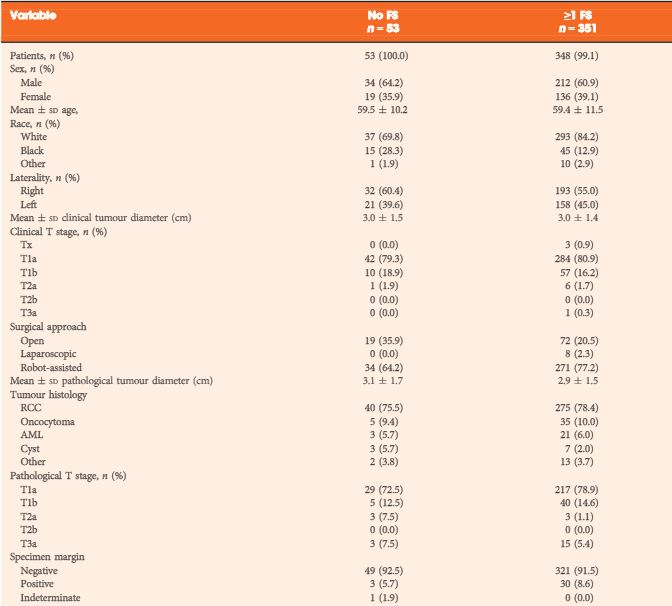
A total of 576 intra-operative FSs were performed in 351 cases to assess the PN tumour bed margin, 19 (5.4%) of which also had a mass sent for FS to assess the tumour type. The concordance rate between the FS diagnosis and the FS control diagnosis was 98.3%. There were 30 (8.5%) final positive specimen margins, of which four (13.3%) were classified as atypia, 17 (56.7%) as negative and nine (30%) as positive on FS diagnosis. Intra-operative management was influenced in six of nine cases with a positive FS diagnosis and in one of nine cases with an FS diagnosis of atypia.

CONCLUSIONS

The relatively high false-negative rate, controversy over the prognosis of a positive margin, and inconsistency in influencing intra-operative management are arguments against the routine use of FS in PN cases.

Read more articles of the week

Editorial: Frozen section during partial nephrectomy: an unreliable test that changes nothing

A core goal of oncological surgery is complete removal of the neoplastic mass. Conventional wisdom with regards to partial nephrectomy (PN) is that a minimal tumour-free margin is sufficient to achieve adequate cancer clearance, minimises loss of normal renal parenchyma and avoids local tumour recurrence [1]. Does this maximisation of nephron preservation and reported positive surgical margin rates after PN ranging from 0% to 7% [2] make intraoperative frozen sections a prerequisite? The results of the paper by Gordetsky et al. [3] in this month’s issue of BJU International suggest that frozen section results from the tumour bed of patients undergoing a PN may be both unreliable and result in subsequent inconsistent management decisions by the operating surgeon.

A recent survey of 197 American urologists revealed that up to 69% (‘always’ or ‘sometimes’) undertake a frozen section during PN [4]. In view of such high penetrance of this test with a resulting high workload for the uro-pathologist, it is critical that the test is reliable and the results are positively and consistently acted upon by the operating urologist.

Gordetsky et al. [3] present interesting data from an expert uro-pathology service, on a consecutive cohort of patients undergoing PN. Reassuringly the pathologist’s skill at making the correct call on the frozen-section specimen was extremely high with a 98% concordance between the actual frozen section and the subsequently created formalin-fixed paraffin-embedded tissue block from the very same piece of tissue. However, despite this high level of accuracy, the sensitivity of the tumour bed frozen section in predicting the actual presence of a positive surgical margin in the resected tumour was only 30% (in other words, of all the patients who actually had positive surgical margins only 30% were identified by frozen section analysis of the tumour bed). As tumour bed biopsies only represent a small fraction of the resection margin this is perhaps unsurprising.

The second issue addressed by Gordetsky et al. [3] is the matter of an inconsistent response of the surgeon to a positive frozen section. In five cases no action was taken, in three cases the tumour bed was re-resected and in a single case a subsequent radical nephrectomy was performed. These results can be compared with those of Sidana et al. [4], where there was a similar inconsistency of management strategies. This inconsistency can be explained by the controversy surrounding the oncological importance of a positive surgical. There is evidence that a positive margin may be associated with an increased risk of recurrence; however, it does not appear to infer a poorer long-term oncological outcome for the patient [5]. It is intriguing that at the time of a completion nephrectomy following a positive surgical margin, residual malignant cells were not found in any of the patients who underwent a re-resection or nephrectomy in this and other studies [3, 6]. It should, however, be noted that published series of conservative management of positive margins are few with only medium-term follow-up. As we know that the natural history of the small renal mass is one of slow growth, any microscopic residual disease may take several years to become clinically apparent and these studies are therefore underpowered.

We think there are several practical arguments against routine use of frozen section. Whilst waiting for the frozen section result some surgeons have been known to keep the kidney ischaemic (16%) resulting in consequent loss of renal function [4]. To avoid this many urologists will undertake the renorrhaphy whilst waiting for the result, a practice becoming increasingly more common with the move towards laparoscopic and robot-assisted PN, where tumour extraction is usually the final step. This practice inevitably influences the subsequent enthusiasm of the surgeon to go back and perform a re-resection and re-do renorrhaphy. It is known that the surgeons’ gross interpretation of the surgical margin approaches the sensitivity of the permanent section and has low false-negative rates, apparently superior to tumour bed frozen section [7]. Routine cautery of the resection bed may also provide an additional safety margin and render any microscopic positive margin clinically insignificant.

It is our opinion that this work by Gordetsky et al. [3] adds credence to the stand that there is no need for a routine tumour bed frozen section in PN and that careful examination of the resected tumour with selected frozen section analysis of suspicious areas is a safe strategy, saves time and provides adequate information for intraoperative decision making.

Read the full article
Grant D. Stewart, *† and Grenville Oades


*Clinical Senior Lecturer, Edinburgh Urological Cancer GroupUniversity of Edinburgh,Honorary Consultant in Urological Surgery, Department of Urology, NHS Lothian, Edinburgh, and ‡Consultant Urological Surgeon, Department of Urology, NHS Greater Glasgow and Clyde, Glasgow, UK


References


1 Sutherland SE, Resnick MI, Maclennan GT, Goldman HB. Does the size of the surgical margin in partial nephrectomy for renal cell cancer really matter? J Urol 2002; 167: 614


2 Marszalek M, Carini M , Chlosta P et al. Positive surgical margins after nephron-sparing surgery. Eur Urol 2012; 61: 75763


3 Gordetsky J, Gorin M, Canner J et al. Frozen section during partial nephrectomy: does it predict positive margins? BJU Int 2015; 116: 86872


Paraganglioma of the paratesticular area: an extremely rare location

We report a case of solitary primary paraganglioma in the paratesticular region, presenting with scrotal swelling and dull pain as the only symptoms. 

Authors: Dr. Sufia Husain MBBS, MD, FRCPath, Senior Registrar (Histopathology Unit). Dept of Pathology

Dr. Emad Raddaoui MD, FCAP, FASC, Consultant Histopathologist (first author), King Khalid University Hospital, College of Medicine, King Saud University, Riyadh, Saudi Arabia.

 
Corresponding Author: Dr. Sufia Husain MBBS, MD, FRCPath, Senior Registrar (Histopathology Unit). Dept of Pathology, P.O. Box 2925, King Khalid University Hospital, College of Medicine, King Saud University, Riyadh-11472, Saudi Arabia. Tel: 00-9661-4671892. Email: [email protected]
 

Introduction
Paraganglia are aggregates of neuroendocrine cells, distributed throughout the body. Paragangliomas are unique neuroendocrine neoplasms arising from these specialized cells of neural crest origin. These tumours are usually located in the carotid body, the jugulo-tympanic body, the mediastinal vessels, or the abdomen, with the majority being benign. Although they have been described in virtually every organ, a paraganglioma at a paratesticular location is extremely rare. Only 8 cases have been reported to date. The clinical features of these tumours are variable, including hypertension, palpitations, headache, sweating, and other symptoms associated with increased catecholamine levels. We report a case of solitary primary paraganglioma in the paratesticular region, presenting with scrotal swelling and dull pain as the only symptoms. We also review in brief the clinical differential diagnosis, possible histogenesis, light microscopic features, immunohistochemistry and electron microscopy results, and general prognosis of the tumour.


Case report 
A 35-year-oldSaudi man presented with an 11-month history of dull ache and dragging sensation in the scrotum, accompanied by a scrotal swelling. There was no history of fever, genital trauma, or genital infection. The patient denied having experienced nausea, vomiting, diarrhea, flushing, palpitations, or weight loss. His vital signs were normal, and the findings of a general physical examination were unremarkable. Examination of the scrotum revealed a right-sided non-tender mass with 3.0 cm being its largest diameter. There were no changes in the overlying skin, and no enlargement of the inguinal lymph nodes. The left side of the scrotum was normal. The complete blood count was normal, as were the levels of tumour markers such as alphafetoprotein, beta human chorionic gonadotrophin, and lactate dehydrogenase. Ultrasonography performed to further define the mass showed a well-circumscribed right paratesticular mass at the upper poleof the right testis, near the head of the epididymis. There was an associated mild varicocoele. The lesion did not involve the epididymis and testis. The right and left testicles were normal in size and echotexture. A chest radiograph and abdominal computed tomography scan showed no significant abnormalities. A provisional clinical diagnosis of a benign paratesticular mass was made, and the patient was counseled before exploratory surgery was performed under general anaesthesia. Tissue from the lesion was submitted for intra-operative frozen section pathologic analysis to enable a decision about testicular preservation. When the specimen was reported as a benign soft tissue tumourwith features consistent with paraganglioma, a testis-sparing operation was chosen. The mass was subsequently excised, but the right testicle and epididymis were not resected. Postoperative recovery was uneventful. The patient was discharged 5 days after surgery, with instructions for regular periodic follow-up with the urologist.
The specimen received in the laboratory was a well-encapsulated mass measuring 3.0 × 2.5 × 2.0 cm. Its external surface was smooth and slightly lobulated, with thin-walled blood vessels running across it. The cut surface was solid and yellowish with focal haemorrhagic areas and delicate septae coursing through it. The tissue was fixed in 10% neutral buffered formalin. Multiple sections were taken from the tissue and embedded in paraffin, cut into 5 micron thick slices, transferred to slides, and stained with haematoxylin and eosin. Unstained slides were also prepared for immunohistochemical study. Light microscopy revealed a tumour comprising cohesive lobules or nests of cells bound by delicate fibrovascular stroma arranged in a Zellballen pattern (Fig.1a). These nests of tumour cells were peripherally encircled by spindle-shaped sustentacular cells. The tumour cells were round to oval with abundant granular eosinophilic cytoplasm, speckled nuclear chromatin, and conspicuous nucleoli. The cells showed mild to moderate nuclear pleomorphism. Scattered multinucleated giant cells were present. Features like intracytoplasmic hyaline globules and eosinophilic intranuclear pseudoinclusions (Fig.1b) were also noted. Mitotic activity, although present, was no more frequent than 1 mitotic figure per 20 high power fields. Necrosis and lymphovascular invasion were absent.
The unstained slides were subjected to immunohistochemical analysis in the Leica Bond Max immunostainer (Vision BioSystems, Newcastle, UK) using the Bond Polymer Refine detection kit (a biotin-free, polymeric horseradish peroxidase-linker antibody conjugate system). The tissue was stained using antibodies against S-100, chromogranin, synaptophysin, CD 56, cytokeratin (CK), and vimentin. The tumour cells exhibited diffuse cytoplasmic positivity forsustentacular cells labeled with chromogranin (Fig.1c), synaptophysin, and CD 56. The sustentacular cells were labeled with S-100;therewere stellate and spindle-shaped cells found cuffing the Zellballen nests (Fig.1d). The tumour cells did not react with CK or vimentin. The Ki67/MIB-1 index was less than 2%.


Figure 1. (a) Photomicrograph showing a low power view of the tumor mass, exhibiting the classical nesting pattern called Zellballen architecture (hematoxylin-eosin, 200× magnification). (b) A higher magnification of the tumor shows cells with abundant eosinophilic cytoplasm and moderate nuclear pleomorphism. An intranuclear pseudoinclusion (arrow) is also noted (hematoxylin-eosin, 400× magnification). (c) The tumor cells reveal strong cytoplasmic positivity for chromogranin (400× magnification). (d)The sustentacular cells rimming the Zellballen nests are highlighted by the S-100 stain (400× magnification).

 
A small amount of paraffin-embedded tissue was submitted to the electron microscopy unit. On ultrastructural evaluation, numerous small dense secretory cytoplasmic granules measuring approximately 150 microns were identified (Fig. 2). The light microscopic, immunohistochemical, and electron microscopic features were thus all characteristic of paraganglioma. The absence of any other lesion elsewhere in the body, both clinically and radiologically, led to a final diagnosis of primary paratesticular paraganglioma.


Figure 2a and 2b. FIG. 2(a) Electron micrograph showing the characteristic multiple dark neurosecretory granules (black arrow) in the cytoplasm of the tumor cells. These are membrane-bound granules with an electron-dense core. (transmission electron microscope, 9000× magnification). (b) The neurosecretory granules are seen around the nucleus at a higher magnification. Also visible, is the rough endoplasmic reticulum (arrowhead) (transmission electron microscope, 28000× magnification).


Discussion
Scrotal paratesticular masses present a diagnostic dilemma to urologists. most paratesticular tumours are benign, about 30% are malignant [1]. Any scrotal mass therefore has to be adequately evaluated in order to rule out malignancy. Exploratory surgery with intraoperative histologic analysis of a frozen section is useful in surgical decision-making. Once the benign nature of the neoplasm is determined on frozen section, a testis-sparing surgical procedure can be performed, with the benefits of retaining the cosmetic and hormonal function of the organ. Common benign tumours of the paratesticular region include lipomas, adenomatoid tumours, leiomyomas, fibromas, papillary mesotheliomas, and cystadenomas [1]. Paragangliomas are extraordinarily rare at this site.
Paragangliomas are neuroendocrine neoplasms of neural crest origin composed of neuroepithelial chief cells. They occur in the fourth and fifth decades and are equally prevalent in both sexes [2]. Most paragangliomas are located in the adrenal gland, where they are referred to as pheochromocytomas. Extra-adrenal paragangliomas are found along the sympathetic and parasympathetic chains, where paraganglia are normally distributed. The common sites for extra-adrenal paragangliomas include the carotid body, vagal body, middle ear, abdomen (organ of Zuckerkandl), and the aortic-pulmonary and laryngeal areas [2].They have also been reported in certain unusual sites like the orbit [3], chambers of the heart, [4] and bone [5]. Paragangliomas have occasionally been reported in the urogenital tract, in the urinary bladder[6], and the prostate [7]. To our knowledge, only 8 cases have been reported worldwide in theliterature so far [8,9], with the first documented case of paratesticular paraganglioma described in 1971 by Eusebi et al. [10].
The histogenesis of paratesticular paragangliomas is a matter of debate. Paraganglia have been observed in the paratesticular tissue around the epididymis and spermatic cord in infancy [11, 12]. Rarely, heterotopic adrenal glands with medullae have also been documented along the route of descent of the testis into the scrotum [11,12]. As a rule, both the heterotopic gland and paraganglial tissue involute during childhood. In exceptional cases, they may persist and eventually harbour a neoplasm. It is our hypothesis that this may be the case in our patient.
These lesions can be functional or non-functional. In a functional lesion, the tumour secretes catecholamines and the presenting symptoms such as headache, sweating, palpitations, and hypertension are secondary to elevated levels of these hormones. Abdominal paragangliomas are more likely to be functional. Although our patient was not tested for catecholamine levels prior to surgery, his was clearly a case of a non-functional paraganglioma, since he displayed none of the symptoms associated with elevated catecholamines. Paragangliomas can also be hereditary or be seen in conjunction with neurofibromatosis type I, Von Hippel-Lindau disease, and multiple endocrine neoplasia type II. Our case had no such associations.
Most paragangliomas are benign, indolent tumours. Malignant lesions are rare, occurring in the fifth to seventh decade, and they tend to be more symptomatic than benign tumours. The diagnosis of malignancy is essentially based on the presence of distant metastasis. Capsular or lymphovascular invasion, a diffuse pattern, confluent tumour necrosis, atypical mitosis, increased mitotic activity (>3 mitotic figures/10 high power fields), fewer S-100 protein-positive sustentacular cells, and a high Ki-67 (MIB-1) index have all been postulated as indicators for predicting malignancy [2]. These indicators, however, are not reliable and cannot be used as well-established histologic criteria for malignancy. Therefore, the treatment of choice for a paraganglioma is complete surgical resectioncoupled with prolonged regular follow-up to rule out a recurrence or the development of metastases. Although there is no specific recommended follow-up strategy, our patient underwent 6 monthly follow-up examinations for the first 3 years and annual ones in the following 2 years. There was no clinical, biochemical, or radiological evidence of recurrence or metastases of the tumour in the 5 years after surgery.
In conclusion, we report a very rare case of non-functional primary paratesticular paraganglioma. We believe that despite their rareincidence in this region, they ought to be considered in the differential diagnosis of a paratesticular neoplasm. Radiological imaging alone is insufficient to determine the nature of a paratesticular mass. Scrotal exploration coupled with intraoperative frozen section analysis can be used as diagnostic tools to rule out the possibility of malignancy, and thus prevent overtreatment with orchidectomy. A diagnosis of paraganglioma is ultimately dependent on a complete and careful histopathological evaluation, in conjunction with consideration of the immunohistochemical findings. Electron microscopy can also be used to confirm the neuroendocrine nature of the neoplasm.


References
1. Khoubehi B, Mishra V, AliM, Motiwala H, Karim O. Adult paratesticulartumours. BJU International 2002;90:707-15.
2. Hunt J. Diseases of the paraganglia system.In: Thompson LDR, editor.Endocrine Pathology. In: Goldblum JR, series ed. Foundations in Diagnostic Pathology. 1st edn. Philadelphia, Churchill Livingstone Elsevier; 2006. p. 327-8.
3. Makhdoomi R, Nayil K, Santosh V, Kumar S. Orbital paraganglioma- a case report and review of literature. Clinical Neuropath. 2010 Mar-Apr;29(2):100-4.
4. Khalid TJ, Zuberi O, Zuberi L, Khalid I. A rare case of cardiac paraganglioma presenting as anginal pain: A case report.Cases J. [serial on the internet]. 2009 Jan 21; 2(1): [about 4 p.]. Available from:https://www.casesjournal.com/content/2/1/72.
5. Laufer I, Edgar MA, Hartl R. Primary intraosseous paraganglioma of the sacrum: a case report. Spine J. 2007; 7(6): 733-8.
6. Cheng L, Beltran AL, MacLennan GT, Montironi R, Bostwick DG.Neoplasms of the urinary bladder. In Bostwick D, Liang C,editors.Urologic Surgical Pathology, 2nd edn.  Edinburgh: Mosby, Elsevier; 2008. P. 315-7.
7. Bostwick DG, Meiers I. Neoplasms of the prostate. In Bostwick D, Liang C, editors.Urologic Surgical Pathology, 2nd edn. Edinburgh: Mosby, Elsevier; 2008.p. 538.
8. Gupta R, Howell RS, Amin MB. Paratesticular paraganlioma: A rare cause of an intrascrotal mass. Arch Pathol Lab Med. 2009; 133(5): 811-3.
9. Garaffa G, Muneer A, Freeman A, et al. Paraganglioma of the spermatic cord: case report and review of the literature. Scientific World Journal 2008; 8: 1256-8.
10. Eusebi V, Massarelli G. Pheochromocytoma of the spermatic cord: report of a case. J Pathol. 1971; 105(4): 283-4.
11. Nistal M, Paniagua R. Non-neoplastic disease of the testis. In Bostwick D, Liang C, editors: Urologic Surgical Pathology, 2nd edn. Edinburgh: Mosby Elsevier; 2008. p. 622-3.
12. Petersen R, Sesterhenn IA, Davis CJ:Urologic Pathology, 3rd edn. Philadelphia: Lippincott Williams & Wilkins;2009. p. 413-4.


Date added to bjui.org: 17/05/2011 


DOI: 10.1002/BJUIw-2011-036-web


© 2024 BJU International. All Rights Reserved.