Tag Archive for: magnetic resonance imaging

Posts

Editorial: Systematic transperineal and MRI‐targeted biopsies: the resolution of uncertainty

The paper published in this issue of the BJUI titled ‘Multicentre evaluation of magnetic resonance imaging supported transperineal biopsy in biopsy‐naïve men with suspicion of prostate cancer’ is timely and helps to resolve some of the uncertainty inherent within the diagnostic pathway 1.

The publication of the PROstate MRI Imaging Study (PROMIS) study, although demonstrating that 25% of patients might avoid prostate biopsy with a normal MRI (Prostate Imaging Reporting and Data System [PI‐RADS] 1–2) and that MRI could identify 90% of patients with high‐risk disease (PI‐RADS 5), did not resolve the issue of what to do with equivocal PI‐RADS 3 scans, uncertainty remained 2. The recent publication of the PRECISION trial (Prostate Evaluation for Clinically Important Disease: Sampling Using Image Guidance or Not?) has only contributed to the uncertainty of systematic TRUS biopsy and has shown that targeted biopsies resolve the issue for <50% of the patients overall and only 12% of those with PI‐RADS 3 lesions had a diagnosis of cancer on targeted biopsy only 3. The study has shown that in the face of an identifiable lesion a MRI‐targeted biopsy is non‐inferior to a blind systematic TRUS biopsy, which was positive in only 28% and implies that a systematic biopsy may be unnecessary, so where does that leave us? The uncertainty within MRI remains at the PI‐RADS 3 level, and particularly with a TRUS biopsy that is not a systematic biopsy of the peripheral zone. The authors of the paper highlighted in this issue of the BJUI 1 help to resolve the issue because they describe a more systematic biopsy.

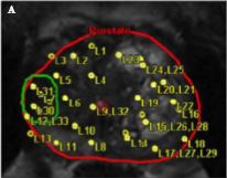
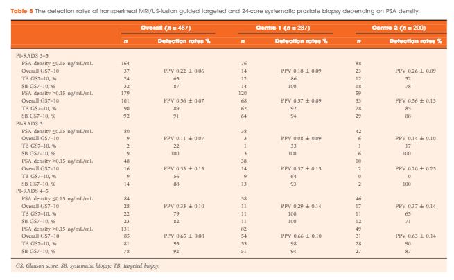
The transperineal (TP) biopsy approach for systematic and targeted biopsy they use is that which was adopted by the Ginsburg Study Group on Enhanced Prostate Diagnostics 4. It is a systematic biopsy that preferentially targets the peripheral zone in a sectoral fashion. It avoids the oversampling inherent in template‐mapping biopsy and the under‐sampling of the non‐systematic transrectal biopsy. Their paper evaluates the combination of an MRI‐targeted biopsy with a systematic TP biopsy. It confirms, as suggested by the PROMIS study, that patients with PI‐RADS 1 or 2 prostates on MRI with a low PSA density <0.1 ng/mL/mL could safely avoid biopsy, based upon a negative predictive value of 0.91 on systematic biopsy. However, in 418 patients with PI‐RADS 4–5 lesions, it was the combination of a targeted and systematic TP biopsy that achieved an overall cancer detection rate of 71%, but that MRI‐targeted biopsies alone had a detection rate of 59% vs 61% for systematic TP biopsies. In the PI‐RAD 3 equivocal group the combined biopsy identified 30% with Gleason score 7–10, whereas targeted biopsy only was positive in 21% vs 27% with systematic biopsies.

The message is clear.

An appropriate systematic biopsy targeted to the peripheral zone remains an essential component of prostate diagnosis even in the MRI era, as indeed it did before MRI was available. In the pre‐MRI days, about one‐third of patients that had negative TRUS biopsies had cancer on TP biopsies and a third of those thought suitable for AS on TRUS biopsy had more significant disease. I suspect in the modern era that figure remains unchanged for those with PI‐RADS 1, 2 or 3, particularly with a PSA density >0.15 ng/mL/mL. As urologists we have always been criticised for over diagnosing and over treating prostate cancer but I suspect that the more heinous crime is that of under treatment of significant disease, it is the very reason why I started doing TP biopsies, to resolve uncertainty. I consider that MRI, for all its benefits in the diagnostic algorithm, cannot yet resolve that uncertainty.

Probably the only patients that merit a target‐only biopsy are those with the high‐PSA, large‐volume disease, easily visible on MRI and usually palpable. Prostate biopsy can be avoided or at least deferred in the PI‐RADS 1–2 group with low PSA density; the rest should be offered a systematic biopsy along with a targeted biopsy. This may be less important in those proceeding to whole gland treatment or surgical extirpation but remains essential in those considering active surveillance, brachytherapy, or any one of the myriad of unproven focal treatments becoming available. The authors should be congratulated for bringing some certainty to uncertainty.

Rick Popert
Urology Centre, Guys Hospital, London, UK


References
  • Hansen NL, Barrett T, Kesch C et al. Multicentre evaluation of magnetic resonance imaging supported transperineal prostate biopsy in biopsy‐naïve men with suspicion of prostate cancerBJU Int 2018122: 40–9


  • Ahmed HU, El‐Shater Bosaily A, Brown LC et al. Diagnostic accuracy of multi‐parametric MRI and TRUS biopsy in prostate cancer (PROMIS): a paired validating confirmatory studyLancet 2017389: 815–22


  • Kuru TH, Wadhwa K, Chang RT et al. Definitions of terms, processes and a minimum dataset for transperineal prostate biopsies: a standardization approach of the Ginsburg Study Group for Enhanced Prostate DiagnosticsBJU Int 2013112: 568–77


Article of the Week: Evaluation of targeted and systematic biopsies using MRI and US image-fusion guided transperineal prostate biopsy

Every Week the Editor-in-Chief selects an Article of the Week from the current issue of BJUI. The abstract is reproduced below and you can click on the button to read the full article, which is freely available to all readers for at least 30 days from the time of this post.

In addition to the article itself, there is an accompanying editorial written by a prominent member of the urological community. This blog is intended to provoke comment and discussion and we invite you to use the comment tools at the bottom of each post to join the conversation.

If you only have time to read one article this week, it should be this one.

Multicentre evaluation of targeted and systematic biopsies using magnetic resonance and ultrasound image-fusion guided transperineal prostate biopsy in patients with a previous negative biopsy


Nienke L. Hansen*†‡, Claudia Kesch§, Tristan Barrett, Brendan Koo, Jan P. Radtke§**, David Bonekamp** , Heinz-Peter Schlemmer**, Anne Y. Warren‡††, Kathrin Wieczorek‡‡Markus Hohenfellner§, Christof Kastner§§ and Boris Hadaschik§


*Department of Diagnostic and Interventional Radiology, University Hospital RWTH Aachen, Aachen, Germany, CamPARI Clinic, Addenbrookes Hospital and University of Cambridge, Cambridge, UK, Department of Diagnostic and Interventional Radiology, University Hospital Cologne, Cologne§Department of Urology, University Hospital Heidelberg, Heidelberg, Germany, Department of Radiology, Addenbrookes Hospital and University of Cambridge, Cambridge, UK, **Department of Radiology, DKFZ, Heidelberg, Germany, ††Department of Pathology, AddenbrookeHospital and University of Cambridge, Cambridge, UK, ‡‡Institute of Pathology, University of Heidelberg, Heidelberg, Germany, and §§Department of Urology, Addenbrookes Hospital and University of Cambridge, Cambridge, UK


Read the full article

Abstract

Objectives

To evaluate the detection rates of targeted and systematic biopsies in magnetic resonance imaging (MRI) and ultrasound (US) image-fusion transperineal prostate biopsy for patients with previous benign transrectal biopsies in two high-volume centres.

Patients and Methods

A two centre prospective outcome study of 487 patients with previous benign biopsies that underwent transperineal MRI/US fusion-guided targeted and systematic saturation biopsy from 2012 to 2015. Multiparametric MRI (mpMRI) was reported according to Prostate Imaging Reporting and Data System (PI-RADS) Version 1. Detection of Gleason score 7–10 prostate cancer on biopsy was the primary outcome. Positive (PPV) and negative (NPV) predictive values including 95% confidence intervals (95% CIs) were calculated. Detection rates of targeted and systematic biopsies were compared using McNemar’s test.

Results

The median (interquartile range) PSA level was 9.0 (6.7–13.4) ng/mL. PI-RADS 3–5 mpMRI lesions were reported in 343 (70%) patients and Gleason score 7–10 prostate cancer was detected in 149 (31%). The PPV (95% CI) for detecting Gleason score 7–10 prostate cancer was 0.20 (±0.07) for PI-RADS 3, 0.32 (±0.09) for PI-RADS 4, and 0.70 (±0.08) for PI-RADS 5. The NPV (95% CI) of PI-RADS 1–2 was 0.92 (±0.04) for Gleason score 7–10 and 0.99 (±0.02) for Gleason score ≥4 + 3 cancer. Systematic biopsies alone found 125/138 (91%) Gleason score 7–10 cancers. In patients with suspicious lesions (PI-RADS 4–5) on mpMRI, systematic biopsies would not have detected 12/113 significant prostate cancers (11%), while targeted biopsies alone would have failed to diagnose 10/113 (9%). In equivocal lesions (PI-RADS 3), targeted biopsy alone would not have diagnosed 14/25 (56%) of Gleason score 7–10 cancers, whereas systematic biopsies alone would have missed 1/25 (4%). Combination with PSA density improved the area under the curve of PI-RADS from 0.822 to 0.846.

Conclusion

In patients with high probability mpMRI lesions, the highest detection rates of Gleason score 7–10 cancer still required combined targeted and systematic MRI/US image-fusion; however, systematic biopsy alone may be sufficient in patients with equivocal lesions. Repeated prostate biopsies may not be needed at all for patients with a low PSA density and a negative mpMRI read by experienced radiologists.

Read more articles of the week

Editorial: Getting to the right biopsy in the right patient at the right time

Guidelines now recommend performing multiparametric MRI (mpMRI) and targeted prostate biopsies in men with a history of prior negative biopsy and continued concern for significant cancer. This new approach to prostate re-biopsy is aimed at improving prostate cancer detection. However, several important clinical factors may help clinicians’ fine-tune the process of repeated prostate biopsy. In this month’s issue of the BJUI, Hansen et al. [1] present a multicentre study of patients with prior negative TRUS biopsy undergoing MRI/TRUS-fusion transperineal biopsy.

In the study, 487 men undergo mpMRI and transperineal biopsy with detection of clinically significant (Gleason score 7–10) cancer as the primary outcome. Several factors are evaluated to compare cancer detection rates, including systematic biopsies, targeted biopsies, PSA density (PSAD), and Prostate Imaging Reporting and Data System (PI-RADS) version 1 score. From their cohort, a suspicious lesion (PIRADS 3–5) was identified in 343 (70%) patients. Prostate cancer was detected in 249 (51%), with 149 (31%) having Gleason score 7–10 cancer. Potentially missed significant cancers from the anterior prostate were found in 27% (40/149). Cancer was detected in 28% (40/144) of patients with PI-RADS 1–2 lesions, with 8% (11/144) being Gleason score 7–10. For patients with PI-RADS 3–5 lesions, cancer was identified in 61% (209/343) with 40% (138/343) being Gleason score 7–10. For patients with PI-RADS 3–5 lesions, systematic biopsies alone failed to detect 13/138 significant cancers, while targeted biopsies missed 24/138 cancers. The combination of systematic and targeted biopsies was significantly better for Gleason score 7–10 prostate cancer detection than either alone. The addition of a PSAD threshold of 0.15 ng/mL/mL for the detection of Gleason score 7–10 resulted in a significant improvement in the area under the curve (0.846) of the receiver operating characteristic curve for PSAD groups and PI-RADS score.

Getting the right biopsy: In this study [1], patients with a prior negative TRUS biopsy underwent TRUS-fusion transperineal biopsy. Having two approaches to prostate biopsy can be advantageous when evaluating men with prior negative biopsies. Historical studies have found comparable prostate cancer detection between transrectal and transperineal biopsies for men undergoing both initial biopsy [2] and saturation re-biopsy [3]. However, the detection of anterior lesions has remained a persistent challenge from the transrectal approach. As in the current study [1], use of transperineal biopsy can detect cancer in up to 30% of tumours that would otherwise be missed on extended template TRUS biopsy [4]. Although attempts to reach anterior lesions from the transrectal approach may be feasible [5], the transperineal approach is felt to provide better sampling in comparison [6].

Getting the right patient: Patient-specific factors such as PI-RADS lesions 3–5 and PSAD have become increasing utilised for stratifying patients who may benefit from additional biopsies using image guidance. As the authors suggest, patients with negative imaging may consider deferring repeat biopsy, particularly those with reassuring PSADs (<0.15 ng/mL/mL). In their study [1], only 4% (6/144) of men with negative mpMRI and a PSAD of <0.15 ng/mL/mL harboured clinically significant cancer (five Gleason score 3 + 4 and one Gleason score 8). Patients with concerning PSAD, but negative mpMRI and those with lesions identified in the peripheral zone could have the option to undergo repeated, fusion-directed TRUS or transperineal biopsy. For patients with lesions identified in the anterior prostate, a transperineal prostate biopsy may provide the highest detection rate.

At the right time: Now that high quality prostate MRI is becoming more widely available; men with a prior negative biopsy should strongly consider the benefit of repeated biopsy after prostate imaging. In addition to identifying suspicious lesions, calculating PSAD has been found to improve the likelihood of detecting clinically significant prostate cancer. Without additional testing, a personalised biopsy plan can be created.

A thorough discussion of the prescribed biopsy approach and the likelihood of detecting a significant cancer is the final step to the right biopsy in the right patient at the right time.

Kelly Stratton


Department of Urology, University of Oklahoma College of Medicine, Oklahoma City, OK, USA


Read the full article


1 Hansen NL, Kesch C, Barrett T et al. Multicentre evaluation of target and systematic biopsies using magnetic resonance and ultrasound image-fusion guided transperineal prostate biopsy in patients with a previous negative biopsy. BJU Int 2016; 120: 6318


2 Hara R, Jo Y, Fujii T et al. Optimal approach for prostate cancer detection as initial biopsy: prospective randomized study comparing transperineal versus transrectal systematic 12-core biopsy. Urology 2008; 71: 1915


3 Abdollah F, Novara G, Briganti A et al. Trans-rectal versus trans- perineal saturation rebiopsy of the prostate: is there a difference in cancer detection rate? Urology 2011; 77: 9215


4 KomaiY, Numao N, Yoshida S et al. High diagnostic ability of multiparametric magnetic re onance imaging to detect anterior prostate cancer miss ed by transrectal 1 2-core biopsy. JUrol2013; 190: 867 7


5 Volkin D, Turkbey B, Hoang AN et al. Multiparametric magnetic resonance imaging (MRI) and subsequent MRI/ultrasonography fusion-guided biopsy increase the detection of anteriorly located prostate cancers. BJU Int 2014; 114: E439


6 Borkowetz A, Platzek I, Toma M et al. Direct comparison of multiparametric magnetic resonance imaging (MRI) results with nal histopathology in patients with proven prostate cancer in MRI/ ultrasonography-fusion biopsy. BJU Int 2016; 118: 21320


Article of the Week: High PCA3 score, PI-RADS grade and Gleason score in patients with elevated PSA undergoing MRI/US fusion TBx

Every Week the Editor-in-Chief selects an Article of the Week from the current issue of BJUI. The abstract is reproduced below and you can click on the button to read the full article, which is freely available to all readers for at least 30 days from the time of this post.

In addition to the article itself, there is an accompanying editorial written by a prominent member of the urological community. This blog is intended to provoke comment and discussion and we invite you to use the comment tools at the bottom of each post to join the conversation.

If you only have time to read one article this week, it should be this one.

High prostate cancer gene 3 (PCA3) scores are associated with elevated Prostate Imaging Reporting and Data System (PI-RADS) grade and biopsy Gleason score, at magnetic resonance imaging/ultrasonography fusion software-based targeted prostate biopsy after a previous negative standard biopsy

Stefano De Luca*, Roberto Passera, Giovanni Cattaneo*, Matteo Manfredi*, Fabrizio Mele*, Cristian Fiori*, Enrico Bollito, Stefano Cirillo§ and Francesco Porpiglia*


*Departments of Urology, San Luigi Gonzaga Hospital, University of Torino, Orbassano, Nuclear Medicine, San Giovanni Battista Hospital, University of Torino, Torino, Pathology, San Luigi Gonzaga Hospital, University of Torino, Orbassano, and §Department of Radiology, Mauriziano Hospital, Torino, Italy


Read the full article

Objective

To determine the association among prostate cancer gene 3 (PCA3) score, Prostate Imaging Reporting and Data System (PI-RADS) grade and Gleason score, in a cohort of patients with elevated prostate-specific antigen (PSA), undergoing magnetic resonance imaging/ultrasonography fusion software-based targeted prostate biopsy (TBx) after a previous negative randomised ‘standard’ biopsy (SBx).

Patients and Methods

In all, 282 patients who underwent TBx after previous negative SBx and a PCA3 urine assay, were enrolled. The associations between PCA3 score/PI-RADS and PCA3 score/Gleason score were investigated by K-means clustering, a receiver operating characteristic analysis and binary logistic regression.

aotwnov4

Results

The PCA3 score difference for the negative vs positive TBx cohorts was highly statistically significant. A 1-unit increase in the PCA3 score was associated to a 2.4% increased risk of having a positive TBx result. A PCA3 score of >80 and a PI-RADS grade of ≥4 were independent predictors of a positive TBx. The association between the PCA3 score and PI-RADS grade was statistically significant (the median PCA3 score for PI-RADS grade groups 3, 4, and 5 was 58, 104, and 146, respectively; P = 0.006). A similar pattern was detected for the relationship between the PCA3 score and Gleason score; an increasing PCA3 score was associated with a worsening Gleason score (median PCA3 score equal to 62, 105, 132, 153, 203, and 322 for Gleason Score 3+4, 4+3, 4+4, 4+5, 5+4, and 5+5, respectively; P < 0.001).

Conclusion

TBx improved PCA3 score diagnostic and prognostic performance for prostate cancer. The PCA3 score was directly associated both with biopsy Gleason score and PI-RADS grade: notably, in the ‘indeterminate’ PI-RADS grade 3 subgroup.

Read more articles of the week


Editorial: PCA3 assay in the MRI/US fusion TBx era: a future to believe in

Men with persistently elevated serum PSA levels after a negative first TRUS-guided systematic prostate biopsy (SBx) represent a great diagnostic challenge. To meet this challenge, urologists need new imaging methods and biomarkers for use in daily clinical practice. The study by De Luca et al. [1] in the present issue of BJUI contributes further data to a growing body of literature addressing the role of prostate cancer gene 3 (PCA3) score and MRI/ultrasonography fusion-targeted prostate biopsy (TBx) in the detection of prostate cancer (PCa) in men who had undergone a previous negative SBx.

De Luca et al. retrospectively analysed data from 282 men undergoing a TBx after a previous negative SBx and PCA3 urine assay for an ongoing suspicion of PCa. They found that the PCA3 score was significantly higher in patients with a positive TBx as compared to those with a negative TBx (121 vs 56; P < 0.001). Futhermore, PCA3 was significantly associated with Prostate Imaging Reporting and Data System (PI-RADS) group (The median PCA3 scores for PI-RADS groups 3, 4 and 5 were 58, 104 and 146, respectively; P = 0.006). Similarly, an increasing PCA3 score was associated with a worse Gleason score (GS) after TBx. These findings are not necessarily novel, but rather are consistent with some of the previous literature [2, 3]. Conversely, Kaufmann et al. [4] did not find any association between PCA3 score and either PI-RADS group or GS. Importantly from a clinical perspective, the authors of the present study observed a statistically significant association between PCA3 score and the ‘indeterminate’ PI-RADS grade III subgroup, thus allowing for the possibility that the combined use of these two diagnostic tools could prevent unnecessary biopsies.

Prostate biopsies are associated with discomfort, anxiety and severe complications. Repeated SBx also results in a greater economic cost and has been associated with overall low PCa detection rates (being negative in almost 80% of examined men). The recent literature has therefore focused on additional tests with the goal of preventing unnecessary biopsies and increasing the probability of detecting PCa during a repeat biopsy. Since its introduction in clinical practice, the urinary PCA3 assay has shown promising results for PCa detection, staging and prognosis; however, recent studies have shown high variability in PCA3 sensitivity and specificity, which can be explained by the low diagnostic performance of SBx in detecting PCa. MRI-guided TBx has shown higher detection rates than SBx and thus could be of clinical utility in improving PCA3 prognostic accuracy in detecting PCa in men with previous negative SBx. De Luca et al. [1], using a univariate logistic regression model to estimate the effect of PCA3 score on TBx results, found that a 1-unit increase in PCA3 score was associated with a 2.4% increase in the odds of having a positive TBx result (odds ratio [OR] 1.024; P < 0.001). The accuracy of this model was 76.2%, with a sensitivity of 82.3% and specificity of 68.5%. A multivariate logistic regression model showed that PI-RADS group ≥4 (OR 10.85) and a PCA3 score >80 (OR 7.17) were independent risk factors for a positive TBx (all P < 0.001). The authors concluded that TBx improved the diagnostic and prognostic performance of the PCA3 score for PCa. Importantly, considering only the ‘indeterminate’ PI-RADS grade III subgroup, a 1-unit increase in PCA3 score was associated with a 2.2% increase in the odds of having a positive TBx (OR 1.022; P < 0.001). These findings are consistent with recent literature showing that the use of multiparametric (mp)MRI to direct biopsies can significantly improve PCA3 score sensitivity [5]. Similarly, Busetto et al. [2] estimated the sensitivity and specificity for PCA3 test and mpMRI to be 68 and 49%, and 74 and 90%, respectively, for cancer detection after an initial negative biopsy, and concluded that mpMRI increased PCA3 score test accuracy.

Repeated prostate biopsy strategies for the suspicion of PCa remain one of the most controversial dilemmas in urology. The results of the present trial help strengthen the evidence in favour of the diagnostic role of the PCA3 score, which could aid the selection of patients for mpMRI. Large prospective trials are needed to confirm the association between PCA3, PI-RADS group and GS in men with PCa. Interestingly, De Luca et al. believe that PCA3 score could be part of a ‘fusion-biopsy era’ in the not-so-distant future. Should the positive correlation between PCA3 and GS be confirmed, PCA3 could have a role in the selection of candidates for active surveillance, and in predicting disease progression during active surveillance follow-up.

Read the full article
Bernardo Rocco*† and Luca Boeri*


*Department of Urology, University of Milan Fondazione IRCCS Ca Granda-Ospedale Maggiore Policlinico, Milan, Italy and Urology, Global Robotics Institute, Florida Hospital Celebration Health, Celebration, FL, USA


References


Article of the Week: TRUS-Guided RB PCa Detection – Reasons for Targeted Biopsy Failure

Every Week the Editor-in-Chief selects an Article of the Week from the current issue of BJUI. The abstract is reproduced below and you can click on the button to read the full article, which is freely available to all readers for at least 30 days from the time of this post.

In addition to the article itself, there is an accompanying editorial written by a prominent member of the urological community. This blog is intended to provoke comment and discussion and we invite you to use the comment tools at the bottom of each post to join the conversation.

Finally, the third post under the Article of the Week heading on the homepage will consist of additional material or media. This week we feature a video from Hannes Cash and Patrick Asbach, discussing their paper.

If you only have time to read one article this week, it should be this one.

Prostate cancer detection on transrectal ultrasonography-guided random biopsy despite negative real-time magnetic resonance imaging/ultrasonography fusion-guided targeted biopsy: reasons for targeted biopsy failure

Hannes Cash*, Karsten Gunzel*, Andreas Maxeiner*, Carsten Stephan*, Thomas Fischer, Tahir Durmus, Kurt Miller*, Patrick Asbach, Matthias Haas† and Carsten Kempkensteffen*


*Department of Urology, and Department of Radiology, ChariteUniversity of Medicine Berlin, Berlin, Germany M. H. and C.K. contributed equally to the study.


Read the full article

Objective

To examine the value of additional transrectal ultrasonography (TRUS)-guided random biopsy (RB) in patients with negative magnetic resonance imaging (MRI)/ultrasonography (US) fusion-guided targeted biopsy (TB) and to identify possible reasons for TB failure.

Patients and Methods

We conducted a subgroup analysis of 61 men with prostate cancer (PCa) detected by 10-core RB but with a negative TB, from a cohort of 408 men with suspicious multiparametric magnetic resonance imaging (mpMRI) between January 2012 and January 2015. A consensus re-reading of mpMRI results (using Prostate Imaging Reporting and Data System [PI-RADS] versions 1 and 2) for each suspicious lesion was performed, with the image reader blinded to the biopsy results, followed by an unblinded anatomical correlation of the lesion on mpMRI to the biopsy result. The potential reasons for TB failure were estimated for each lesion. We defined clinically significant PCa according to the Epstein criteria and stratified patients into risk groups according to the European Association of Urology guidelines.

JulAOTW3Results

Results

Our analysis showed that RB detected significant PCa in 64% of patients (39/61) and intermediate-/high-risk PCa in 57% of patients (35/61). The initial mpMRI reading identified 90 suspicious lesions in the cohort. Blinded consensus re-reading of the mpMRI led to PI-RADS score downgrading of 45 lesions (50%) and upgrading of 13 lesions (14%); thus, negative TB could be explained by falsely high initial PI-RADS scores for 32 lesions (34%) and sampling of the target lesion by RB in the corresponding anatomical site for 36 out of 90 lesions (40%) in 35 of 61 patients (57%). Sampling of the target lesion by RB was most likely for lesions with PI-RADS scores of 4/5 and Gleason scores (GS) of ≥7. A total of 70 PCa lesions (67% with GS 6) in 44 patients (72%) were sampled from prostatic sites with no abnormalities on mpMRI.

Conclusion

In cases of TB failure, RB still detected a high rate of significant PCa. The main reason for a negative TB was a TB error, compensated for by positive sampling of the target lesion by the additional RB, and the second reason for TB failure was a falsely high initial PI-RADS score. The challenges that arise for both MRI diagnostics and prostate lesion sampling are evident in our data and support the integration of RB into the TB workflow.

Read more articles of the week

Editorial: MRI-Fusion Biopsy – Behind the Scenes

MRI information of the prostate is increasingly used for improving the diagnostic yield of prostate biopsies [1]. However, increasing complexity of a procedure makes it prone to errors at multiple technical and human levels. Incorporating MRI information and ultrasonography (US) images for MRI-fusion biopsies is a technically challenging task. It involves various steps such as the acquisition and fusion of MRI and US images, the needle guidance during biopsy, and the diligence of the pathological evaluation of biopsy specimens. These different steps and interfaces between different medical professions influence the diagnostic performance of MRI-fusion biopsies.

For example, in daily clinical practice, MRIs from different institutions still harbour a great variance of sequences and reporting, despite the European Society of Urogenital Urology (ESUR) recently introducing acquisition and imaging protocols and a new and advanced version of the Prostate Imaging Reporting and Data System (PIRADS) version 2.0 [2]. The usefulness of such reporting schemes is evidenced by a moderate-to-good interobserver agreement between uro-radiologists for tumour lesion interpretation and corresponding κ values ranging from 0.55 to 0.80 [3]. Important pitfalls of image interpretation are benign lesions such as prostatitis, BPH and fibrosis, which might score similarly to prostate cancer lesions. This problem is further aggravated by a high proportion of patients that receive their first multiparametric MRI (mpMRI) of the prostate in the repeat-biopsy setting with a high burden of post-biopsy artefacts (haemorrhage, capsular irregularity) and lower overall cancer detection rate. Also, during MRI-fusion biopsy patient movement, prostate deformation by the US probe, and mismatch of image planes can lead to a biopsy error exceeding 4 mm. Moreover, targeting error might be aggravated by MRI underestimation of the tumour volume compared with final pathology [4]. After various authors reported the advantages and accuracy of MRI/US-fusion biopsy approaches, Cash et al. [5] address potential reasons for targeted biopsy failure to detect prostate cancer compared with random biopsy. Within their analyses the authors address potential limitations and technical considerations. Based on different technical biopsy strategies (with the patient placed within the MRI scanner (‘in-bore’) vs outside) and different technical approaches, these considerations are very important.

In contrast to cognitive fusion, most MRI/US platforms allow needle tracking by archiving the needle orientation, either by an electromagnetic, image-based or stepper-based mechanism [1]. However, lesion targeting by needle guidance is highly dependent on the dimensions of the primary lesion, numbers of relevant lesions, localisation, and overall prostate volume, making MRI-US fusion and cognitive fusion more error prone (i.e. aiming off the mark with the needle) than in-bore biopsies. Moreover, different technical fusion approaches provide different degrees of manual/automated adjustment tools, with for example either rigid or elastic image transformation to facilitate MRI/US image alignment.

In their analyses, Cash et al. [5] found that 34% of negative targeted biopsies could be explained by initially too high estimated PIRADS scores that were downgraded at re-reading. Interestingly, the remaining lesions were without an mpMRI correlate but within this group 92.9% showed a primary Gleason 3 pattern in biopsy pathology, suggesting a high degree of invisibility on mpMRI. Subanalyses did not show an association of targeted biopsy failures in the ventral location. Therefore, the study by Cash et al. [5] is an important precursor for further analyses to address other underlying reasons for targeted biopsy failure. Moreover, it reveals the need for a tight collaboration of radiologists, urologists, and pathologists as interdisciplinary partners involved in MRI-fusion biopsy. Consequently, the optimal diagnostic performance of MRI-fusion biopsies can only be achieved through standardised MRI performance, reading and reporting of MRI findings, as well as final correlation of MRI findings with histopathological work up.

Read the full article
Lars Budaus and Sami-Ramzi Leyh-Bannurah
Martini-Clinic University Hospital Hamburg-Eppendorf, Hamburg, Germany


References


Video: TRUS-Guided RB Prostate Cancer Detection – Reasons for Targeted Biopsy Failure

Prostate cancer detection on transrectal ultrasonography-guided random biopsy despite negative real-time magnetic resonance imaging/ultrasonography fusion-guided targeted biopsy: reasons for targeted biopsy failure

Hannes Cash*, Karsten Gunzel*, Andreas Maxeiner*, Carsten Stephan*, Thomas Fischer, Tahir Durmus, Kurt Miller*, Patrick Asbach, Matthias Haas† and Carsten Kempkensteffen*


*Department of Urology, and Department of Radiology, ChariteUniversity of Medicine Berlin, Berlin, Germany M. H. and C.K. contributed equally to the study.


Read the full article

Objective

To examine the value of additional transrectal ultrasonography (TRUS)-guided random biopsy (RB) in patients with negative magnetic resonance imaging (MRI)/ultrasonography (US) fusion-guided targeted biopsy (TB) and to identify possible reasons for TB failure.

Patients and Methods

We conducted a subgroup analysis of 61 men with prostate cancer (PCa) detected by 10-core RB but with a negative TB, from a cohort of 408 men with suspicious multiparametric magnetic resonance imaging (mpMRI) between January 2012 and January 2015. A consensus re-reading of mpMRI results (using Prostate Imaging Reporting and Data System [PI-RADS] versions 1 and 2) for each suspicious lesion was performed, with the image reader blinded to the biopsy results, followed by an unblinded anatomical correlation of the lesion on mpMRI to the biopsy result. The potential reasons for TB failure were estimated for each lesion. We defined clinically significant PCa according to the Epstein criteria and stratified patients into risk groups according to the European Association of Urology guidelines.

JulAOTW3Results

Results

Our analysis showed that RB detected significant PCa in 64% of patients (39/61) and intermediate-/high-risk PCa in 57% of patients (35/61). The initial mpMRI reading identified 90 suspicious lesions in the cohort. Blinded consensus re-reading of the mpMRI led to PI-RADS score downgrading of 45 lesions (50%) and upgrading of 13 lesions (14%); thus, negative TB could be explained by falsely high initial PI-RADS scores for 32 lesions (34%) and sampling of the target lesion by RB in the corresponding anatomical site for 36 out of 90 lesions (40%) in 35 of 61 patients (57%). Sampling of the target lesion by RB was most likely for lesions with PI-RADS scores of 4/5 and Gleason scores (GS) of ≥7. A total of 70 PCa lesions (67% with GS 6) in 44 patients (72%) were sampled from prostatic sites with no abnormalities on mpMRI.

Conclusion

In cases of TB failure, RB still detected a high rate of significant PCa. The main reason for a negative TB was a TB error, compensated for by positive sampling of the target lesion by the additional RB, and the second reason for TB failure was a falsely high initial PI-RADS score. The challenges that arise for both MRI diagnostics and prostate lesion sampling are evident in our data and support the integration of RB into the TB workflow.

Read more articles of the week

Multiparametric MRI – Is the result convincing for AS patients?

Sir,

We read with interest the recent ‘Article of the Month’ by Park et al. in which they concluded that multi-parametric 3T-MRI can be used to predict adverse pathological features and to assess eligibility of patients for active surveillance (AS), in those initially meeting the PRIAS criteria [1]. Nevertheless, we would urge a degree of caution before widespread adoption of this strategy in patient selection for AS.

Firstly, it is accepted that there are false positives with multi-parametric MRI, with the addition of contrast only leading to a minor increase in accuracy, due to increased sensitivity being offset by reduced specificity [2]. Thus, it is imperative to at least make some effort to correlate tumour site on MRI with site on histopathology, which the authors acknowledge was not performed in their study.

Whilst realising that substantial technical difficulties arise in the correlation of imaging with radical prostatectomy specimens, we believe that, as a minimum, tumour side on MRI should be compared to tumour side on histopathology. This is relatively straightforward and could have been performed by Park et al., since 41% of the patients in the study were pathological stage T2a/b.

For example, an audit of 76 patients suitable for AS at our unit (Wirral University Teaching Hospital, UK), but electing for mapping transperineal template guided saturation biopsy, revealed that 53 patients had undergone MRI with diffusion weighted and 23 patients full multi-parametric dynamic contrast enhanced imaging, using a 1.5 T scanner. When analysed without correlation to tumour side the sensitivity was 83%, specificity was 68% and positive predictive value was 79%. However, when analysed with respect to tumour side on MRI with tumour side on histopathology the result becomes 73%, 61%, 79% respectively.

Park et al. concludes that MRI can be used to assess the eligibility of patients with PCa for AS, which is not backed up by their data. With only 11.7% exhibiting no tumour visible on imaging, the investigation will only exclude a relatively small proportion of patients from AS, whereas in the 88.3% with visible cancer on imaging, 50.2% did not have their cancer upgraded and 47.9% had favourable disease on final histology of the whole specimen. Thus, the authors have demonstrated a statistical significant correlation between identification of a lesion on MRI and the risks of upgrading and unfavourable disease, but not demonstrated that multi-parametric 3T MRI is a clinically useful investigation in this setting. In essence, introduction of MP-MRI would be likely to exclude more patients suitable for AS than those with adverse pathology. It seems more likely that it can only be built into a nomogram, rather than a stand-alone assessment tool.

Read the article

Debashis Sarkar*, Nijel J Parr**
*Research Fellow Urology, **Consultant Urologist, Wirral University Teaching Hospital, Upton, UK

Correspondence: Debashis Sarkar, Research Fellow Urology, Wirral University Teaching Hospital,
Upton, UK. e-mail: [email protected]

References

  1. Park BH, Jeon HG, Choo SH et al. Role of multiparametric 3.0-Tesla magnetic resonance imaging in patients with prostate cancer eligible for active surveillance. BJU Int 2014; 113: 864–870
  2. Tanimoto A, Nakashima J, Kohno H, Shinmoto H and Kuribayashi S. Prostate cancer screening: The clinical value of diffusion-weighted imaging and dynamic MR imaging in combination with T2-weighted imaging. J Magn Reson Imaging 2007; 25: 146–152


Article of the month: MRI and active surveillance for prostate cancer

Every week the Editor-in-Chief selects the Article of the Week from the current issue of BJUI. The abstract is reproduced below and you can click on the button to read the full article, which is freely available to all readers for at least 30 days from the time of this post.

In addition to the article itself, there is an accompanying editorial written by a prominent member of the urological community. This blog is intended to provoke comment and discussion and we invite you to use the comment tools at the bottom of each post to join the conversation.

Finally, the third post under the Article of the Week heading on the homepage will consist of additional material or media. This week we feature a video from Dr. Hyun M. Lee discussing his paper.

If you only have time to read one article this week, it should be this one.

Role of multiparametric 3.0-Tesla magnetic resonance imaging in patients with prostate cancer eligible for active surveillance

Bong H. Park, Hwang G. Jeon, Seol H. Choo, Byong C. Jeong, Seong I. Seo, Seong S. Jeon, Han Y. Choi and Hyun M. Lee

Department of Urology, Samsung Medical Center, Sungkyunkwan University School of Medicine, Seoul, Korea
Current address: Bong H. Park, Department of Urology, Incheon St. Mary’s Hospital, The Catholic University of Korea College of Medicine, Seoul, Korea

Read the full article
OBJECTIVE

• To evaluate predictors of more aggressive disease and the role of multiparametric 3.0-T magnetic resonance imaging (MRI) in selecting patients with prostate cancer for active surveillance (AS).

PATIENTS AND METHODS

• We retrospectively assessed 298 patients with prostate cancer who met the Prostate Cancer Research International: Active Surveillance (PRIAS) criteria, defined as T1c/T2, PSA level of ≤10 ng/mL, PSA density (PSAD) of <0.2 ng/mL2, Gleason score <7, and one or two positive biopsy cores.

• All patients underwent preoperative MRI, including T2-weighted, diffusion-weighted, and dynamic contrast-enhanced imaging, as well as radical prostatectomy (RP) between June 2005 and December 2011.

• Imaging results were correlated with pathological findings to evaluate the ability of MRI to select patients for AS.

RESULTS

• In 35 (11.7%) patients, no discrete cancer was visible on MRI, while in the remaining 263 (88.3%) patients, a discrete cancer was visible.

• Pathological examination of RP specimens resulted in upstaging (>T2) in 21 (7%) patients, upgrading (Gleason score >6) in 136 (45.6%), and a diagnosis of unfavourable disease in 142 (47.7%) patients.

• The 263 patients (88.3%) with visible cancer on imaging were more likely to have their cancer status upgraded (49.8% vs 14.3%) and be diagnosed with unfavourable disease (52.1% vs 14.3%) than the 35 patients (11.7%) with no cancer visible upon imaging, and these differences were statistically significant (P < 0.001 for all).

• A visible cancer lesion on MRI, PSAD, and patient age were found to be predictors of unfavourable disease in multivariate analysis.

CONCLUSION

• MRI can predict adverse pathological features and be used to assess the eligibility of patients with prostate cancer for AS.

Read more articles of the week


© 2024 BJU International. All Rights Reserved.