Tag Archive for: magnetic resonance imaging

Posts

Editorial: Multiparametric MRI and active surveillance for prostate cancer: future directions

A growing body of data exists suggesting an important role of MRI in selecting men with prostate cancer for active surveillance (AS). In the present study, Park et al. [1] show that a suspicious lesion on MRI was independently predictive of adverse pathology after radical prostatectomy (RP). This finding supports existing data suggesting that suspicious lesions on MRI confer an increased risk of disease reclassification among men enrolled in AS [2]. Indeed, in our institutional AS experience we found that men with a suspicious lesion on MRI were more likely to have biopsy reclassification with extended follow-up [3].While these data are provocative, much work remains to be done before the adoption of MRI as a standard screening tool for entry into AS for men with very low-risk prostate cancer.

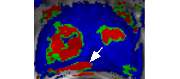
Introduction of functional sequence imaging into multiparametric MRI protocols has resulted in improved detection and characterisation of clinically localised prostate cancer. However, before widespread implementation into AS protocols can occur, increased rigor and standardisation in image interpretation is needed. As in the present study, 5-point Likert scales have become an increasingly popular method of quantifying a lesions likelihood of representing cancer [1]. Still other authors have quantified a lesions level of suspicion using both weighted and non-weighted scoring systems based on the number of positive MRI sequences [3,4]. While useful for statistical analysis, these reporting methods are fraught with concerns of inter-observer variability and generalizability. Additionally, a recent report by Lee et al. [5] found that a simple measurement of lesion diameter on diffusion-weighted MRI was predictive of insignificant disease after RP. Combining the plethora of functional and morphological data obtained by multiparametric MRI into a standardised, reproducible tool will greatly facilitate implementation of MRI into current AS screening protocols.

As a step in the right direction, Stamatakis et al. [4] recently generated a nomogram for predicting biopsy reclassification in men on AS after taking into consideration both functional and morphological characteristics of MRI lesions. Adding an additional layer of complexity, they also assessed the utility of calculated values, e.g. lesion density (lesion volume/prostate volume), in predicting biopsy reclassification. Briefly, their analysis showed that the number of lesions, lesion suspicion, and lesion density were predictive of biopsy reclassification. While nomogram validation and testing of its predictive value on pathological outcomes is needed, this represents a major advance in the standardised application of MRI to AS cohorts.

Despite great strides in the application of multiparametric MRI to AS cohorts, a significant concern about the false-negative rate exists. Considering the present report, of the 35 men with no visible lesion on MRI, 14.3% men had unfavourable pathology after RP [1]. This is similar to previous studies reporting disease reclassification rates of <18% [2,6]. These men with normal imaging and high-grade cancer highlight the importance of incorporating imaging and clinical data when selecting men for AS. Better defining the false-negative rate of multiparametric MRI, and effectively identifying men with a normal MRI and high-grade disease remain major challenges.

Considering all of the available data, it is becoming increasingly clear that MRI has the potential for improving the identification of patients for whom AS would be safe. It is currently the practice at our institution to refer eligible men for multiparametric MRI before enrolment in AS. Our future scholarly efforts should be directed at the standardisation of reporting MRI data and the development of user-friendly AS criteria that synthesise MRI results with clinicopathological data.

Jeffrey K. Mullins and H. Ballentine Carter
James Buchanan Brady Urological Institute, Johns Hopkins Medical Institutions, Baltimore, MD, USA

References

  1. Park BH, Jeon HG, Choo SH et al. Role of multiparametric 3.0-Tesla magnetic resonance imaging in patients with prostate cancer eligible for active surveillance. BJU Int 2014; 113: 864–70
  2. Margel D, Yap SA, Lawrentschuk N et al. Impact of multiparametric endorectal coil prostate magnetic resonance imaging on disease reclassification among active surveillance candidates: a prospective cohort study. J Urol 2012; 187: 1247–52
  3. Mullins JK, Bonekamp D, Landis P et al. Multiparametric magnetic resonance imaging findings in men with low-risk prostate cancer followed using active surveillance. BJU Int 2013; 111: 1037–45
  4. Stamatakis L, Siddiqui MM, Nix JW et al. Accuracy of multiparametric magnetic resonance imaging in confirming eligibility for active surveillance for men with prostate cancer. Cancer 2013; 119: 3359–66
  5. Lee DH, Koo KC, Lee SH et al. Tumor lesion diameter on diffusion weighted magnetic resonance imaging could help predict insignificant prostate cancer in patients eligible for active surveillance: preliminary analysis. J Urol 2013; 190: 1213–7
  6. Guzzo TJ, Resnick MJ, Canter DJ et al. Endorectal T2-weighted MRI does not differentiate between favorable and adverse pathologic features in men with prostate cancer who would qualify for active surveillance. Urol Oncol 2012; 30: 301–5


Video: MRI can assess the eligibility of patients with prostate cancer for AS

Role of multiparametric 3.0-Tesla magnetic resonance imaging in patients with prostate cancer eligible for active surveillance

Bong H. Park, Hwang G. Jeon, Seol H. Choo, Byong C. Jeong, Seong I. Seo, Seong S. Jeon, Han Y. Choi and Hyun M. Lee

Department of Urology, Samsung Medical Center, Sungkyunkwan University School of Medicine, Seoul, Korea
Current address: Bong H. Park, Department of Urology, Incheon St. Mary’s Hospital, The Catholic University of Korea College of Medicine, Seoul, Korea

OBJECTIVE

• To evaluate predictors of more aggressive disease and the role of multiparametric 3.0-T magnetic resonance imaging (MRI) in selecting patients with prostate cancer for active surveillance (AS).

PATIENTS AND METHODS

• We retrospectively assessed 298 patients with prostate cancer who met the Prostate Cancer Research International: Active Surveillance (PRIAS) criteria, defined as T1c/T2, PSA level of ≤10 ng/mL, PSA density (PSAD) of <0.2 ng/mL2, Gleason score <7, and one or two positive biopsy cores.

• All patients underwent preoperative MRI, including T2-weighted, diffusion-weighted, and dynamic contrast-enhanced imaging, as well as radical prostatectomy (RP) between June 2005 and December 2011.

• Imaging results were correlated with pathological findings to evaluate the ability of MRI to select patients for AS.

RESULTS

• In 35 (11.7%) patients, no discrete cancer was visible on MRI, while in the remaining 263 (88.3%) patients, a discrete cancer was visible.

• Pathological examination of RP specimens resulted in upstaging (>T2) in 21 (7%) patients, upgrading (Gleason score >6) in 136 (45.6%), and a diagnosis of unfavourable disease in 142 (47.7%) patients.

• The 263 patients (88.3%) with visible cancer on imaging were more likely to have their cancer status upgraded (49.8% vs 14.3%) and be diagnosed with unfavourable disease (52.1% vs 14.3%) than the 35 patients (11.7%) with no cancer visible upon imaging, and these differences were statistically significant (P < 0.001 for all).

• A visible cancer lesion on MRI, PSAD, and patient age were found to be predictors of unfavourable disease in multivariate analysis.

CONCLUSION

• MRI can predict adverse pathological features and be used to assess the eligibility of patients with prostate cancer for AS.

Read more articles of the week


Article of the week: Using MRI to select and monitor active surveillance CaP patients

Every week the Editor-in-Chief selects the Article of the Week from the current issue of BJUI. The abstract is reproduced below and you can click on the button to read the full article, which is freely available to all readers for at least 30 days from the time of this post.

In addition to the article itself, there is an accompanying editorial written by a prominent member of the urological community. This blog is intended to provoke comment and discussion and we invite you to use the comment tools at the bottom of each post to join the conversation.

If you only have time to read one article this week, it should be this one.


Multiparametric magnetic resonance imaging findings in men with low-risk prostate cancer followed using active surveillance

Jeffrey K. Mullins*, David Bonekamp, Patricia Landis*, Hosne Begum, Alan W. Partin*, Jonathan I. Epstein*, H. Ballentine Carter* and Katarzyna J. Macura*

*James Buchanan Brady Urological Institute, Russell H. Morgan Department of Radiology, and Department of Pathology, Johns Hopkins Medical Institutions, Baltimore, MD, USA

Read the full article
OBJECTIVE

• To assess the performance of multiparametric magnetic resonance imaging (MRI) in identifying pathological-index (path-index) lesions, defined as cancer present in the same prostate sextant in two separate surveillance biopsies, in men followed within an active surveillance (AS) programme for low-risk prostate cancer (CaP) with extended follow-up.

MATERIALS AND METHODS

• A total of 50 men, representing >215 person-years of follow-up in an AS programme, who were referred for prostate MRI were randomly chosen to have their images reviewed by a radiologist with expertise in prostate MRI, who was blinded to biopsy results.

• Index lesions on MRI were defined as a single suspicious lesion ≥10 mm or >2 lesions in a given prostate sextant. Lesions on MRI were considered suspicious if ≥2 abnormal parameters co-registered anatomically. Path-index lesions were defined as cancer present in a given prostate sextant on two separate biopsy sessions.

• Sensitivity and specificity were calculated to test the performance of MRI for identifying path-index lesions.

• Clinical and pathological features were compared between men with and without a MRI-index lesion.

RESULTS

• A total of 31 path-index and 13 MRI-index lesions were detected in 22 and 10 patients, respectively.

• Multiparametric MRI demonstrated excellent specificity and negative predictive value (0.974 and 0.897, respectively) for the detection of path-index lesions. Sensitivity (0.19) and positive predictive value (0.46) were considerably lower.

• Patients with an index lesion on MRI were younger and less likely to have met the ‘Epstein’ criteria for very low-risk CaP.

• Compared with men without an MRI lesion, a significant increase in biopsy reclassification was noted for men with a MRI lesion (40 vs 12.5%, P = 0.04).

CONCLUSIONS

• A non-suspicious MRI was highly correlated with a lack of path-index lesions in an AS population.

• Multiparametric MRI may be useful in both the selection and monitoring of patients undergoing AS.


Read Previous Articles of the Week


Editorial: Multiparametric MRI in active surveillance – time to rethink our current strategy?

Active surveillance for low-risk prostate cancer is gaining increasing acceptance. Indeed, many would argue that it is now the primary management strategy for men who have little to gain from radical therapy but who may incur some harms. However, active surveillance is far from a perfect pathway. First, many men and their physicians find it unacceptable to not treat a known cancer. Second, the burden of follow-up with clinical examinations and serum PSA testing on both men and healthcare systems is far from cost-neutral. Third, the need for repeat transrectal biopsies, which many advocate, carries harms of complications and the difficulties of inaccuracy. Fourth, there is some concern that the window of curability may be lost when men eventually go on to have radical therapy, although overall and disease-specific survival is in fact reassuringly high in the medium term.

Mullins et al. have attempted to address some of these issues by evaluating the role of multi-parametric MRI (mpMRI) in men followed using active surveillance. The results, albeit preliminary, are very encouraging. The ability of mpMRI to exclude clinically significant prostate cancer found on repeat biopsies reflects those results we have seen from other groups (J Urol 2012, BJU Int 2011). Further, they show that the presence of a lesion on mpMRI more often predicts reclassification on repeat biopsy. This has been supported by others who have demonstrated that the inclusion of mpMRI findings into a nomogram was able to predict clinically insignificant prostate cancer better than models without imaging. Mullins et al. have been appropriately guarded about their own results and point out the weaknesses of their cohort in an open manner so readers can judge the external validity of their findings; however, the significance of these results for the urological community cannot be underestimated, particularly as they point us in the direction of important research questions and clinical trials that need to be formulated to give us the answers we need to improve patient care.

There is an increasing body of evidence pointing to TRUS-guided prostate biopsy as being one of the major problems in the current prostate cancer pathway. As a test, it is both inaccurate, unreliable and has harms. It is inaccurate because about one-third of men with low-risk disease have grade or burden reclassification when a better test (template biopsy) is used. It is unreliable because the status of ‘cancer’ and ‘no cancer’ fluctuates from one biopsy to the next. It is harmful not only because it can cause complications (bleeding, sepsis and pain), but also because it detects clinically insignificant disease the treatment of which the man gains little benefit from. So, the problems with active surveillance do not stem from the fact that surveillance per se is flawed, but rather from its heavy reliance on a deeply flawed diagnostic test.

So, what are the key questions for the field of active surveillance that require a coordinated effort to deliver in a timely fashion? First, could the use of mpMRI before biopsy avoid unnecessary diagnosis of clinically insignificant prostate cancer? Second, if low grade and low-volume lesions were found on an accurate biopsy (template mapping and/or MRI-targeted), could we re-designate these lesions as something other than ‘cancer’? Combined, these two changes could in effect, make active surveillance unnecessary. Third, if mpMRI has a predilection for detecting clinically significant lesions, should the presence of a lesion on imaging lead to a man being excluded from active surveillance? Thus, should all men who are considering active surveillance undergo mpMRI and possibly template mapping biopsies? Fourth, can repeat mpMRI, as opposed to repeat transrectal biopsy, detect disease progression in men on active surveillance, and how is progression defined on imaging? Fifth, is the tissue-preserving strategy of focal therapy an alternative for men suitable for active surveillance or an alternative for those men with intermediate- and high-risk disease who stand to benefit from treatment but wish to minimise the harms of treatment?

It is clear that amongst all of these elements of research we will need to embed health economics to ensure that novel strategies are both clinically and cost-effective. Nonetheless, these are exciting times for those of us who work to innovate in clinical practice and research and improve the care of men with localised prostate cancer.


Hashim U. Ahmed
MRC Clinician Scientist and Clinical Lecturer in Urology, Division of Surgery and Interventional Science, University College London, London, UK

Read the full article
© 2024 BJU International. All Rights Reserved.