
MALIGNANT PRIAPISM DUE TO PRIMARY CARCINOMA OF THE PENIS – AN UNREPORTED ENTITY
Priapism is defined as full or partial erection persisting beyond four hours after sexual stimulation and orgasm. Malignant priapism caused by invasion of malignant cells into the cavernosal sinuses and their associated venous systems has been reported to be caused by penile metastasis but malignant priapism due to primary squamous cell carcinoma of the penis has not been reported in literature so far. Treatment of malignant priapism due to penile metastasis is palliative and the prognosis is poor. However, carcinoma of the penis presenting with priapism is principally a locoregional disease and aggressive multimodality treatment can be curative. Hence, we report this unique case of primary squamous cell carcinoma of the penis with malignant priapism, to illustrate both the significance of diagnosing this rare condition and that aggressive treatment can improve survival in these patients.
Authors: Karthikeyan, Vilvapathy Senguttuvan ; Sistla, Sarath; Manikandan, R; Ali, Sheik Manwar; Rajkumar, Nagarajan; Sasi, Sajith P
Corresponding Author: Karthikeyan, Vilvapathy Senguttuvan
Introduction
The term malignant priapism (MP) describes persistent, nonsexual erections caused by invasion of malignant cells into the cavernosal sinuses and their associated venous systems. Penile metastases commonly arise from the genitourinary tract, but MP due to primary squamous cell carcinoma of the penis has not been reported.1,2 Treatment of secondary penile lesions with MP has generally been aimed at palliation and improved quality of life.3 However, carcinoma of the penis with priapism is essentially a locoregional disease and aggressive multimodality treatment can be curative. Hence, we report this unique case of primary squamous cell carcinoma of the penis with malignant priapism to highlight the need for diagnosis and aggressive treatment to improve survival in these patients.
Case summary
A 37 year old man presented with progressive swelling and ulceration of the penis of two years duration, with serosanguinous discharge and bilateral inguinal swelling for four months. On examination, he was emaciated, with an indurated growth of 4 x 4 cm2 in size at the tip of the penis extending up to the pubic symphysis and also a priapism. The growth was fixed to the pubic bone and he had bilateral non-tender, mobile, hard, inguinal lymph nodes (Figure 1). Biopsy of the ulcer revealed poorly differentiated squamous cell carcinoma with metastasis in the left inguinal nodes on fine needle aspiration cytology. There were no distant metastases on chest x ray and abdominal ultrasound. The patient developed acute urinary retention during investigation and underwent insertion of a suprapubic catheter. He was to undergo neoadjuvant chemotherapy, which was subsequently deferred due to his poor general condition and he received palliative external beam radiotherapy instead. He gradually deteriorated over the next two months and succumbed to his illness.
Discussion
Priapism is defined as a full or partial erection that persists beyond four hours after sexual stimulation and orgasm, or is unrelated to sexual stimulation. It may be of ischaemic or non-ischaemic types.4 Ischaemic (veno-occlusive or low-flow) priapism (IP) is characterized by persistent erection marked by rigidity of the corpora cavernosa with little or no cavernous arterial inflow. Non ischaemic (arterial or high-flow) priapism (NIP) is defined as persistent erection due to unregulated cavernous arterial inflow. The corpora are typically tumescent but not rigid and there is no associated pain in NIP. Cavernous blood gases demonstrate hypoxia, hypercarbia, and acidosis in IP but there are absent in NIP.4 The most common etiology for NIP is a straddle injury to the crura.4 Other mechanisms include trauma, complications of penile diagnostics and vascular erosions complicating metastatic infiltration of the corpora.2-6
Peacock in 1938 introduced the term malignant priapism (MP) to describe persistent, nonsexual erections caused by invasion of malignant cells into the cavernosal sinuses and their associated venous systems.2,5 Penile secondaries were first described by Eberth in 1870.1 This condition usually starts with penile tumefaction, which may sometimes be painful.7 Approximately 460 cases of secondary penile malignancy have been reported, with 20% to 50% presenting with priapism, however there is no report of primary malignancy of the penis presenting with priapism.1 MP must be differentiated from disorders presenting with an induration of the corpus cavernosum, such as Peyronie’s disease, thrombosis of the corpus cavernosum or of the deep dorsal artery of the penis and idiopathic priapism.7
MP is usually of low-flow type and is believed to be due to a complete blockage of cavernous sinuses and venous system following neoplastic invasion and a consequent unrelenting erection.1 The most frequent primary sites are the genitourinary tract (prostate, bladder, testis) in 70% of cases and the gastrointestinal system in 23% of cases.3,5 The other rare primary sites include lung,3 mandibular chondrosarcoma, malignant melanoma, Burkitt’s lymphoma and nasopharyngeal cancer and it is seen also in leukemoid reactions and paraneoplastic reactions.7 The other mechanisms for low-flow priapism include retrograde venous or lymphatic transport, arterial embolism, obstruction or thrombosis of the corpora cavernosa and irritation of the neural pathways caused by the metastatic tumor.1-3 Additionally, arterial rupture due to tumor invasion3 or high flow in the cavernosal arteries with diastolic reversal of flow may result in high-flow MP.1,3,5 Our patient had a poorly differentiated squamous cell carcinoma histology, which probably due to rapid growth resulted in blockage of the cavernous sinuses resulting in low-flow priapism.
Corporal biopsies are considered to be the most direct method of evaluating the underlying cause as well as the primary site of neoplasm in all cases of malignant priapism.1-3,8 Penile ultrasonography is a sensitive method to show the metastatic lesions and duplex ultrasonography differentiates high flow from low-flow priapism.7 Magnetic resonance imaging may differentiate corporal metastasis mimicking priapism from true ischemic priapism caused by obstruction of venous outflow.4
The treatment of penile metastases depends on the size and location of the lesion, the presence of priapism, and the prognosis of the primary neoplasm.3 The various treatment options include local excision, partial or total penectomy, radiotherapy, chemotherapy and a conservative approach.3,4,7 Treatment of penile metastases with MP is aimed at palliation and improving quality of life, as prognosis is poor with an overall survival of less than 18 months and conservative treatment is preferred.1,3
There are no treatment guidelines for the treatment of MP due to squamous cell carcinoma of the penis. Our patient had extensive disease with involvement of the entire penis extending up to the pubic symphysis with fixity, making it inoperable. A diagnosis of primary penile malignancy must be considered in MP because it is essentially a locoregional disease as opposed to secondaries causing priapism, and aggressive multimodality therapy can be curative.
Conclusion
Though malignant priapism is commonly caused by corporal metastases, primary malignancy of the penis can rarely present with priapism. Corporal biopsy is imperative as the prognosis and life expectancy in penile secondaries is poor, but primary carcinoma with malignant priapism is principally a locoregional disease and aggressive multimodality treatment can salvage these patients.
References
1. Lin YH, Kim JJ, Stein NB, Khera M. Malignant priapism secondary to metastatic prostate cancer: a case report and review of literature. Rev Urol 2011;13(2):90-94.
2. Chan PT, Bégin LR, Arnold D, Jacobson SA, Corcos J, Brock GB. Priapism secondary to penile metastasis: a report of two cases and a review of the literature. J Surg Oncol 1998;68(1):51-9.
3. Guvel S, Kilinc F, Torun D, Egilmez T, Ozkardes H. Malignant priapism secondary to bladder cancer. J Androl 2003;24(4):499-500.
4. Broderick GA. Priapism. In: Wein AJ, Kavoussi LR, Novick AC, Partin AW, Peters CA eds. Campbell-Walsh Urology Tenth Edition. China: Elsevier Saunders, 2011;749-69.
5. Dubocq FM, Tefilli MV, Grignon DJ, Pontes JE, Dhabuwala CB. High flow malignant priapism with isolated metastasis to the corpora cavernosa. Urology 1998;51(2):324-6.
6. Kotake Y, Gohji K, Suzuki T, Watsuji T, Kusaka M, Takahara K, et al. Metastases to the penis from carcinoma of the prostate. Int J Urol 2001;8(2):83-6.
7. Eguíluz Lumbreras P, Palacios Hernández A, Heredero Zorzo O, Cañada de Arriba F, García García J, Gómez Zancajo VR, et al. Malignant priapism and secondary bladder cancer. Arch Esp Urol 2009;62(3):239-42.
8. Kvarstein B. Bladder cancer complicated with priapism. A case report. Scand J Urol Nephrol Suppl 1996;179:155-6.
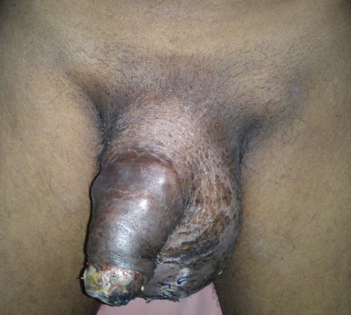
Figure 1: Photograph showing the extensive penile growth extending up to the pubic symphysis causing malignant priapism with bilateral enlarged inguinal lymphnodes.
Date added to bjui.org: 18/11/2012
DOI: 10.1002/BJUIw-2012-089-web