Tag Archive for: MRI

Posts

The PROMIS of MRI

Hashim AhmedThe prostate cancer pathway is controversial and views are often polarized. For a researcher, this is the perfect melting pot for innovation and practice-changing studies. It is clear that we need to reduce the harms of treatment, not only by treating very few low-risk cancers but also by innovations in surgery. It is pleasing to see Grasso et al. [1] systematic review of surgical innovation that may potentially lead to improvements in urinary incontinence after radical prostatectomy. This was a diligently conducted systematic review and points to the need for a randomized trial, which the authors tell us is currently being conducted.

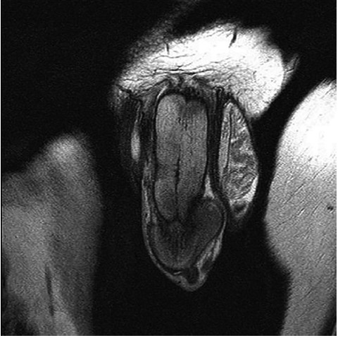
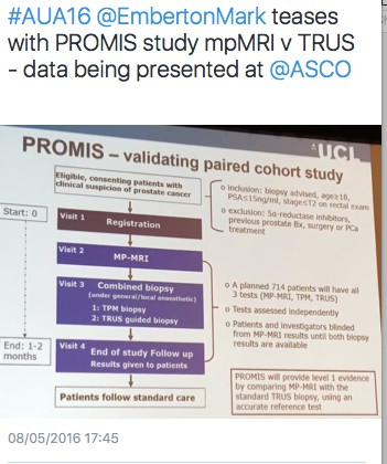
The era of multiparametric MRI (mpMRI) for prostate cancer diagnosis is upon us. Few of us will live through such a wholesale change in the entire pathway for diagnosis and treatment of a cancer, and a common one at that. Whilst a few of us have been using mpMRI prior to first biopsy, there can be no doubting that we now have level 1b evidence to support the adoption of mpMRI prior to a first prostate biopsy as the standard care. The NIHR-HTA/MRC-CTU/UCL PROstate MR Imaging Study (or PROMIS) has been long awaited, and its initial results were presented at ASCO last month [2]. mpMRI performed better than expectations in a multicentre setting across 11 NHS trusts and just over a dozen radiologists. Sensitivity was 93% (95% CI 88–96) and the negative predictive value was 89% (95% CI 83–94). Although the focus, quite rightly, has been on mpMRI, equally significant has been the discovery of how bad a test TRUS-guided biopsy really was, with a sensitivity for clinically significant prostate cancer of only 48% (95% CI 42–55).

These findings answer several criticisms of mpMRI. First, that it is not as accurate as retrospective data suggest. It is, provided you do not expect it to find every millimetre of significant disease. Second, it is not reproducible outside of expert centres. It is, provided you quality assure every scanner, optimize the sequences iteratively, quality control scans and have robust training for radiologists. Third, it cannot be carried out on 1.5-Tesla scanners. It can; all the PROMIS scans were 1.5 Tesla without an endorectal coil. Fourth, it misses lots of clinically significant prostate cancers. It does not, but this depends on your definition of clinical significance. In this respect, the study by Cash et al. [3] is pertinent. They evaluated the rates of subsequent cancer found on ‘negative’ mpMRIs and, using the very conservative Epstein definition, found a high rate of missed ‘significant’ cancers. The rate of Gleason 7 disease missed was lower and some missed cancers were attributable to interobserver variability in mpMRI reporting. All centres should evaluate their own data to determine where their own negative predictive value sits and then strive to improve upon this through a constant iterative dialogue between urology and radiology. PROMIS shows that mpMRI has very high performance characteristics that should be possible across the board.

There is considerable work still to be done. Cost-effectiveness analyses are under way; with these data, NICE will need to consider their clinical recommendations, having laboured the point that they wished to await PROMIS. The challenge of dissemination and maintenance of quality standards is not to be underestimated. Work on determining what is out there, who is capable of performing such scans and reporting them, whether there is enough capacity in the NHS and whether all centres are capable of carrying out targeted biopsies are all legitimate health policy issues.

Similar to mammography standards laid down centrally, we will need to insist on: independent (not self-) accreditation; independent scan and report audits, with outliers (too many negatives, too many positives, too many equivocals) reviewed to determine whether further standardization training is required; rates of clinically significant and insignificant cancers detected on subsequent biopsy; rates of repeat biopsies; and rates of unnecessary radical therapy on low risk cases. We should all look within our centres to ensure we can meet these expectations.


Hashim U. Ahmed, BJUI Consulting Editor – Imaging Division of Surgery and Interventional Sciences, UCL, and
Department of Urology, UCL Hospital NHS Foundation Trust, London, UK

References

1. Grasso AAC, Mistretta FA, Sandri M et al. Posterior musculofascial reconstruction after radical prostatectomy: an updated systematic review and a meta-analysis. BJU Int 2016; 118: 2034 Wiley Online Library

2. Ahmed HU. The PROMIS study: a paired-cohort, blinded confirmatory study evaluating the accuracy of multi-parametric MRI and TRUS biopsy in men with an elevated PSA. J Clin Oncol 2016; 34: (suppl; abstr 5000)

3. Cash H, Günzel K, Maxeiner A et al. Prostate cancer detection on transrectal ultrasonography-guided random biopsy despite negative real-time magnetic resonance imaging/ ultrasonography fusion-guided targeted biopsy: reasons for targeted biopsy failure. BJU Int 2016; 118: 3543 Wiley Online Library


West Coast Urology: Highlights from the AUA 2016 in San Diego… Part 2

By Ben Challacombe (@benchallacombe) and Jonathan Makanjuola (@jonmakurology)


The AUA meeting was starting to hot up with the anticipation of the Crossfire sessions, PSA screening and the MET debate that appeared to rumble on.  We attended the MUSIC (Michigan Urological Surgery Improvement Collaborative) session. It is a fantastic physician led program including >200 urologists, which aims to improve the quality of care for men with urological diseases. It is a forum for urologists across Michigan, USA to come together to collect clinical data, share best practices and implement evidence based quality improvement activities. One of their projects is crowd reviewing of RALP by international experts for quality of the nerve spare in order to improve surgical outcomes.

AUA2.1 AUA2.2


The MET debate continues to cause controversy. In the UK there has been almost uniform abandonment of the use of tamsulosin for ureteric stones following The Lancet SUSPEND RCT.

AUA2.4 AUA2.3


The MET crossfire debate was eagerly awaited. The debate was led by James N’Dow (@NDowJames) arguing against and Philipp Dahm (@EBMUrology) in favour of MET. Many have criticised the SUSPEND paper for lack of CT confirmation of stone passage. Dr Matlaga (@BrianMatlaga) stated that comparing previous studies of MET to SUSPEND is like comparing apples to oranges due to different outcome measures. He recommended urologists continue MET until more data is published. More conflicting statements were made suggesting that MET is effective in all patients especially for large stones in the ureter. The AUA guidelines update was released and stated that MET can be offered for distal ureteric stones less than 10mm.

AUA2.5 AUA2.6


In a packed Endourology video session there were many high quality video presentations. One such video was a demonstration of the robotic management for a missed JJ ureteric stent. Khurshid Ghani (@peepeeDoctor) presented a video demonstrating the pop-corning and pop-dusting technique with a 100w laser machine.

AUA2.7


One of the highlights of the Sunday was the panel discussion plenary session, Screening for Prostate Cancer: Past, Present and Future. In a packed auditorium Stacy Loeb (@LoebStacy), gave an excellent overview of PSA screening with present techniques including phi, 4K and targeted biopsies. Freddie Hamdy looked into the crystal ball and gave a talk on future directions of PSA testing and three important research questions that still needed to be answered. Dr. Catalona presented the data on PSA screening and the impact of the PLCO trial. He argued that due to inaccurate reporting, national organisations should restore PSA screening as he felt it saved lives.

AUA2.8 AUA2.9 AUA2.10

There was a twitter competition for residents and fellows requiring participants to  tweet an answer to a previously tweeted question including the hashtag #scopesmart and #aua16. The prize was Apple Watch. Some of the questions asked included; who performed the 1st fURS? And what is the depth of penetration of the Holmium laser?

UK trainees picked up the prizes on the first two days.

AUA2.11

The British Association of Urological Surgeons (BAUS) / BJU International (BJUI) / Urological Society of Australia and New Zealand (USANZ) session was a real highlight of day three of the AUA meeting. There were high quality talks from opinion leaders in their sub specialities. Freddie Hamdy from Oxford University outlined early thoughts from the protecT study and the likely direction of travel for management of clinically localised prostate cancer. Prof Emberton (@EmbertonMark) summarised the current evidence for the role of MRI in prostate cancer diagnosis including his thoughts on the on going PROMIS trial. Hashim Ahmed was asked if HIFU was ready for the primetime and bought us up to speed with the latest evidence.

AUA2.12 AUA2.13


The eagerly awaited RCT comparing open prostatectomy vs RALP by the Brisbane group was summarised with regards to study design and inclusion criteria. It is due for publication on the 18th May 2016 so there was a restriction of presenting results.  Dr Coughlin left the audience wanting more despite Prof. Dasgupta’s best effort to get a sneak preview of the results!  We learnt from BAUS president Mark Speakman (@Parabolics) about the UK effort to improve the quality of national outcomes database for a number of index urological procedures.

AUA2.14 AUA2.15


Oliver Wiseman (@OJWiseman) gave us a flavour of outcomes from the BAUS national PCNL database and how they are trying drive up standards to improve patient care. A paediatric surgery update was given by Dr Gundeti. The outcomes of another trial comparing open vs laparoscopic vs RALP was presented. There was no difference in outcomes between the treatment modalities but Prof. Fydenburg summarised by saying that the surgeon was more important determinant of outcome than the tool. Stacy Loeb closed the meeting with an excellent overview of the use of twitter in Urology, followed by a drinks reception.

AUA2.16 AUA2.17


It was not all about stones and robots. The results of the Refractory Overactive Bladder: Sacral NEuromodulation vs. BoTulinum Toxin Assessment (ROSETTA) trial results were presented. Botox came out on top against neuromodulation in urgency urinary incontinence episodes over 6 months, as well as other lower urinary tract symptoms.

AUA2.18


The late breaking abstract session presented by Stacy Loeb highlighted a paper suggesting a 56% reduction in high-grade prostate cancer for men on long term testosterone. This was a controversial abstract and generated a lot of discussion on social media.


AUA2.20 AUA2.21

AUA2.22

AUA2.23


It has been an excellent meeting in San Diego and we caught up with old and met new friends. It was nice to meet urologists from across the globe with differing priorities and pressures. There was a good British, Irish and Australian contingent flying the flag for their respective countries. It was another record-breaking year for the #AUA16 on twitter. It surpassed the stats for #AUA15 with over 30M impressions, 16,659 tweets 2,377 participants. See you all in Boston for AUA 2017.


West Coast Urology : Highlights from the AUA 2016 in San Diego… Part 1

By Ben Challacombe (@benchallacombe) and Jonathan Makanjuola (@jonmakurology)


The 2016 AUA returned to the beautiful city of San Diego set on the shores of the Pacific in an excellent conference centre located in the centre of the town adjacent to the Gaslamp district. For a change the wifi was excellent and allowed enhanced levels of social media interaction and urological discussion. Opening these interactions were 2 key sessions which provoked much debate. Firstly the announcement that after over 10 years of trying the FDA has approved HIFU treatment although it seemed to get there through a slightly “de novo” pathway. Apparently the FDA approved it as an ablation tools but not for prostate cancer.

AUA16.1

Although not directly approved for use in prostate cancer, that is exactly what it is going to be used for. A packed house saw a debate with evidence from both sides. Dr Nathan Lawrentschuk promoted the 4 Ds of HIFU. His key point was that 56/101 had a post treatment biopsy of which 51 where biopsy positive!

AUA16.2

The second big session focussed on the AUA/SAR consensus statement  document on prostate cancer diagnostics. This recommended a “High Quality” MRI should be strongly considered if patient has a rising PSA with a previous negative biopsy, has persistent clinical suspicion for prostate cancer or is undergoing a repeat biopsy. There was no mention of MRI for all at the pre-biopsy stage which many had hoped for and only 2 lines on trans-perineal biopsy as an option. This is of course related to health resources and the outpatient office-based nature of most USA urologists.

AUA16.3

A welcome innovation was the Crossfire Sessions which pitted 2 well known advocates of one treatment against 2 with the opposite views. It was hardly debating of the Oxbridge variety but none the less did provoke some useful discussions. Topics included radical prostatectomy vs radiotherapy, endoscopic vs nephro-ureterectomy management of upper tract TCC, and enucleation at partial nephrectomy vs formal resection. Standing room only at the back of the halls but no real audience interaction or voting which was a shame. 

AUA16.4

The session which really woke everyone up was Rene Sotalo’s wonderful complication horror show. Bleeding, bleeding and more bleeding in a variety of ways. How would you handle this he asked? Pray I thought! But this and similar sessions clearly show the benefits of recording all cases and reviewing these DVDs if something goes wrong. The cause of some complications were only identified by review of the intra-operative tapes. Some clinical titbits learn’t included  using only a horizontal incision for the camera port at RARP to reduce hernias and turning off pneumocompression stockings if there is a major venous injury to prevent excessive venous bleeding.

AUA16.5

From a SoME perspective there was both good and bad. One poster showed that 40% of graduating US residents had publicly accessible unprofessional content on social media. Food for thought at the consultant interview no doubt, but on the other side SoMe ranks third in the acquisition of urological knowledge (and climbing…). One hack produced this tweeting guideline for all to reflect on.

AUA16.6

Prof Prokar Dasgupta had the honour of presenting the widely anticipated session on emerging robotic technology . At last there appears to be some real competition to Intuitive’s dominance on the way. There are at least 3 credible robotic systems on the way. He finished with an intriguing slide on Dr Google being the most powerful doctor in the world!

AUA16.7

Despite Europe and Asia moving towards the use of PMSA PET , the USA is not moving in this direction due to reimbursement issues if the PMSA molecule.

AUA16.8

There was a lot of interest in a packed auditorium to see live surgery for a single use disposable fURS “Lithovue” with some reporting superior vision , optics and deflection.

AUA16.9AUA16.10


There were some sceptics amongst the stone community with the environmental impact and cost effectiveness a concern.

AUA16.11 AUA16.12


With the popular Gaslamp district a stones throw away many delegates went after the conference for a meal and drinks. The local baseball team San Diego Padres was a popular destination with may watching baseball for the 1st time whist others had gone for a run along the harbour and even caught a sighting of some seals!

AUA16.13AUA16.14

RE: Opportunity of widening the resort to multiparametric MRI/transrectal ultrasound fusion imaging-guided prostate cancer brachytherapy

Sir,

Thank you for your interest in our article regarding whole-gland brachytherapy to the prostate for prostate cancer (1). Your letter is highlighting the expanding role of brachytherapy to that of focal therapy (2). We agree that multiparametric magnetic resonance imaging (mpMRI) scans have expanded the ability to localise tumours and indeed that they may be useful in carefully selected men wishing to undergo focal therapy. However, other  advances such as the use of fiducial markers and spacers have also allowed better dosimetry and for a reduction in side effects (3).  The safety and performance of brachytherapy in whole gland treatment means we should have faith in it as a modality to destroy cancer on the focal therapy setting. There are new trials being developed with focal brachytherapy and we look forward to the results in the coming years.


References

  1. Chao MW, Grimm P, Yaxley J, Jagavkar R, Ng M, Lawrentschuk N. Brachytherapy: state-of-the-art radiotherapy in prostate cancer. BJU Int  2015; 116(S3): 80-8. doi: 10.1111/bju.13252.
  1. Nguyen PL, Trachtenberg J, Polascik TJ. The role of focal therapy in the management of localised prostate cancer: a systematic review. Eur Urol. 2014 Oct;66(4):732-51. doi: 10.1016/j.eururo.2013.05.048. Epub 2013 Jun 6. Review.
  1. Ng M1, Brown E, Williams A, Chao M, Lawrentschuk N, Chee R. BJU Int. 2014 Mar;113 Suppl 2:13-20. doi: 10.1111/bju.12624. Fiducial markers and spacers in prostate radiotherapy: current applications.


Letter to the Editor

Opportunity of widening the resort to multiparametric MRI/transrectal ultrasound fusion imaging-guided prostate cancer brachytherapy  

Sir,

I have recently read, with high interest, the review article “Brachytherapy: state-of-the-art radiotherapy in prostate cancer”, by Chao et al.[1].  The authors made extremely clear the advanced technologies of computerized treatment planning and imaging-guided delivery modalities to reach a tailored ablative prostate tumor target dose by resorting to either low-dose-rate (LDR) or high-dose-rate (HDR) different brachytherapy procedures as regards three basic – low, intermediate, high – disease risk classificative conditions.   

It is today proven that focal instrumental procedures inside the prostate gland – from biopsy to various prostate cancer focused ablative strategies, among which laser interstitial thermal therapy and particularly the prostate cancer brachytherapy – might require the resort to proper software digital overlay-mediated fusion of both beforehand multiparametric magnetic resonance imaging (mpMRI) scans and later real-time transrectal 3D ultrasound findings.  Like this, indeed, intriguing developments  in software modelling techniques have led to reach, by a mpMRI-ultrasound image fusion approach, more accurate targeted prostate cancer biopsies than those by transrectal ultrasound imaging alone achieved [2,3].

If the transperineal focal laser prostate tumor ablation  usually occurs only with the guidance of mpMRI (T2-weighted, diffusion-weighted, dynamic contrast material) [4], as regards the prostate cancer brachytherapy, instead, it is more and more timely, for just targeting the tumor “index-dominant lesion”, the resort to mpMRI/transrectal real-time ultrasound fusion imaging.  Quite recently, mpMRI/real-time transrectal ultrasound software-mediated digital co-registration has allowed to properly carry-out, in patients suffering from intermediate/high risk prostate carcinoma with mpMRI visible “index- dominant” intraprostatic nodule, the HDR ¹⁹² Ir transperineal temporary implant-brachytherapy  as accurate partial prostate radiation dose escalation supplemental to hypofractionated external beam radiotherapy [5,6].   

Given the interesting, even rare, reports on this subject, it would be advisable to widen the resort to the above-outlined mpMRI/transrectal  ultrasound fusion imaging-guided prostate cancer brachytherapy, particularly for a suitably targeted dominant tumor nodule detection/ablation.


Contardo Alberti

L D of Surgical Semeiotics, University of Parma, Parma, Italy


 References

1  Chao MW, Grimm P, Yaxley J, Jagavkar R, Ng M, Lawrentschuk N. Brachytherapy: state-of-the-art radiotherapy in prostate cancer. BJU Int  2015; 116(S3): 80-8. doi: 10.1111/bju.13252.

2  Shoji S, Hiraiwa S, Endo J, Hashida K, Tomonaga T, Nakano M et al. Manually controlled targeted prostate biopsy with real-time fusion imaging of multiparametric magnetic resonance imaging and stransrectal ulrasound :an early experience. Int J Urol 2015; 22(2): 173-8. doi: 10.1111/iju.12643.

 3  Marks L, Young S, Natarajan S.  MRI-ultrasound fusion for guidance of targeted prostate biopsy. Curr Opin Urol 2013; 23(1): 43-50. doi: 10.1097/MOU.0b013e32835ad3ee.

4  Woodrum DA, Kawashima A, Gorny KR, Mynderse LA. Magnetic resonance-guided thermal therapy for localized and recurrent prostate cancer. Magn Reson Imaging Clin N Am.2015; 23(4): 607-19. doi:10.1016/j.mric.2015.05.014

5  Bubley GJ, Bloch BN, Vazquez C, Genega E, Holupka E, Rofsky N, Kaplan I.  Accuracy of endorectal magnetic resonance/transrectal ultrasound fusion for detection of prostate cancer during brachytherapy. Urology 2013;81(6): 1284-9. doi: 10.1016/j.urology.2012.12.051.

6  Gomez-Iturriaga A, Casquero F, Urresola A, Ezquerro A, Lopez JI, Espinosa JM et al. Dose escalation to dominant intraprostatic lesions with MRI-transrectal  ultrasound fusion high-dode-rate prostate brachytherapy. Radiother Oncol 2016 Feb 15. doi: 10.1016/j.radonc.2016.02.004 (Epub ahead of print).


Article of the Week: Combination of mpMRI and TTMB of the prostate to identify candidates for hemi-ablative FT

Every Week the Editor-in-Chief selects an Article of the Week from the current issue of BJUI. The abstract is reproduced below and you can click on the button to read the full article, which is freely available to all readers for at least 30 days from the time of this post.

In addition to the article itself, there is an accompanying editorial written by a prominent member of the urological community. This blog is intended to provoke comment and discussion and we invite you to use the comment tools at the bottom of each post to join the conversation.

Finally, the third post under the Article of the Week heading on the homepage will consist of additional material or media. This week we feature a video from Mr. Mark Emberton, discussing his paper. 

If you only have time to read one article this week, it should be this one.

Combination of multi-parametric magnetic resonance imaging (mp-MRI) and transperineal template-guided mapping biopsy (TTMB) of the prostate to identify candidates for hemi-ablative focal therapy

Minh Tran*†‡, James Thompson*§, Maret Bohm†, Marley Pulbrook, Daniel Moses¶, Ron Shnier**, Phillip Brenner*§, Warick Delprado††, Anne-Maree Haynes†, Richard Savdie§ and Phillip D. Stricker*§


*St Vincents Prostate Cancer CentreGarvan Institute of Medical Research & The Kinghorn Cancer Centre, DarlinghurstSchool of Medicine, University of Sydney§School of Medicine, University of New South Wales, SydneySpectrum Medical Imaging , **Southern Radiology, Randwick, and†† Douglass Hanly Moir Pathology, Darlinghurst, NSW, Australia


Read the full article
OBJECTIVE

To evaluate the accuracy of combined multiparametric magnetic resonance imaging (mpMRI) and transperineal template-guided mapping biopsy (TTMB) for identifying lobes with significant prostate cancer (PCa) for the application of hemi-ablative focal therapy (FT).

PATIENTS AND METHODS

From January 2012 to January 2014, 89 consecutive patients, aged ≥40 years, with a PSA level ≤15 ng/mL, underwent in sequential order: mpMRI, TTMB and radical prostatectomy (RP) at a single centre. Analysis was performed on 50 patients who met consensus guidelines for FT. Lobes were stratified into lobes with significant cancer (LSC), lobes with insignificant cancer and lobes with no cancer. Using histopathology at RP, the predictive performance of combined mpMRI + TTMB in identifying LSC was evaluated.

RESULTS

The sensitivity, specificity and positive predictive value for mpMRI + TTMB for LSC were 97, 61 and 83%, respectively. The negative predictive value (NPV), the primary variable of interest, for mpMRI + TTMB for LSC was 91%. Of the 50 patients, 21 had significant unilateral disease on mpMRI + TTMB. Two of these 21 patients had significant bilateral disease on RP not identified on mpMRI + TTMB.

CONCLUSIONS

In the selection of candidates for FT, a combination of mpMRI and TTMB provides a high NPV in the detection of LSC.

Read more articles of the week

Editorial: Doubling our precision of risk stratification in early prostate cancer? Too good to be true?

It is difficult to over-estimate the enormity of the revolution involved in transitioning away from an organ-based system of care in prostate cancer (prostatectomy or radiotherapy to prostate cancer of any grade, number, location or volume) to one in which the target condition is defined and verified, and then subsequently treated with a margin. Since Hugh Hampton-Young undertook the first radical prostatectomy more than 100 years ago, an organ-based system of care has been the only plausible strategy for men with early-stage prostate cancer. This is because our risk stratification methods could certainly tell us who had prostate cancer when it was indeed identified, but they could not tell us with any degree of precision how much cancer was present (number of foci and volume of cancer), what grade it was and where within the prostate the cancer resided. Surgery or radiotherapy to the whole gland, it was thought, was a reasonable mitigation (at the cost of over-treatment in a significant proportion of men) to the systematic under-estimation of risk to the patient. The degree to which our attempts at risk stratification failed our patients was emphasized in a recent UK report that showed an increase of >50% in histological grade (the most important determinant of risk) at radical prostatectomy compared with the risk the patient was told at the time of diagnosis[1]. ‘Upgrading’ is an inverse measure of the quality of our risk stratification.

With this background it should be of little surprise that there remains considerable skepticism about our ability to localize disease within the prostate, define its volume with some degree of precision and identify the worst histological grade within the tumour most of the time. These conditions, which most patients, quite reasonably, might expect of a modern cancer diagnostic programme, need to be fulfilled if we are to move away from an organ-based strategy for all towards a system of care that risk stratifies with precision, treats only those who are likely to benefit and tries to preserve tissue and function when we are able to do so.

The technologies and developments that have permitted a reduction in risk stratification error from 50 to ~5% are the subject of a paper by Tran et al. [2] in the present issue of BJUI. This group from Australia exploited a cohort of men that underwent a series of tests that culminated in a radical prostatectomy. These comprised: high-quality multiparametric MRI at a range of magnet strengths; formal scoring of the MRI using an ordinal scale of risk; a subsequent 5-mm transperineal template-guided biopsy modified from Winston Barzell’s original account; and additional sampling of prostate sectors that corresponded to a high likelihood of clinically significant cancer based on the MRI reading, a process otherwise known as ‘targeting’. The outputs of these tests were compared with the presence or absence of clinically significant prostate cancer in the 100 prostate lobes evaluated from the 50 eligible men who underwent surgery. The authors’ a priori threshold for declaring clinically significant disease in the tests was the presence or a PIRADS 4–5 MRI lesion (with or without concordant pathology) and/or the presence of exclusive Gleason pattern 3 amounting to ≥4 mm maximum cancer core length or the presence of any Gleason pattern 4 or 5 [3]. At radical prostatectomy slightly different criteria were employed. Clinically significant prostate cancer constituted an exclusive Gleason pattern 3 lesion provided it was ≥1.3 mL. The presence of patterns 4 and 5 within the lesion triggered ‘significance’, as did evidence of capsular invasion or extraprostatic extension.

Although it was a small study, the men included were exposed to the best diagnostic profile that it was possible to have and were subjected to a reference test which most of us would trust. What did they find? Just how well did a modern diagnostic panel rule in or rule out prostate cancer that exceeded a minimum threshold of 4 mm of Gleason pattern 3? Within the hundred lobes that were evaluated, 21 clinically significant cancers were identified in the diagnostic process. At radical prostatectomy two of these were at the midline and therefore attributed, within the rules, to both sides of the gland. In one of these cases there was a 5-mm diameter (0.1 mL) Gleason 3+4 lesion with 10% Gleason pattern 4 that was overlooked by the combined diagnostic process. A lesion of this volume can evade a well-applied sampling strategy based on a 5-mm sampling frame, especially if the lesion is non-spherical. The relatively low component of pattern 4 combined with the relatively low volume means that MRI would also have a hard time detecting it. Under the conditions described in the present paper, the authors concluded that combined MRI and intensive biopsy conferred a sensitivity of 97% and a negative predictive value of 91% for a fairly conservative definition of clinically significant disease. This level of accuracy, which is nearly twice as good as that of which we were previously capable, will result in major benefits for patients in terms of communicating risk with an order of precision that was hitherto not possible. This should translate to more appropriate treatment allocation, both avoiding unnecessary treatment and having treatment when it is likely to be beneficial. It should also result in a significant proportion of patients being offered the option of a tissue-preserving therapy when this is an option [4]. Most importantly, this new precision opens up an opportunity for greater involvement of the patient in the process of informed decision-making [5]. Something that was not really possible in the face of yesterday’s diagnostic uncertainty.

Read the full article
Mark Emberton
Division of Surgery and Interventional Science, University College London, London, UK


References


Video: Combination of mpMRI and TTMB of the prostate to identify candidates for hemi-ablative FT

Combination of multi-parametric magnetic resonance imaging (mp-MRI) and transperineal template-guided mapping biopsy (TTMB) of the prostate to identify candidates for hemi-ablative focal therapy

Minh Tran*†‡, James Thompson*§, Maret Bohm†, Marley Pulbrook, Daniel Moses¶, Ron Shnier**, Phillip Brenner*§, Warick Delprado††, Anne-Maree Haynes†, Richard Savdie§ and Phillip D. Stricker*§


*St Vincents Prostate Cancer CentreGarvan Institute of Medical Research & The Kinghorn Cancer Centre, DarlinghurstSchool of Medicine, University of Sydney§School of Medicine, University of New South Wales, SydneySpectrum Medical Imaging , **Southern Radiology, Randwick, and†† Douglass Hanly Moir Pathology, Darlinghurst, NSW, Australia


Read the full article
OBJECTIVE

To evaluate the accuracy of combined multiparametric magnetic resonance imaging (mpMRI) and transperineal template-guided mapping biopsy (TTMB) for identifying lobes with significant prostate cancer (PCa) for the application of hemi-ablative focal therapy (FT).

PATIENTS AND METHODS

From January 2012 to January 2014, 89 consecutive patients, aged ≥40 years, with a PSA level ≤15 ng/mL, underwent in sequential order: mpMRI, TTMB and radical prostatectomy (RP) at a single centre. Analysis was performed on 50 patients who met consensus guidelines for FT. Lobes were stratified into lobes with significant cancer (LSC), lobes with insignificant cancer and lobes with no cancer. Using histopathology at RP, the predictive performance of combined mpMRI + TTMB in identifying LSC was evaluated.

RESULTS

The sensitivity, specificity and positive predictive value for mpMRI + TTMB for LSC were 97, 61 and 83%, respectively. The negative predictive value (NPV), the primary variable of interest, for mpMRI + TTMB for LSC was 91%. Of the 50 patients, 21 had significant unilateral disease on mpMRI + TTMB. Two of these 21 patients had significant bilateral disease on RP not identified on mpMRI + TTMB.

CONCLUSIONS

In the selection of candidates for FT, a combination of mpMRI and TTMB provides a high NPV in the detection of LSC.

Read more articles of the week
© 2024 BJU International. All Rights Reserved.