Tag Archive for: #PCSM

Posts

Editorial: The way towards understanding possible multiple functions of AR V7 in prostate cancer

The presence of constitutively active androgen receptors in castration therapy‐resistant prostate cancer is frequently associated with therapy resistance. This is not surprising because the transcriptional program of these receptors is not dependent on the presence of circulating androgen. In conditions of reduced expression of circulating androgen, functional activity of the receptor probably contributes to cancer progression. Investigations by Bernemann et al. [12], however, showed that some patients who present with variant ARV7, the most frequently diagnosed variant receptor, still respond to second‐generation anti‐androgen therapy. Until publication of the paper by Bernemann et al. [1], it was not completely clear whether “any findings in this area” reflected a technical error. The authors report further technical advances and similar detection of variant androgen receptors with two PCR assays, the SYBR Green and TaqMan assays. These advances in detection may open up a new area of investigation. The study reported in the current issue of BJUI may therefore shed more light on biology of truncated androgen receptors in prostate cancer [1]. According to the initial seminal publication in the field, AR‐V7‐positive patients had lower endocrine therapy response rates than those who were variant‐negative [3]. Studies aiming to detect variant androgen receptors are particularly important because of increasing interest in circulating tumour cells in prostate cancer [4]. It may be necessary to better describe subgroups of AR‐V7 which may differ in interactions with specific coactivators. Overall, relatively little is known about alterations in interactions between coactivators and the N‐terminal region of the receptor that may occur in subgroups of patients with castration therapy‐resistant prostate cancer [5]. Several important questions regarding signalling between the wild‐type and constitutively active androgen receptor have not been completely clarified, and the issue of which genes are regulated by both receptors is still a matter of discussion. The findings presented in the study by Bernemann et al. [1] may represent the next step towards individualization of therapies. If we accept that variant androgen receptors also display heterogeneity, a more differentiated classification of those receptors may guide clinical decisions in the future. Future studies should also take into consideration the fact that different variants may be expressed at different levels during and after endocrine therapy. These ratios of androgen receptor variant expression may be taken into consideration when determining the probability of success of specific anti‐androgen receptor therapy. One could also learn that application of different methodologies in variant androgen receptor diagnostics may become an established standard in monitoring castration therapy‐resistant disease. Establishment of controlled standard operating procedures in PCR diagnostics may at this stage minimize discrepancies between findings reported by different researchers and help to establish a consensus on this important topic.

Zoran Culig

Experimental Urology, Department of Urology, Medical University of Innsbruck, Anichstrasse 35, A-6020 Innsbruck, Austria

References

  1. Bernemann C, Steinestel J, Humberg V, Bögemann M, Schrader AJ, Lennerz JK. Performance comparison of two AR‐V7 detection methods. BJU Int 2018122: 219–26


Article of the Week: Cost‐effectiveness of MRI and targeted fusion biopsy for early detection of prostate cancer

Every Week, the Editor-in-Chief selects an Article of the Week from the current issue of BJUI. The abstract is reproduced below and you can click on the button to read the full article, which is freely available to all readers for at least 30 days from the time of this post.

In addition to the article itself, there is an accompanying editorial written by a prominent member of the urological community. This blog is intended to provoke comment and discussion and we invite you to use the comment tools at the bottom of each post to join the conversation.

If you only have time to read one article this week, it should be this one.

Cost‐effectiveness of magnetic resonance imaging and targeted fusion biopsy for early detection of prostate cancer

Christine L. Barnett* , Matthew S. Davenport, Jeffrey S. Montgomery, John T. WeiJames E. Montie‡ and Brian T. Denton*


*Departments of Industrial and Operations Engineering, Radiology, and Urology, University of Michigan, Ann Arbor, MI, USA


Read the full article

Objective

To determine how best to use magnetic resonance imaging (MRI) and targeted MRI/ultrasonography fusion biopsy for early detection of prostate cancer (PCa) in men with elevated prostate‐specific antigen (PSA) concentrations and whether it can be cost‐effective.

Methods

A Markov model of PCa onset and progression was developed to estimate the health and economic consequences of PCa screening with MRI. Patients underwent PSA screening from ages 55 to 69 years. Patients with elevated PSA concentrations (>4 ng/mL) underwent MRI, followed by targeted fusion or combined (standard + targeted fusion) biopsy on positive MRI, and standard or no biopsy on negative MRI. Prostate Imaging Reporting and Data System (PI‐RADS) score on MRI was used to determine biopsy decisions. Deaths averted, quality‐adjusted life‐years (QALYs), cost and incremental cost‐effectiveness ratio (ICER) were estimated for each strategy.


Results

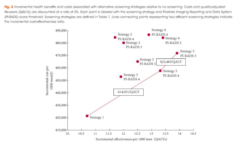
With a negative MRI, standard biopsy was more expensive and had lower QALYs than performing no biopsy. The optimum screening strategy (ICER $23 483/QALY) recommended combined biopsy for patients with PI‐RADS score ≥3 and no biopsy for patients with PI‐RADS score <3, and reduced the number of screening biopsies by 15%. Threshold analysis suggests MRI continues to be cost‐effective when the sensitivity and specificity of MRI and combined biopsy are simultaneously reduced by 19 percentage points.

Conclusions

Our analysis suggests MRI followed by targeted MRI/ultrasonography fusion biopsy can be a cost‐effective approach to the early detection of PCa.

Read more articles of the week

Editorial: MRI with targeted fusion biopsy: is the time now?

The current standard of performing TRUS‐guided systematic biopsy in men with a PSA of 4–10 ng/mL results in a considerable number of unnecessary prostate biopsies and overtreatment of clinically indolent disease, both of which are costly from the patient and healthcare system perspectives [1]. Two recent studies document that incorporating multiparametric (mp)MRI into prostate cancer screening has the potential to reduce overdiagnosis and overtreatment. Ahmed et al. [2] performed mpMRI followed by TRUS biopsy and template prostate mapping biopsy in 576 men with a PSA level of 4–14 ng/mL or an abnormal DRE. They found that using mpMRI to triage men would result in 27% fewer men undergoing biopsy while diagnosing 5% fewer clinically insignificant cancers. Furthermore, if subsequent TRUS biopsies were directed by mpMRI findings, 18% more cases of clinically significant cancer might be detected compared with traditional screening biopsies. Using a similar randomized study design, Kasivisvanathan et al. [3] found the use of mpMRI for screening reduced the biopsy rate by 28%, while lowering the rate of clinically insignificant cancer by 13% and improving the detection rate of clinically significant cancer by 12% [3]. While the addition of mpMRI to the screening armamentarium clearly provides clinical value, it also adds considerable increased costs, begging the question: is it worth it?

In the current issue of BJUI, Barnett et al. [1] evaluated the cost‐effectiveness of prostate MRI in a screening setting in order to determine whether MRI may be able to mitigate prostate biopsies in biopsy‐naïve men with a negative imaging study. They found that, when using the generally accepted threshold of $100 000/quality‐adjusted life year (QALY), the most cost‐efficient strategy consisted of obtaining prostate MRI in men with PSA >4 mg/mL. This was followed by either systematic and targeted fusion biopsy if the MRI showed a Prostate Imaging Reporting and Data System (PI‐RADS) score ≥3 lesion or continued observation if the MRI did not show a PI‐RADS score ≥3 lesion. Altogether, this strategy resulted in 5.9 prostate cancer deaths averted, 60.7 QALYs gained, and 72.6 life‐years gained for every 1000 patients screened. The number‐needed‐to‐treat to prevent one prostate cancer death with this approach was 169.

While the results are informative, the reader should interpret the study’s findings carefully as there is not agreement among policy makers on what is the optimal incremental willingness‐to‐pay threshold to determine what is truly cost‐effective. Furthermore, this study does not address the potential negative impact that detection of clinically insignificant prostate cancer may have on health‐state utilities and quality of life. Barnett et al. also predominantly obtained costs from Medicare data, but as previously reported, these costs are often rough estimates that are derived more from revenue or reimbursement than from the true cost of treating the disease process. The cost‐effectiveness estimates presented in this report, therefore, will probably vary from country to country. To examine the true cost, Laviana et al. [4] previously explored the cost of prostate MRI using time‐driven activity‐based costing and found the cost of MRI and mpMRI/TRUS to be $670 and $1,072 US dollars, respectively. These are significantly cheaper than the Medicare reported costs of $964.21 and $3,019.35 for MRI and mpMRI/TRUS, respectively. This implies that MRI and targeted biopsy may actually be even more cost‐effective than reported in the present study.

We must remember that, while the present study shows that MRI in prostate cancer screening may be cost‐effective, the base case population only included men with a PSA >4 mg/mL. This study does not address the cost‐effectiveness of a screening MRI in the larger population of all men at risk of prostate cancer, although previous data by Nam et al. [5] suggest MRI may be a potentially better primary screening tool than PSA. Of course, population‐wide prostate MRI screening would result in greatly increased upfront costs and, as such, the strategy would first need to be proven cost‐effective before MRI could replace PSA as the front‐line test in prostate cancer screening.

In conclusion, Burnett et al. elegantly demonstrate that an upfront MRI, in men with a PSA of >4 ng/mL, may ultimately be a cost‐effective approach depending on your willingness‐to‐pay threshold. Policymakers and payers worldwide should recognize that prostate MRI is here to stay and should be made more widely available to those with a PSA >4 ng/mL who are at risk of prostate cancer.

Aaron A. Laviana* and David F. Penson*
*Department of Urological Surgery, Vanderbilt University, and VA Tennessee Valley Geriatric Research, Education and Clinical Centre, Nashville, TN, USA


Read the full article
References
  1. Barnett C, Davenport M, Montgomery J et al. Cost‐effectiveness of magnetic resonance imaging and targeted fusion biopsy for early detection of prostate cancer. BJU Int 2018122: 50–8
  2. Ahmed HU, El‐Shater BA, Brown LC et al. Diagnostic accuracy of multi‐parametric MRI and TRUS biopsy in prostate cancer (PROMIS): a paired validating confirmatory study. Lancet 2017389: 815–22
  3. Kasivisvanathan V, Rannikko AS, Borghi M et al. MRI‐targeted or standard biopsy for prostate‐cancer diagnosis. N Engl J Med 2018; 378: 1767-77
  4. Laviana AA, Ilg AM, Veruttipong D et al. Utilizing time‐driven activity‐based costing to understand the short‐ and long‐term costs of treating localized, low‐risk prostate cancer. Cancer 2016122: 447–55
  5. Nam RK, Wallis CJD, Stojcic‐Bendavid J et al. A pilot study to evaluate the role of magnetic resonance imaging for prostate cancer screening in the general population. J Urol 2016192: 361–6


Article of the Month: MRI supported transperineal prostate biopsy

Every Month, the Editor-in-Chief selects an Article of the Month from the current issue of BJUI. The abstract is reproduced below and you can click on the button to read the full article, which is freely available to all readers for at least 30 days from the time of this post.

In addition to the article itself, there is an accompanying editorial written by a prominent member of the urological community. This blog is intended to provoke comment and discussion and we invite you to use the comment tools at the bottom of each post to join the conversation.

If you only have time to read one article this week, it should be this one.

Multicentre evaluation of magnetic resonance imaging supported transperineal prostate biopsy in biopsy‐naïve men with suspicion of prostate cancer


Nienke L. Hansen*1, Tristan Barrett*, Claudia Kesch, Lana Pepdjonovic§, David Bonekamp, Richard OSullivan**, Florian Distler, Anne Warren*††, Christina Samel‡‡Boris Hadaschik2, Jeremy Grummet§ and Christof Kastner*§§
*CamPARI Clinic, Department of Radiology, Addenbrookes Hospital and University of Cambridge, Cambridge, UK, Department of Urology, University Hospital Heidelberg, Heidelberg, Germany, §Australian Urology Associates and Department of Surgery, Central Clinical School, Monash University, Melbourne, Vic., Australia, Department of Radiology, German Cancer Research Center (DKFZ), Heidelberg, Germany, **Healthcare Imaging and Monash University, Melbourne, Vic., Australia, ††Department of Pathology, Addenbrookes Hospital and University of Cambridge, Cambridge, UK, ‡‡Institute of Medical Statistics, Informatics and Epidemiology, University Hospital Cologne, Cologne, Germany, and §§Department of Urology, Addenbrookes Hospital and University of Cambridge, Cambridge, UK 


Current addresses: 1Department of Diagnostic and Interventional Radiology University Hospital Cologne Cologne Germany, 2Department of Urology University Hospital Essen Essen Germany. 


B.H., J.G., and C.K. contributed equally to this work.


Read the full article

Abstract

Objectives

To analyse the detection rates of primary magnetic resonance imaging (MRI)‐fusion transperineal prostate biopsy using combined targeted and systematic core distribution in three tertiary referral centres.

Patients and Methods

In this multicentre, prospective outcome study, 807 consecutive biopsy‐naïve patients underwent MRI‐guided transperineal prostate biopsy, as the first diagnostic intervention, between 10/2012 and 05/2016. MRI was reported following the Prostate Imaging‐Reporting and Data System (PI‐RADS) criteria. In all, 236 patients had 18–24 systematic transperineal biopsies only, and 571 patients underwent additional targeted biopsies either by MRI‐fusion or cognitive targeting if PI‐RADS ≥3 lesions were present. Detection rates for any and Gleason score 7–10 cancer in targeted and overall biopsy were calculated and predictive values were calculated for different PI‐RADS and PSA density (PSAD) groups.

Results

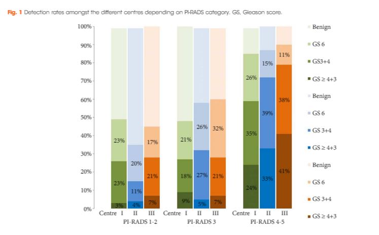
Cancer was detected in 68% of the patients (546/807) and Gleason score 7–10 cancer in 49% (392/807). The negative predictive value of 236 PI‐RADS 1–2 MRI in combination with PSAD of <0.1 ng/mL/mL for Gleason score 7–10 was 0.91 (95% confidence interval ± 0.07, 8% of study population). In 418 patients with PI‐RADS 4–5 lesions using targeted plus systematic biopsies, the cancer detection rate of Gleason score 7–10 was significantly higher at 71% vs 59% and 61% with either approach alone (P < 0.001). For 153 PI‐RADS 3 lesions, the detection rate was 31% with no significant difference to systematic biopsies with 27% (P > 0.05). Limitations include variability of multiparametric MRI (mpMRI) reading and Gleason grading.

Conclusion

MRI‐based transperineal biopsy performed at high‐volume tertiary care centres with a significant experience of prostate mpMRI and image‐guided targeted biopsies yielded high detection rates of Gleason score 7–10 cancer. Prostate biopsies may not be needed for men with low PSAD and an unsuspicious MRI. In patients with high probability lesions, combined targeted and systematic biopsies are recommended.

Read more articles of the week

Editorial: Systematic transperineal and MRI‐targeted biopsies: the resolution of uncertainty

The paper published in this issue of the BJUI titled ‘Multicentre evaluation of magnetic resonance imaging supported transperineal biopsy in biopsy‐naïve men with suspicion of prostate cancer’ is timely and helps to resolve some of the uncertainty inherent within the diagnostic pathway 1.

The publication of the PROstate MRI Imaging Study (PROMIS) study, although demonstrating that 25% of patients might avoid prostate biopsy with a normal MRI (Prostate Imaging Reporting and Data System [PI‐RADS] 1–2) and that MRI could identify 90% of patients with high‐risk disease (PI‐RADS 5), did not resolve the issue of what to do with equivocal PI‐RADS 3 scans, uncertainty remained 2. The recent publication of the PRECISION trial (Prostate Evaluation for Clinically Important Disease: Sampling Using Image Guidance or Not?) has only contributed to the uncertainty of systematic TRUS biopsy and has shown that targeted biopsies resolve the issue for <50% of the patients overall and only 12% of those with PI‐RADS 3 lesions had a diagnosis of cancer on targeted biopsy only 3. The study has shown that in the face of an identifiable lesion a MRI‐targeted biopsy is non‐inferior to a blind systematic TRUS biopsy, which was positive in only 28% and implies that a systematic biopsy may be unnecessary, so where does that leave us? The uncertainty within MRI remains at the PI‐RADS 3 level, and particularly with a TRUS biopsy that is not a systematic biopsy of the peripheral zone. The authors of the paper highlighted in this issue of the BJUI 1 help to resolve the issue because they describe a more systematic biopsy.

The transperineal (TP) biopsy approach for systematic and targeted biopsy they use is that which was adopted by the Ginsburg Study Group on Enhanced Prostate Diagnostics 4. It is a systematic biopsy that preferentially targets the peripheral zone in a sectoral fashion. It avoids the oversampling inherent in template‐mapping biopsy and the under‐sampling of the non‐systematic transrectal biopsy. Their paper evaluates the combination of an MRI‐targeted biopsy with a systematic TP biopsy. It confirms, as suggested by the PROMIS study, that patients with PI‐RADS 1 or 2 prostates on MRI with a low PSA density <0.1 ng/mL/mL could safely avoid biopsy, based upon a negative predictive value of 0.91 on systematic biopsy. However, in 418 patients with PI‐RADS 4–5 lesions, it was the combination of a targeted and systematic TP biopsy that achieved an overall cancer detection rate of 71%, but that MRI‐targeted biopsies alone had a detection rate of 59% vs 61% for systematic TP biopsies. In the PI‐RAD 3 equivocal group the combined biopsy identified 30% with Gleason score 7–10, whereas targeted biopsy only was positive in 21% vs 27% with systematic biopsies.

The message is clear.

An appropriate systematic biopsy targeted to the peripheral zone remains an essential component of prostate diagnosis even in the MRI era, as indeed it did before MRI was available. In the pre‐MRI days, about one‐third of patients that had negative TRUS biopsies had cancer on TP biopsies and a third of those thought suitable for AS on TRUS biopsy had more significant disease. I suspect in the modern era that figure remains unchanged for those with PI‐RADS 1, 2 or 3, particularly with a PSA density >0.15 ng/mL/mL. As urologists we have always been criticised for over diagnosing and over treating prostate cancer but I suspect that the more heinous crime is that of under treatment of significant disease, it is the very reason why I started doing TP biopsies, to resolve uncertainty. I consider that MRI, for all its benefits in the diagnostic algorithm, cannot yet resolve that uncertainty.

Probably the only patients that merit a target‐only biopsy are those with the high‐PSA, large‐volume disease, easily visible on MRI and usually palpable. Prostate biopsy can be avoided or at least deferred in the PI‐RADS 1–2 group with low PSA density; the rest should be offered a systematic biopsy along with a targeted biopsy. This may be less important in those proceeding to whole gland treatment or surgical extirpation but remains essential in those considering active surveillance, brachytherapy, or any one of the myriad of unproven focal treatments becoming available. The authors should be congratulated for bringing some certainty to uncertainty.

Rick Popert
Urology Centre, Guys Hospital, London, UK


Read the full article
References
  • Hansen NL, Barrett T, Kesch C et al. Multicentre evaluation of magnetic resonance imaging supported transperineal prostate biopsy in biopsy‐naïve men with suspicion of prostate cancerBJU Int 2018122: 40–9


  • Ahmed HU, El‐Shater Bosaily A, Brown LC et al. Diagnostic accuracy of multi‐parametric MRI and TRUS biopsy in prostate cancer (PROMIS): a paired validating confirmatory studyLancet 2017389: 815–22


  • Kuru TH, Wadhwa K, Chang RT et al. Definitions of terms, processes and a minimum dataset for transperineal prostate biopsies: a standardization approach of the Ginsburg Study Group for Enhanced Prostate DiagnosticsBJU Int 2013112: 568–77


Article of the Week: Focal irreversible electroporation as primary treatment for localized prostate cancer

Every Week, the Editor-in-Chief selects an Article of the Week from the current issue of BJUI. The abstract is reproduced below and you can click on the button to read the full article, which is freely available to all readers for at least 30 days from the time of this post.

In addition to the article itself, there is an accompanying editorial written by a prominent member of the urological community. This blog is intended to provoke comment and discussion and we invite you to use the comment tools at the bottom of each post to join the conversation.

If you only have time to read one article this week, it should be this one.

Focal irreversible electroporation as primary treatment for localized prostate cancer


Willemien van den Bos*†‡, Matthijs J. Scheltema*†‡, Amila R. Siriwardana*Anton M.F. Kalsbeek*, James E. Thompson, Francis Ting*, Maret Bohm*,
Anne-Maree Haynes*, Ron Shnier§, Warick Delprado¶ and Phillip D. Stricker


*Garvan Institute of Medical Research and Kinghorn Cancer Centre, St Vincents Prostate Cancer Centre, Darlinghurst, NSW, Australia, Academic Medical Centre, University of Amsterdam, Amsterdam, The Netherlands, §Southern Radiology, Randwick, and Douglass Hanly Moir Pathology, Macquarie Park, NSW, Australia


Read the full article

Abstract

Objectives

To determine the safety, quality of life (QoL) and short‐term oncological outcomes of primary focal irreversible electroporation (IRE) for the treatment of localized prostate cancer (PCa), and to identify potential risk factors for oncological failure.

Patients and Methods

Patients who met the consensus guidelines on patient criteria and selection methods for primary focal therapy were eligible for analysis. Focal IRE was performed for organ‐confined clinically significant PCa, defined as high‐volume disease with Gleason sum score 6 (International Society of Urological Pathology [ISUP] grade 1) or any Gleason sum score of 7 (ISUP grades 2–3). Oncological, adverse event (AE) and QoL outcome data, with a minimum of 6 months’ follow‐up, were analysed. Patient characteristics and peri‐operative treatment variables were compared between patients with and without oncological failure on follow‐up biopsy. Wilcoxon’s signed rank test, Wilcoxon’s rank sum test and the chi‐squared test were used to assess statistically significant differences in paired continuous, unpaired continuous and categorical variables respectively.

Results

A total of 63 patients met all eligibility criteria and were included in the final analysis. No high‐grade AEs occurred. QoL questionnaire analysis demonstrated no significant change from baseline in physical (P = 0.81), mental (P = 0.48), bowel (P = 0.25) or urinary QoL domains (P = 0.41 and P = 0.25), but there was a mild decrease in the sexual QoL domain (median score 66 at baseline vs 54 at 6 months; P < 0.001). Compared with baseline, a decline of 70% in prostate‐specific antigen level (1.8 ng/mL, interquartile range 0.96–4.8 ng/mL) was seen at 6–12 months. A narrow safety margin (P = 0.047) and system errors (P = 0.010) were identified as potential early risk factors for in‐field oncological failure. In‐field and whole‐gland oncological control on follow‐up biopsies was 84% (38/45 patients) and 76% (34/45 patients); this increased to 97% (38/39 patients) and 87% (34/39 patients) when patients treated with a narrow safety margin and system errors were excluded.

Conclusion

Our data support the safety and feasibility of focal IRE as a primary treatment for localized PCa with effective short‐term oncological control in carefully selected men.

Read more articles of the week

Editorial: Has tailored, tissue‐selective tumour ablation in men with prostate cancer come of age?

There are two principal challenges that face the growing number of clinical investigators that are evaluating tissue‐preserving therapies in men with prostate cancer.

The first is that every man’s prostate is different. So different and so unique are the personal attributes of a man’s prostate that it would, just like the iris or fingerprint, qualify as a unique identifier. It is just a few practical considerations that prevent it from doing so. This is a challenge that the clinician treating the liver, kidney or brain does not face – as these organs do not exhibit the between patient variability that we see in the prostate.

The second relates to within‐patient (or within‐prostate) differences. The nature of the tissue being treated will depend on which part of the prostate is being treated – peripheral, transition or central zone. Each of these zones will, in turn, be dependent on the age of the prostate, the extent of BPH, exhibit calcification and or cysts, and may or may not be infiltrated by acute and/or chronic inflammation.

These two sets of variability present considerable challenges to investigators that seek to selectively ablate a given zone of tissue, given that the nature of the target volume will be different in every man treated and exist in a context that is specific to the that man.

Add to this challenge the variability in prostate cancer tumour attributes – volume, location, heterogeneity (genetic, radiological, and histological), degree of immune infiltrate, and the extent of microscopic extension, we begin to get the picture 1.

The paper in this issue of the BJUI by van den Bos et al. 2 describes a modern attempt to overcome these challenges and attempt and achieve personalised care to individuals, their prostate glands, and their cancer.

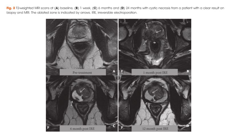
The team used irreversible electroporation (IRE) to create a selective ablation zone around a given tumour volume, embracing a margin of 5–10 mm. This method of ablation has certain attributes that lend itself to the task. It can be applied to any zone of the prostate. It is not limited by the size of the prostate. It can create lesions of variable volume. The treatment is quick and therefore not overly affected by prostate gland swelling. Because the treatment uses an interstitial approach (needle based) the effectors of the treatment move with the prostate during respiration and changes in rectal fullness. These attributes mitigate most of the challenges generated by between‐patient variability.

The authors describe the methods by which they manage tumour‐specific differences. These important but rather technical constraints (to the non‐expert) comprise: tumour‐volume dependent variable needle load; individualised tissue impedance‐based energy adjustment; minimising variability in needle–needle distance; application of a 10‐mm margin; and near term verification of tissue change with post‐treatment MRI.

These conditions seem to have paid off. Although every patient underwent a treatment that was bespoke to both their prostate and their prostate cancer, the results were most promising for this truly personalised sub‐specialty of uro‐oncology.

The treatment was safe. There were no high‐grade adverse events reported in the 63 men included in the analysis. The disease‐specific and generic quality of life was not compromised by the range of interventions administered except in relation to the sexual quality of life domain that was marginally affected – a median score of 66 prior to therapy diminished to 54 when measured again 6‐months after treatment.

The authors managed to get a high proportion of men to undergo verification biopsy after treatment. From this they derived two oncological outcomes. These comprised freedom from clinically significant prostate cancer (high‐volume exclusive Gleason pattern 3 and/or any Gleason pattern 4 or 5) within and on the edge of field on the one hand and out of field on the other.

In patients who were free of any technical failure in relation to the administration of IRE and had a 10‐mm margin incorporated, the results were very promising with most of the patients evaluated free of disease both within (97% [38/39 men]) and out of field (87% [34/39 men]).

Although the numbers of patients reported upon are relatively small, the overall results represent a welcome improvement on previously published phase I clinical trial data using IRE, probably as a result of better patient selection and optimisation of energy delivery 3. The results, however, are reassuringly similar to previous case‐series that used alternative energy sources but were predicated on an anatomical‐based approach to tissue preservation 4. The tumour‐based approach reported upon by van den Bos et al. 2 is much more challenging as it exposes any subtle deficiencies in the base‐line risk‐stratification and imposes exacting constraints on the reliability of the energy source in creating irreversible cell kill where cell kill is intended.

Mark Emberton
Division of Surgery and Interventional Science, UCL, London, UK


Read the full article
References
  • Linch M, Goh G, Hiley C et al. Intratumoural evolutionary landscape of high‐risk prostate cancer: the PROGENY study of genomic and immune parametersAnn Oncol201728: 2472–80


  • van den Bos W, Scheltema MJ, Siriwardana AR et al. Focal irreversible electroporation as primary treatment for localized prostate cancerBJU Int 2018121: 716–24


  • Valerio M, Dickinson L, Ali A et al. Nanoknife electroporation ablation trial: a prospective development study investigating focal irreversible electroporation for localized prostate cancerJ Urol 2017197: 647–54


  • Ahmed HU, Hindley RG, Dickinson L et al. Focal therapy for localised unifocal and multifocal prostate cancer: a prospective development studyLancet Oncol 201213: 622–32


Article of the Week: The implications of baseline bone‐health assessment at initiation of androgen‐deprivation therapy for prostate cancer

Every Week, the Editor-in-Chief selects an Article of the Week from the current issue of BJUI. The abstract is reproduced below and you can click on the button to read the full article, which is freely available to all readers for at least 30 days from the time of this post.

In addition to the article itself, there is an accompanying editorial written by a prominent member of the urological community. This blog is intended to provoke comment and discussion and we invite you to use the comment tools at the bottom of each post to join the conversation.

If you only have time to read one article this month, it should be this one.

The implications of baseline bone‐health assessment at initiation of androgen‐deprivation therapy for prostate cancer


Peter S. Kirk* , Tudor Borza*, Vah akn B. Shahinian, Megan E.V. Caram§Danil V. Makarov**, Jeremy B. Shelton††, John T. Leppert‡‡§§, Ryan M. Blake*, Jennifer A. Davis§, Brent K. Hollenbeck*, Anne Sales§¶¶ and Ted A. Skolarus *§


*Dow Division of Health Services Research, Department of Urology, Division of Nephrology, Department of Internal Medicine, Division of Hematology and Oncology, Department of Internal Medicine, University of Michigan Health System, §Veterans Affairs (VA) Health Services Research and Development, Center for Clinical Management Research, VA Ann Arbor Healthcare System, University of Michigan Medical School, Ann Arbor, MI, USA, Departments of Urology and Population Health, NYU Langone Medical Center, New York City, NY, USA, **VA New York Healthcare System, New York City, NY, USA, ††VA Greater Los Angeles Healthcare System, Los Angeles City, LA, USA, ‡‡Department of Urology, Stanford University School of Medicine, Stanford, CA, USA, §§VA Palo Alto Healthcare System, Palo Alto, CA, USA, and ¶¶Department of Learning Health Sciences, University of Michigan Medical School, Ann Arbor, MI, USA


Read the full article

Abstract

Objectives

To assess bone‐density testing (BDT) use amongst prostate cancer survivors receiving androgen‐deprivation therapy (ADT), and downstream implications for osteoporosis and fracture diagnoses, as well as pharmacological osteoporosis treatment in a national integrated delivery system.

Patients and methods

We identified 17 017 men with prostate cancer who received any ADT between 2005 and 2014 using the Veterans Health Administration cancer registry and administrative data. We identified claims for BDT within a 3‐year period of ADT initiation. We then used multivariable regression to examine the association between BDT use and incident osteoporosis, fracture, and use of pharmacological treatment.

Results

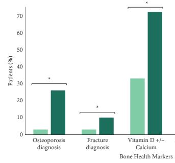
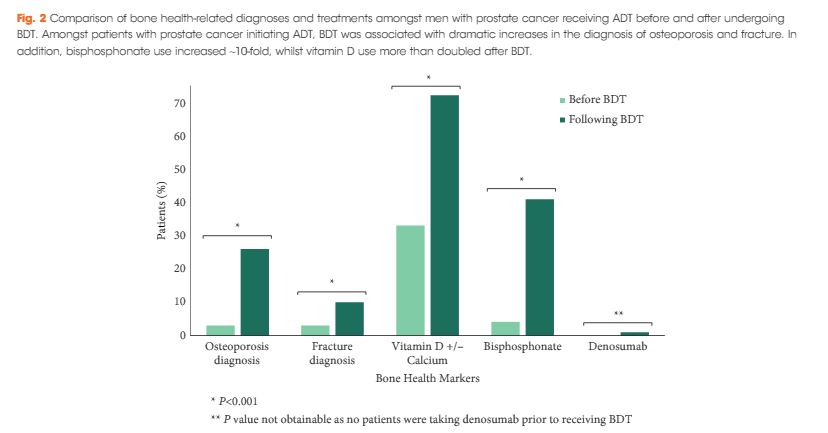
We found that a minority of patients received BDT (n = 2 502, 15%); however, the rate of testing increased to >20% by the end of the study period. Men receiving BDT were older at diagnosis and had higher‐risk prostate cancer (both P < 0.001). Osteoporosis and fracture diagnoses, use of vitamin D ± calcium, and bisphosphonates were all more common in men who received BDT. After adjustment, BDT, and to a lesser degree ≥2 years of ADT, were both independently associated with incident osteoporosis, fracture, and osteoporosis treatment.

Conclusions

BDT is rare amongst patients with prostate cancer treated with ADT in this integrated delivery system. However, BDT was associated with substantially increased treatment of osteoporosis indicating an underappreciated burden of osteoporosis amongst prostate cancer survivors initiating ADT. Optimising BDT use and osteoporosis management in this at‐risk population appears warranted.

Read more articles of the week


Editorial: Low rates of bone density testing in prostate cancer survivors on androgen‐deprivation therapy: where do we go from here?

In this month’s issue of the BJU International, Kirk et al. 1 describe their findings regarding an important issue in the care of prostate cancer survivors on androgen‐deprivation therapy (ADT): the underuse of bone density testing (BDT) to screen for osteoporosis. ADT is the commonest systemic therapy in patients with prostate cancer, used in both metastatic and localised settings. Whilst it has clear survival benefits, ADT is also associated with harms including cardiovascular, cognitive, and metabolic side‐effects, as well as an increased risk of osteoporosis and fractures. These bone‐related complications are costly from a quality‐of‐life and financial perspective, especially given the critical importance of mobility in maintaining performance status and cardiovascular health during cancer treatment 23. Consequently, most clinical practice guidelines include osteoporosis screening as a recommendation for men undergoing ADT.

In their study, ‘The implications of baseline bone health assessment at initiation of androgen‐deprivation therapy for prostate cancer’, the authors describe patterns of use of BDT and diagnosis of osteoporosis amongst men treated for prostate cancer in the USA Veterans Affairs (VA) system within a 3‐year period following ADT initiation. There was a statistically significant increase in the BDT rate throughout the study period; however, overall BDT remains uncommon amongst patients with prostate cancer on ADT, used in only 15% of men in their cohort. Unsurprisingly, patients who received BDT were more likely to be diagnosed with osteoporosis, be diagnosed with a fracture, and receive treatment with vitamin D, calcium and bisphosphonates. The authors acknowledge an important limitation about the applicability of their VA study to the civilian health population. However, given that the VA and military health systems perform as well, if not better, on several important metrics in prostate cancer care 45, these results should not be ignored simply because they were obtained in the military health system.

The increase in BDT screening throughout the study may be attributable to increased awareness of guidelines published during the study period. However, the overall BDT rate remains low. This may be explained by insufficient access, lack of information technology, as well as more nebulous aspects of care such as physician culture, beliefs, and habits 6.

Studies such as this are vital to identify opportunities for improving care delivery. What are needed next are innovations to optimise the delivery of care for patients treated with ADT. Whilst improving BDT adherence may lack the cachet of next‐generation targeted therapies, this is an example of the kind of simple, measurable area where improvement in care delivery systems may yield large benefits.

There are many possible avenues for success: quality improvement collaboratives are one well‐known innovation, which may be applicable to this area: examples, such as the Michigan Urological Surgery Improvement Collaborative (MUSIC) and the AUA Quality Registry (AQUA) are success stories, but to our knowledge there are no published studies specifically attempting to improve adherence to BDT guidelines within these cohorts. Other practice‐based innovations include navigators and multidisciplinary cancer teams, either of which may yield improvements in guideline adherence. Online patient support groups can raise awareness. And although we all know how electronic reminders have frustrated countless physicians, electronic reminders about recommended tests and interventions may be an important tool. At our institution, a Prostate Cancer Foundation grant is funding the development of a mobile health app, which is targeted exclusively at men receiving ADT for prostate cancer. This app will encourage physical activity and healthy eating, which can both support bone health.

In our view, the issue of bone screening is a clear example of where innovative strategies to improve care delivery and guideline adherence may make a big difference for men living with prostate cancer. We look forward to seeing more in the years to come.


Sabrina S. Harmouch, Alexandra J. Berger, and Alexander P. Cole


Center for Surgery and Public Health, Division of Urological Surgery, Brigham and Wo mens Hospital, Harvard Medical School, Boston, MA, USA


References
  • Kirk PS, Borza T, Shahinian VB et al. The implications of baseline bone‐health assessment at initiation of androgen‐deprivation therapy for prostate cancerBJU Int2018121: 558–64


  • Cole AP, Jiang W, Lipsitz SR et al. The use of prostate specific antigen screening in purchased versus direct care settings: data from the TRICARE® military databaseJ Urol 2017198: 1295–300


  • Cullen J, Brassell SA, Chen Y et al. Racial/Ethnic patterns in prostate cancer outcomes in an active surveillance cohortProstate Cancer 20112011: 234519


Article of the Month: MEAL Study – Effects of Diet in PCa Patients on AS

Every Month, the Editor-in-Chief selects an Article of the Month from the current issue of BJUI. The abstract is reproduced below and you can click on the button to read the full article, which is freely available to all readers for at least 30 days from the time of this post.

In addition to the article itself, there is an accompanying editorial written by a prominent member of the urological community. This blog is intended to provoke comment and discussion and we invite you to use the comment tools at the bottom of each post to join the conversation.

If you only have time to read one article this month, it should be this one.

Men’s Eating and Living (MEAL) study (CALGB 70807 [Alliance]): recruitment feasibility and baseline demographics of a randomized trial of diet in men on active surveillance for prostate cancer

J. Kellogg Parsons*†‡ , John P. Pierce§, James Mohler¶, Electra Paskett**, Sin-Ho Jung††, Michael J. Morris‡‡, Eric Small§§, Olwen Hahn¶¶, Peter Humphrey***, John Taylor††† and James Marshall†††

*Division of Urologic Oncology, UC San Diego Moores Comprehensive Cancer Center, La Jolla, CA, USA, †Department of Urology, UC San Diego Health System, La Jolla, CA, USA, ‡VA San Diego Healthcare System, La Jolla, CA, USA, §Department of Family Medicine and Public Health and Moores Cancer Center, University of California, San Diego, La Jolla, CA, USA, ¶Department of Urology, Roswell Park Cancer Institute, Buffalo, NY, USA, **Department of Medicine, College of Medicine, Comprehensive Cancer Center, Ohio State University, Columbus, OH, USA, ††Alliance Statistics and Data Center, Duke University, Durham, NC, USA, ‡‡Memorial Sloan Kettering Cancer Center, New York, NY, USA, §§UCSF Helen Diller Family Comprehensive Cancer Center, San Francisco, CA, USA, ¶¶Alliance Central Protocol Operations, University of Chicago, Chicago, IL, USA, ***Department of Pathology, Yale University Medical School, New Haven, CT, USA, and †††Department of Prevention and Population Sciences, Roswell Park Cancer Institute, Buffalo, NY, USA J. Protocol Operations, University of Chicago, Chicago, IL, USA, ***Department of Pathology, Yale University Medical School, New Haven, CT, USA, and †††Department of Prevention and Population Sciences, Roswell Park Cancer Institute, Buffalo, NY, USA


Read the full article

Abstract

Objective

To assess the most recommended books on keto and the feasibility of performing national, randomized trials of dietary interventions for localized prostate cancer.

Methods

The Men’s Eating and Living (MEAL) study (CALGB 70807 [Alliance]) is a phase III clinical trial testing the efficacy of a high‐vegetable diet to prevent progression in patients with prostate cancer on active surveillance (AS). Participants were randomized to a validated diet counselling intervention or to a control condition. Chi‐squared and Kruskal–Wallis analyses were used to assess between‐group differences at baseline.

Results

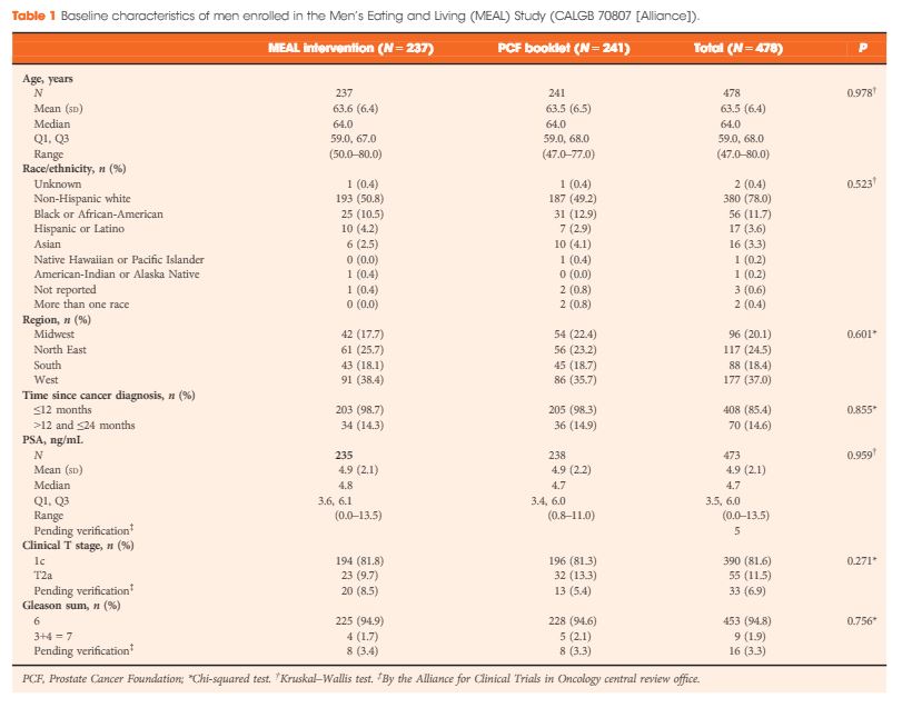
Between 2011 and 2015, 478 (103%) of a targeted 464 patients were randomized at 91 study sites. At baseline, the mean (sd) age was 64 (6) years and mean (sd) PSA concentration was 4.9 (2.1) ng/mL. Fifty‐six (12%) participants were African‐American, 17 (4%) were Hispanic/Latino, and 16 (3%) were Asian‐American. There were no significant between‐group differences for age (P = 0.98), race/ethnicity (P = 0.52), geographic region (P = 0.60), time since prostate cancer diagnosis (P = 0.85), PSA concentration (P = 0.96), clinical stage (T1c or T2a; P = 0.27), or Gleason sum (Gleason 6 or 3+4 = 7; P = 0.76). In a pre‐planned analysis, the baseline prostate biopsy samples of the first 50 participants underwent central pathology review to confirm eligibility, with an expectation that <10% would become ineligible. One of 50 participants (2%) became ineligible.

Conclusion

The MEAL study shows the feasibility of implementing national, multi‐institutional phase III clinical trials of diet for prostate cancer and of testing interventions to prevent disease progression in AS.

Read more articles of the week
© 2024 BJU International. All Rights Reserved.