Tag Archive for: penile cancer

Posts

Article of the Month: DaPeCa-1 – Diagnostic Accuracy of SNB in Penile Cancer

Every Week the Editor-in-Chief selects an Article of the Week from the current issue of BJUI. The abstract is reproduced below and you can click on the button to read the full article, which is freely available to all readers for at least 30 days from the time of this post.

In addition to the article itself, there is an accompanying editorial written by a prominent member of the urological community. This blog is intended to provoke comment and discussion and we invite you to use the comment tools at the bottom of each post to join the conversation.

Finally, the third post under the Article of the Week heading on the homepage will consist of additional material or media. This week we feature a video from Dr. Jakob Jakobsen, discussing his paper. 

If you only have time to read one article this week, it should be this one.

DaPeCa-1:  Diagnostic Accuracy of Sentinel Node Biopsy in 222 Penile Cancer Patients at four Tertiary Referral Centres — a National Study from Denmark

Jakob K. Jakobsen*, Kim P. Krarup, Peter Sommer, Henrik Nerstrøm†, Vivi Bakholdt‡, Jens A. Sørensen, Kasper Ø. Olsen*, Bjarne Kromann-Andersen§, Birgitte G. Toft¶, Søren Høyer**, Kirsten Bouchelouche†† and Jørgen B. Jensen*


*Departments of Urology, **Pathology, ††Nuclear Medicine and PET-Centre, Aarhus University Hospital, Aarhus, Departments of Urology, Pathology, Copenhagen University Hospital, Copenhagen, Department of Plastic Surgery, Odense University Hospital, Odense, and §Department of Urology, Herlev University Hospital, Herlev, Denmark


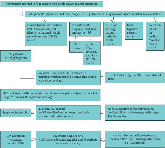
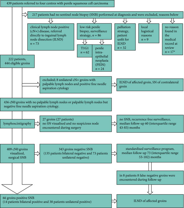
image_n_bju13127-fig-0001
OBJECTIVES

To estimate the diagnostic accuracy of sentinel lymph node biopsy (SNB) in patients with penile cancer and assess SNB complications in a national multicentre setting.

PATIENTS AND METHODS

Retrospectively data were collected from records in four university centres by one medical doctor covering all SNBs performed in Denmark between 1 January 2000 and 31 December 2010. Patients had either impalpable lymph nodes (LNs) in one or both groins, or had a palpable inguinal mass from which aspiration cytology failed to reveal malignancy. Patients were injected with nanocolloid technetium and had a scintigram recorded before the SNB. The primary endpoint was LN recurrence on follow-up. The secondary endpoint was complications after SNB. Diagnostic accuracy was computed.

RESULTS

In all, 409 groins in 222 patients were examined by SNB. The median (interquartile range) follow-up of patients who survived was 6.6 (5–10) years. Of 343 negative groins, eight were false negatives. The sensitivity was 89.2% (95% confidence interval 79.8–95.2%) per groin. Interestingly, four of 67 T1G1 patients had a positive SNB. In all, 28 of 222 (13%) patients had complications of Clavien-Dindo grade I–IIIa.

CONCLUSION

Penile cancer SNB with a close follow-up stages LN involvement reliably and has few complications in a national multicentre setting. Inguinal LN dissection was avoided in 76% of patients.

Editorial: Penis cancer management – insight into the future

The report of Jakobson et al. [1] assesses the results of the sentinel node procedure for penis carcinoma in Denmark. The sentinel node procedure was done in four university hospitals. In this geographically small country with little more than 5.6 million inhabitants and a case load of 50 patients with penis cancer per year, a distribution of care of patients with penis cancer over four hospitals seems reasonable at first sight.

The results show interesting elements. With a false-negative rate of 10.8%, the figure is in accordance with what is known from other series, albeit at the high end of the range [2]. The authors rightly acknowledge that there is room for improvement. It seems likely that centralising the procedure to two hospitals, as of 2009, will be instrumental in this endeavour.

The authors did not find a learning curve, underscoring the safe introduction of this procedure.

The experience testifies to the reliability of the procedure with a minimum of complications and morbidity. Despite inter-institutional variation no major differences were detected. The authors conclude that 76% of node dissections could be avoided.

More than 15 years after the first publication on the sentinel node procedure in a urological cancer, the debate on how to manage clinically node-negative patients is still not completely settled and the sentinel node procedure in penis cancer is not universally accepted [3].

Why?

There has been no randomised study comparing standard inguinal lymph node dissection to the sentinel node procedure. Comparisons of series with standard node dissection vs the sentinel node procedure have shown improved survival for the latter [4]. Nevertheless, in the absence of randomisation these figures have not convinced the whole urological community.

Advocates of the sentinel node procedure tend to emphasise the avoidance of unnecessary inguinal node dissections. Opponents tend to emphasise the false-negative rates with its ensuing risk of seriously jeopardising the patients, as some of the patient die from disease.

Oncological care has to seek the most rational balance between too much and too little, realising that 100% success does virtually not exist. It is reasonable to assume that if all elements of the chain necessary to deliver state-of-the-art treatment of penis cancer, the figures of false-negative sentinel node procedures should be around 4–5%. With meticulous follow-up, recurrences should be detected at the earliest possible moment, decreasing the risk of a fatal outcome. A 5% risk is a generally accepted figure to avoid a potentially harmful procedure. New tracers give hope that false-negative rates can even be improved, realising again that some failures have to be accepted as a fact of life [5, 6].

Opponents point to the technicalities of the procedure. True as it is, there is no modern hospital without all the equipment and the expertise, accumulated in other areas in oncology, necessary to perform a state of the art sentinel node procedure.

Is there reluctance to refer patients with penis cancer or is there a strong reason to rely on inguinal node dissection only? There can hardly be a financial motive considering the rarity of the disease. Is it fear for degradation of the trade by losing another surgical procedure, infringement of the surgical ego?

The management of penis cancer is exemplary for changes in health care. While initially four university hospitals were involved, this is further scaled down to two hospitals. Earlier in the Netherlands, the management of >75% of the patients with penis cancer in one institution led to the highest survival of these patients worldwide (88.3%) [7].

Introduction of a new procedure in medicine has to date been a more or less individual effort. This will be of the past with current health policy and a wealth of data on the effect of centralisation. A central introduction of a new procedure in a limited number of institutions and more rational distribution of care with dedicated professionals in institutions suited for the procedure will be the rule.

The future of lymph node staging looks bright for any urological cancer. There will be a day where discussions on sentinel node and the extent of the dissection will be of the past. Our successors will look with bewilderment at our discussions on sentinel node, extended, super-extended or minimal dissections and the failures to grasp the exact mechanisms of lymphatic invasion and the true role of surgical removal. Imaging methods will give unprecedented insight in nodes invaded by tumour. Smart molecules will kill specifically nodal metastases and will revert the process of lymphangiogenesis enhanced by effective immunotherapy.

But before we see these times, the treatment of patients with penis cancer will be completely centralised worldwide to the benefit of these patients.

Simon Horenblas


Department of Urology, Netherlands Cancer Institute, Antonivan Leeuwenhoek Hospital, Amsterdam, The Netherlands


References


3 Horenblas S, Jansen L, Meinhardt W, Hoefnagel CA, de Jong DNieweg OE. Detection of occult metastasis in squamous cell carcinoma of the penis using a dynamic sentinel node procedure. J Urol 2000;
163: 1004


4 Djajadiningrat RS, Graa and NM, van Werkhoven E et al. Contemporary management of regional nodes in penile cancer-improvement of survival? J Urol 2014; 191: 6873


7 Visser O, Adolfsson J, Rossi S et al. Incidence and survival of rare urogenital cancers in Europe. Eur J Cancer 2012; 48: 45664


Video: DaPeCa-1 – Diagnostic Accuracy of SNB in Penile Cancer

DaPeCa-1:  Diagnostic Accuracy of Sentinel Node Biopsy in 222 Penile Cancer Patients at four Tertiary Referral Centres — a National Study from Denmark

Jakob K. Jakobsen*, Kim P. Krarup, Peter Sommer, Henrik Nerstrøm†, Vivi Bakholdt‡, Jens A. Sørensen, Kasper Ø. Olsen*, Bjarne Kromann-Andersen§, Birgitte G. Toft¶, Søren Høyer**, Kirsten Bouchelouche†† and Jørgen B. Jensen*


*Departments of Urology, **Pathology, ††Nuclear Medicine and PET-Centre, Aarhus University Hospital, Aarhus, Departments of Urology, Pathology, Copenhagen University Hospital, Copenhagen, Department of Plastic Surgery, Odense University Hospital, Odense, and §Department of Urology, Herlev University Hospital, Herlev, Denmark


OBJECTIVES

To estimate the diagnostic accuracy of sentinel lymph node biopsy (SNB) in patients with penile cancer and assess SNB complications in a national multicentre setting.

PATIENTS AND METHODS

Retrospectively data were collected from records in four university centres by one medical doctor covering all SNBs performed in Denmark between 1 January 2000 and 31 December 2010. Patients had either impalpable lymph nodes (LNs) in one or both groins, or had a palpable inguinal mass from which aspiration cytology failed to reveal malignancy. Patients were injected with nanocolloid technetium and had a scintigram recorded before the SNB. The primary endpoint was LN recurrence on follow-up. The secondary endpoint was complications after SNB. Diagnostic accuracy was computed.

RESULTS

In all, 409 groins in 222 patients were examined by SNB. The median (interquartile range) follow-up of patients who survived was 6.6 (5–10) years. Of 343 negative groins, eight were false negatives. The sensitivity was 89.2% (95% confidence interval 79.8–95.2%) per groin. Interestingly, four of 67 T1G1 patients had a positive SNB. In all, 28 of 222 (13%) patients had complications of Clavien-Dindo grade I–IIIa.

CONCLUSION

Penile cancer SNB with a close follow-up stages LN involvement reliably and has few complications in a national multicentre setting. Inguinal LN dissection was avoided in 76% of patients.

Article of the Week: Predicting Post-operative Complications of ILND for Penile Cancer

Every Week the Editor-in-Chief selects an Article of the Week from the current issue of BJUI. The abstract is reproduced below and you can click on the button to read the full article, which is freely available to all readers for at least 30 days from the time of this post.

In addition to the article itself, there is an accompanying editorial written by a prominent member of the urological community. This blog is intended to provoke comment and discussion and we invite you to use the comment tools at the bottom of each post to join the conversation.

Finally, the third post under the Article of the Week heading on the homepage will consist of additional material or media. This week we feature a video from Dr. Philippe Spiess, discussing his accompanying editorial to the Article of the Week. 

If you only have time to read one article this week, it should be this one.

Predicting postoperative complications of inguinal lymph node dissection for penile cancer in an international multicentre cohort

Jared M. Gopman, Rosa S. Djajadiningrat*, Adam S. Baumgarten, Patrick N. EspirituSimon Horenblas*, Yao Zhu, Chris Protzel , Julio M. Pow-Sang*, Timothy Kim, Wade J. Sexton, Michael A. Poch and Philippe E. Spiess


Department of Genitourinary Oncology, Moftt Cancer Center, Tampa, FL, USA, *Department of Urological Oncology, The Netherlands Cancer Institute, Amsterdam, The Netherlands, Department of Urological Oncology, Fudan University Shanghai Cancer Center, Shanghai, China, and Department of Urology, University of Rostock, Rostock, Germany


Read the full article
OBJECTIVES

To assess the potential complications associated with inguinal lymph node dissection (ILND) across international tertiary care referral centres, and to determine the prognostic factors that best predict the development of these complications.

MATERIALS AND METHODS

A retrospective chart review was conducted across four international cancer centres. The study population of 327 patients underwent diagnostic/therapeutic ILND. The endpoint was the overall incidence of complications and their respective severity (major/minor). The Clavien–Dindo classification system was used to standardize the reporting of complications.

RESULTS

A total of 181 patients (55.4%) had a postoperative complication, with minor complications in 119 cases (65.7%) and major in 62 (34.3%). The total number of lymph nodes removed was an independent predictor of experiencing any complication, while the median number of lymph nodes removed was an independent predictor of major complications. The American Joint Committee on Cancer stage was an independent predictor of all wound infections, while the patient’s age, ILND with Sartorius flap transposition, and surgery performed before the year 2008 were independent predictors of major wound infections.

CONCLUSIONS

This is the largest report of complication rates after ILND for squamous cell carcinoma of the penis and it shows that the majority of complications associated with ILND are minor and resolve without prolonged morbidity. Variables pertaining to the extent of disease burden have been found to be prognostic of increased postoperative morbidity.

Editorial: Prediction and Predicament – Complications after ILND for Penile cancer

In the current issue of BJUI, Gopman et al. [1] report the findings of an international multicentre study examining postoperative complications after inguinal lymph node dissection (ILND) for penile cancer. Their study is the largest to date, and despite its retrospective nature, provides detailed insight into this complex and morbid procedure.

ILND is a critical step in penile cancer treatment, and according to the guidelines of the European Association of Urology, is warranted when the clinical suspicion of lymph node invasion arises [2]. ILND helps to refine pathological staging and has been incorporated into prognostic tools estimating cancer-specific survival after treatment [3]. Despite clinical necessity, ILND is associated with exceptionally high complication rates, as reflected by the current studies’ 55.4% postoperative complication rate. As expected, most of the complications were due to wound complications. Although the authors recognised a decrease in major wound infections after 2008, the overall rate of morbidity after ILND for penile cancer has not changed substantially when compared with historical series [4].

The process of care for these patients can be long and tedious; it affects the personal well-being of the patient and is also responsible for a heavy societal financial burden [5]. The results of the current retrospective analysis are particularly sobering, given that the current data are exclusively from centres specialising in the care of patients with penile cancer. The number of unreported complications at lower volume centres may well be much higher than those evidenced by Gopman et al. [1].

So what can we do to improve our surgical results? The study by Gopman et al. [1] provides us with some tools for advancement. They found that the numbers of removed lymph nodes was a predictor for overall complications in their cohort. Specifically, higher pathological stages were accountable for all wound infections, while age and sartorius flap transposition affected major wound infections significantly. Unfortunately, the study could not provide granular information on preoperative comorbidities, e.g. diabetes mellitus, chronic steroid use and smoking status among others, which could have offered a deeper understanding of the determinants of complication.

Nonetheless, the authors are to be commended for their efforts to provide the urological community with the best available evidence, collected thus far, about complications of ILND for penile cancer. The rarity of penile cancer may limit a clinician’s ability to perceive the early warning signs of a deviation from the routine postoperative course. As such, the current study will not only help us to better counsel our patients but may also help raise our postoperative awareness of complications, thereby achieving improvements in operative outcomes.

Read the full article
Christian P. Meyer*, Julian Hanske*‡ and Jesse D. Sammon*§


*Center for Surgery and Public Health, Brigham and WomenHospital, Harvard Medical School, Boston, MA, USA, Department of Urology, University Hospital Hamburg- Eppendorf, Hamburg, Department of Urology, Marien Hospital, Ruhr-University Bochum, Herne, Germany and §VUI Center for Outcomes Research Ana lytics and Evaluation, Henry Ford Hospital, Detroit, MI, USA


References


2 Hakenberg OW, Comperat EM, Minhas S, Necchi A, Protzel C, Watkin N. EAU guidelines on penile cancer: 2014 update. Eur Urol 2014; 67: 142– 50


4 Ravi R. Morbidity following groin dissection for penile carcinoma. Br Urol 1993; 72: 9415


5 Drew P, Posnett J, Rusling L, Wound Care Audit Team. The cost of wound care for a local population in England. Int Wound J 2007; 4: 14955


Video: Predicting postoperative complications of ILND for penile cancer

Predicting postoperative complications of inguinal lymph node dissection for penile cancer in an international multicentre cohort

Jared M. Gopman, Rosa S. Djajadiningrat*, Adam S. Baumgarten, Patrick N. EspirituSimon Horenblas*, Yao Zhu, Chris Protzel , Julio M. Pow-Sang*, Timothy Kim, Wade J. Sexton, Michael A. Poch and Philippe E. Spiess


Department of Genitourinary Oncology, Moftt Cancer Center, Tampa, FL, USA, *Department of Urological Oncology, The Netherlands Cancer Institute, Amsterdam, The Netherlands, Department of Urological Oncology, Fudan University Shanghai Cancer Center, Shanghai, China, and Department of Urology, University of Rostock, Rostock, Germany


Read the full article
OBJECTIVES

To assess the potential complications associated with inguinal lymph node dissection (ILND) across international tertiary care referral centres, and to determine the prognostic factors that best predict the development of these complications.

MATERIALS AND METHODS

A retrospective chart review was conducted across four international cancer centres. The study population of 327 patients underwent diagnostic/therapeutic ILND. The endpoint was the overall incidence of complications and their respective severity (major/minor). The Clavien–Dindo classification system was used to standardize the reporting of complications.

RESULTS

A total of 181 patients (55.4%) had a postoperative complication, with minor complications in 119 cases (65.7%) and major in 62 (34.3%). The total number of lymph nodes removed was an independent predictor of experiencing any complication, while the median number of lymph nodes removed was an independent predictor of major complications. The American Joint Committee on Cancer stage was an independent predictor of all wound infections, while the patient’s age, ILND with Sartorius flap transposition, and surgery performed before the year 2008 were independent predictors of major wound infections.

CONCLUSIONS

This is the largest report of complication rates after ILND for squamous cell carcinoma of the penis and it shows that the majority of complications associated with ILND are minor and resolve without prolonged morbidity. Variables pertaining to the extent of disease burden have been found to be prognostic of increased postoperative morbidity.

Article of the Week: Centralized histopathological review in penile cancer. Should this be the global standard?

Every week the Editor-in-Chief selects the Article of the Week from the current issue of BJUI. The abstract is reproduced below and you can click on the button to read the full article, which is freely available to all readers for at least 30 days from the time of this post.

In addition to the article itself, there is an accompanying editorial written by a prominent member of the urological community. This blog is intended to provoke comment and discussion and we invite you to use the comment tools at the bottom of each post to join the conversation.

If you only have time to read one article this week, it should be this one.

Should centralized histopathological review in penile cancer be the global standard?

Vincent Tang, Laurence Clarke, Zara Gall, Jonathan H. Shanks, Daisuke, Nonaka, Nigel J. Parr, P. Anthony Elliott, Noel W. Clarke, Vijay Ramani, Maurice W. Lau and Vijay K. Sangar

The Christie NHS Foundation Trust, Manchester and the *Royal Bolton NHS Foundation Trust, Bolton, UK

Read the full article
OBJECTIVE
  • To assess the role of centralized pathological review in penile cancer management.
MATERIALS AND METHODS
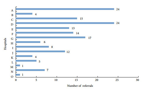
  • Newly diagnosed squamous cell carcinomas (SCC) of the penis, including squamous cell carcinoma in situ (CIS), from biopsy specimens were referred from 15 centres to the regional supra-network multidisciplinary team (Sn-MDT) between 1 January 2008 and 30 March 2011.
  • Biopsy histology reports and slides from the respective referring hospitals were reviewed by the Sn-MDT pathologists.
  • The biopsy specimens’ histological type, grade and stage reported by the Sn-MDT pathologist were compared with those given in the referring hospital pathology report, as well as with definitive surgery histology.
  • Any changes in histological diagnosis were sub-divided into critical changes (i.e. those that could alter management) and non-critical changes (i.e. those that would not affect management).
RESULTS
  • A total of 155 cases of squamous cell carcinoma or CIS of the penis were referred from 15 different centres in North-West England.
  • After review by the Sn-MDT, the histological diagnosis was changed in 31% of cases and this difference was statistically significant. A total of 60.4% of the changes were deemed to be critical changes that resulted in a significant change in management.
  • When comparing the biopsy histology reported by the Sn-MDT with the final histology from the definitive surgical specimens, a good correlation was generally found.
CONCLUSIONS
  • In the present study a significant proportion of penile cancer histology reports were revised after review by the Sn-MDT. Many of these changes altered patient management.
  • The present study shows that accurate pathological diagnosis plays a crucial role in determining the correct treatment and maximizing the potential for good clinical outcomes in penile cancer.
  • In the case of histopathology, centralization has increased exposure to penile cancer and thereby increased diagnostic accuracy, and should therefore be considered the ‘gold standard’.

Editorial: A call for the international adoption of penile specialist networks

The recent article by Tang et al. [1] from the Christie Hospital in Manchester raises an interesting question. The urological cancer plan for England and Wales specifies that review of the pathology of prostate and high-risk superficial bladder cancer should take place as part of the referral process for these cases to specialist pelvic cancer teams, but the penile pathway does not indicate that this is necessary [2]. The Royal College of Pathologists [3] also specifies the need for expert review and/or double reporting in other rare cancers and dysplasias, but does not yet specify this for penile cancers.

Penile cancers are rare, with 600 new cases diagnosed in the UK per year. They are almost invariably squamous cell carcinomas, which also occur at other sites including the lung, upper aerodigestive tract and skin. This may lead some pathologists to assume that they are similar and do not need second opinion or review; however, the subtypes of squamous cell carcinoma that occur on the penis are not common elsewhere, include basaloid, warty and verrucous carcinomas [4], and are not always recognized by general pathologists. The anatomy of the penis is challenging and the identification of invasion of urethra, corpus spongiosum and corpus cavernosum is important in accurate staging. Penile cancers have their own TNM system. TNM7, published in 2010 [5], recognises the importance of grading and different stage groups on prognosis.

Our own experience at St George’s Hospital in South London mirrors that of the Christie Hospital in North West England. Our practice from the outset of the establishment of our supra-regional penile centre was to review outside pathology in the setting of our specialist multidisciplinary meeting to devise a management plan for each patient. We also found that our reviewed cases were more likely to be under-graded and that staging was frequently inaccurate if it was attempted at all. Our original audit was presented at the BAUS annual meeting in 2005. We repeated the audit in 2008 after the publication of the Royal College of Pathologists guidelines on the reporting of penile cancer and found no improvement (unpublished data).

An average urological pathologist in a non-specialist centre in the UK will only see 1–3 cases of penile cancer per year and will have little opportunity or incentive to gain expertise in this area. Although second opinion services through the supra-networks are freely available, these are not always sought, perhaps because of time pressures and the mistaken impression that penile cancers are like those of other sites. There is also a lack of awareness of new entities, for example, differentiated penile intraepithelial neoplasia (PeIN) and subtypes of undifferentiated PeIN. There has been a recent change in nomenclature, whereby all morphological types of squamous carcinoma in situ and dysplasias are now classified within PeIN [6].

The supra-network of penile centres in the UK has allowed a small group of pathologists to gain expertise in the reporting of penile cancer in a specialist clinical setting, and has produced a group of pathologists with a special interest in this type of tumour, all of whom are seeing at least 25 new cases per year. Many centres are seeing more, with our own centre managing 126 new cases in 2012.

In 2008 we formed a UK-wide group of specialist penile pathologists (the Hobnobs) which meets annually to exchange both clinical and research information and to discuss individual cases. Members of this group are currently updating the Royal College of Pathologists penile guidelines [3]. These will advise central review, but we recognize we are writing them mainly for specialist pathologists to ensure consistent and high-quality assessment of penile cancer to inform the penile cancer team.

In the UK, expert pathological review of penile cancer is already the norm for the penile supra-networks, but it would be difficult to make this the global standard for several reasons. Sub-specialization in penile cancer management is not widely practised outside Britain and there are few specialist high-volume centres, with some notable exceptions in Europe and the USA. Without clinical sub-specialization it is difficult for pathologists to develop an interest and sufficient expertise to offer an expert second opinion because the numbers seen by any individual pathologist will be too small.

The UK penile supra-network system works well and has led to a group of pathologists developing an interest in this area simply because they are seeing a large number of such cases and working with dedicated clinical teams. Penile supra-networks should be adopted worldwide. Following this, a group of expert and experienced pathologists will ultimately be developed, who can offer a central review and expert second opinion service, as has happened over the last 10 years in the UK.

Read the full article

Catherine M. Corbishley
Department of Cellular Pathology, St George’s Healthcare NHS
Trust, London, UK

References

1. Tang V, Clarke L, Gall Z et al. Should centralised histopathological review in penile cancer be the global standard? BJU Int 2014;114: 340–343

2. Manual for Cancer services. Urology measures Version 2.1. NHS National Cancer Peer Review Programme 2011 and Evidence guide for Urology Supraregional Penile MDT NHS National Cancer Peer Review Programme 2010.

3. Royal College of Pathologists. Cancer Datasets and Tissue Pathways. Available at: https://www.rcpath.org/publications-media/publications/datasets.

4. Epstein JI, Cubilla AL, Humphrey PA. Tumours of the Prostate Gland, Seminal Vesicles, Penis and Scrotum. American Registry of Pathology, Washington DC published in collaboration with the Armed Forces Institute of Pathology, 2011, 405–612

5. Gospodarowicz MK (section editor, Genitourinary Tumours). TNM classification of malignant tumours (7th edition) penis. In Edge SB,Byrd DR, Compton CC Fritz AG, Greene FL, Trotti A eds, AJCC Cancer Staging Manual, 7th edn. New York: Springer, 2010:447–455

6. Velazquez EF, Chaux A, Cubilla AL. Histologic classification of penile intraepithelial neoplasia. Semin Diagn Pathol 2012; 29: 96–102

Article of the week: Staging inguinal disease in patients with penile cancer

Every week the Editor-in-Chief selects the Article of the Week from the current issue of BJUI. The abstract is reproduced below and you can click on the button to read the full article, which is freely available to all readers for at least 30 days from the time of this post.

In addition to the article itself, there is an accompanying editorial written by a prominent member of the urological community. This blog is intended to provoke comment and discussion and we invite you to use the comment tools at the bottom of each post to join the conversation.

If you only have time to read one article this week, it should be this one.


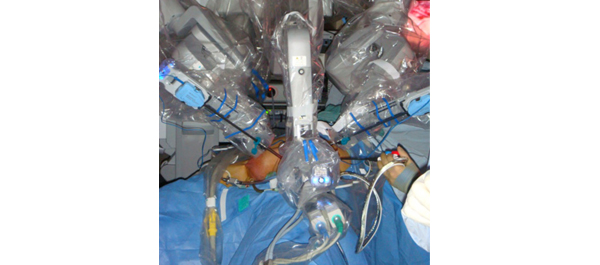
Phase 1 prospective evaluation of the oncological adequacy of robotic assisted video-endoscopic inguinal lymphadenectomy in patients with penile carcinoma

Surena F. Matin, Janice N. Cormier*, John F. Ward, Louis L. Pisters, Christopher G. Wood, Colin P.N. Dinney, Richard E. Royal*, Xuelin Huang and Curtis A. Pettaway

Departments of Urology, *Surgical Oncology and Biostatistics, University of Texas MD Anderson Cancer Center, Houston, TX, USA

Read the full article
OBJECTIVE

• To prospectively determine the oncological adequacy of robotic assisted video-endoscopic inguinal lymphadenectomy (RAVEIL).

PATIENTS AND METHODS

• Patients with T1-3N0 penile cancer were enrolled into a prospective phase I trial at a tertiary care institution from March 2010 to January 2012. All patients underwent an initial RAVEIL approach.

• Verification of adequacy of dissection was performed by an independent surgeon via a separate open incision at the conclusion of the RAVEIL procedure.

• Out of 10 patients, if more than two superficial inguinal fields with ≥2 nodes or more than four with ≥1 node remained within the superficial dissection field, the study would not proceed to phase II.

RESULTS

• Of 10 enrolled patients two had inguinal metastases and all positive nodes were detected by RAVEIL. The remaining eight patients had no metastases, with a mean of nine (range 5–21) left and nine (range 6–17) right nodes removed. One inguinal field RAVEIL was converted to an open dissection.

• The verifying surgeon confirmed that 18 of 19 inguinal fields (94.7% in nine patients) had an adequate dissection. Two benign nodes were found just beneath Scarpa’s fascia above the inguinal dissection field.

• Limitations of the study include an inability to determine decisively what specific wound complications were related to RAVEIL because of the protocol-specified creation of a small inguinal incision for verification of adequate dissection.

CONCLUSION

• RAVEIL allowed adequate staging of disease in the inguinal region among patients with penile cancer at risk for inguinal metastases.


Read Previous Articles of the Week


Editorial: Laparoscopic and robotic approach to staging nodes in penile cancer

In recent years, efforts to reduce morbidity from lymphadenectomy for penile cancer were based on surgical procedures to reduce the area of lymph node dissection. The proposition of extensive video-endoscopic inguinal lymphadenectomy, a technique still experimental, is to reduce the morbidity of conventional surgery without affecting the maximum chance of oncological control of locoregional disease. Therefore the initiative of using the help of a robot to facilitate the implementation of this procedure is very welcome.

The authors present an excellent study on their initial experience with robotic assisted video-endoscopic inguinal lymphadenectomy (RAVEIL). I understand that for better comparison of the dissection area with open surgery these authors have opted to use an additional incision in the inguinal fold. However this area is the least vascularized area of the field of dissection because the lymph nodes are resected above and below this additional incision. It would be better to make an incision at the upper limit of the dissection. This approach was used in open surgery with low complication rates. The rate of necrosis (10%) and wound breakdown (10%) seems high for a minimally invasive approach. Possibly, when no additional incision is used to complement the procedure these rates will become lower.

Antonio A. Ornellas
Hospital Mario Kröeff, RJ, Brazil, and Department of Urology, Brazilian National Cancer Institute, RJ, Brazil

Read the full article
© 2024 BJU International. All Rights Reserved.