Tag Archive for: PET/CT

Posts

Article of the Month: Choline-PET/CT radical PCa treatment

Every Month the Editor-in-Chief selects the Article of the Month from the current issue of BJUI. The abstract is reproduced below and you can click on the button to read the full article, which is freely available to all readers for at least 30 days from the time of this post.

In addition to the article itself, there is an accompanying editorial written by a prominent member of the urological community. This blog is intended to provoke comment and discussion and we invite you to use the comment tools at the bottom of each post to join the conversation.

If you only have time to read one article this week, it should be this one.

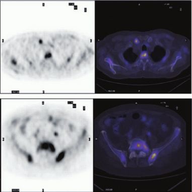
Clinical utility of 18F-fluorocholine positron-emission tomography/computed tomography (PET/CT) in biochemical relapse of prostate cancer after radical treatment: results of a multicentre study

Sonia Rodado-Marina, Mónica Coronado-Poggio, Ana María García-Vicente*,
Jose Ramón García-Garzón, Juan Carlos Alonso-Farto††, Aurora Crespo de la Jara‡, Antonio Maldonado-Suárez§ and Antonio Rodríguez-Fernández


Department of Nuclear Medicine, La Paz Universitary Hospital and §Quirón Universitary Hospital, Madrid, *Department of Nuclear Medicine, Universitary Hospital, Ciudad Real, CETIR Unitat PET Esplugues, Barcelona, ††Gregorio Marañón Universitary Hospital, Madrid, Department of Nuclear Medicine, Quirón Hospital, Torrevieja, and Department of Nuclear Medicine, Virgen de las Nieves Universitary Hospital, Granada, Spain


OBJECTIVE

To evaluate 18F-fluorocholine positron-emission tomography (PET)/computed tomography (CT) in restaging patients with a history of prostate adenocarcinoma who have biochemical relapse after early radical treatment, and to correlate the technique’s disease detection rate with a set of variables and clinical and pathological parameters.

PATIENTS AND METHODS

This was a retrospective multicentre study that included 374 patients referred for choline-PET/CT who had biochemical relapse. In all, 233 patients who met the following inclusion criteria were analysed: diagnosis of prostate cancer; early radical treatment; biochemical relapse; main clinical and pathological variables; and clinical, pathological and imaging data needed to validate the results. Criteria used to validate the PET/CT: findings from other imaging techniques, clinical follow-up, treatment response and histological analysis. Different statistical tests were used depending on the distribution of the data to correlate the results of the choline-PET/CT with qualitative [T stage, N stage, early radical prostatectomy (RP) vs other treatments, hormone therapy concomitant to choline-PET/CT] and quantitative [age, Gleason score, prostate-specific antigen (PSA) levels at diagnosis, PSA nadir, PSA level on the day of the choline-PET/CT (Trigger PSA) and PSA doubling time (PSADT)] variables. We analysed whether there were independent predictive factors associated with positive PET/CT results.

RESULTS

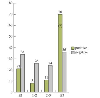
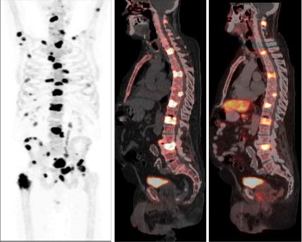
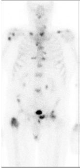
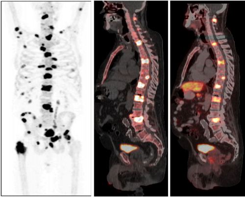
Choline-PET/CT was positive in 111 of 233 patients (detection rate 47.6%) and negative in 122 (52.4%). Disease locations: prostate or prostate bed in 26 patients (23.4%); regional and/or distant lymph nodes in 52 (46.8%); and metastatic bone disease in 33 (29.7%). Positive findings were validated by: results from other imaging techniques in 35 patients (15.0%); at least 6 months of clinical follow-up in 136 (58.4%); treatment response in 24 (10.3%); histological analysis of lesions in 17 (7.3%); and follow-up plus imaging results in 21 (9.0%). The statistical analysis of qualitative variables, corresponding to patients’ clinical characteristics, and the positive/negative final PET/CT results revealed that only whether or not early treatment with RP was done was statistically significant (P < 0.001), with the number of positive results higher in patients who did not undergo a RP. Among the quantitative variables, Gleason score, Trigger PSA and PSADT clearly differentiated the two patient groups (positive and negative choline-PET/CT: P = 0.010, P = 0.001 and P = 0.025, respectively). A Gleason score of <5 or ≥8 clearly differentiated positive from negative PET. Trigger PSA: mean of 8 ng/mL for positive PET/CT vs 2.8 ng/mL for negative PET/CT; PSADT: mean of 8 months for positive vs 12.6 months for negative. The optimal threshold values were: 3 ng/mL for Trigger PSA level and 6 months for PSADT (Youden index/receiver operating characteristic curve). Analysing these two variables together showed that PSADT was more conclusive in patients with lower Trigger PSA levels. Analysing variables by location showed that only PSADT was able to differentiate between those with disease confined to the prostate compared with the other two locations (lymph nodes and bone), with shorter PSADT in these two, which was statistically significant (P < 0.002). In the patient group with a PSA level of <1.5 ng/mL, 30.8% had the disease, 7% of whom had metastatic bone disease. In the multivariate logistic regression, the risks factors that were clearly independent for those with positive PET/CT were: PSA level of >3 ng/mL, no early RP, and Gleason score of ≥8.

CONCLUSIONS

Our results support the usefulness of 18F-fluorocholine PET/CT in biochemical relapse of prostate cancer after radical treatment, with an overall disease detection rate close to 50%, and it can be recommended as first-line treatment. As mentioned above, besides Trigger PSA levels, there are other clinical and pathological variables that need to be considered so as to screen patients properly and thus minimise the number of nodular lesions and increase the diagnostic accuracy of the examination.

Editorial: Choline-PET/CT in relapsing prostate cancer patients

18F-choline positron emission tomography (PET)/C T has become a modern imaging technique in men with prostate cancer and biochemical relapse after local treatment with curative intent (radical prostatectomy, external beam/intensity-modulated radiation therapy, brachytherapy) in order to differentiate between local, locoregional and systemic relapse. Although 18F-choline PET/CT will probably be replaced by prostate-specific membrane antigen-PET/CT in the near future, the present paper by Rodada-Marina et al. [1] is important for daily routine because the authors attempt to define the current role of 18F-choline PET/CT in the diagnostic algorithm of men with relapsing PSA and to define specific patient cohorts in whom 18F-choline PET/CT might have a significant impact in the decision-making process regarding the most appropriate treatment.

Two issues are important to me when discussing the potential indication for performing new imaging studies in my patients with relapsing PSA: (1) whether the method is sensitive enough to detect a metastatic deposit at a given PSA serum concentration and (2) whether a positive finding using this imaging method would change my treatment recommendation. In this context, the current recommendation is 18F-choline PET/CT at a PSA serum concentration >1 ng/mL if a therapeutic consequence will be drawn [2]. If the patient would not be a candidate for a secondary local treatment option, such as salvage radiation therapy or salvage radical prostatectomy, but he would be treated with androgen deprivation therapy anyhow, none of the modern imaging studies would make sense.

In the present paper, a total of 233 patients from six different institutions were included in a retrospective study. One of the most important findings of this paper is that the detection rate was only 47.6%, despite relatively high mean and median trigger PSA serum levels of 5.3 and 2.8 ng/mL, respectively. The detection rates varied between 23.5 and 38.2% in men with PSA serum levels between <1 and 2–3 ng/mL and the detection only increased to 67% in men with PSA levels ≥3 ng/mL. Moreover, the authors identified that the best threshold for the trigger PSA level was 3.5 ng/mL, with a sensitivity and a specificity of 64 and 76%, respectively. With regard to PSA doubling time (PSA-DT), the best threshold was < 6 months, with a sensitivity and a specificity of 58% only. Based on these very high PSA serum levels at the time of imaging studies, which had the potential intent to select the most appropriate therapy, the majority of patients were already beyond the scope of secondary local therapy with curative intent [2, 3]. Furthermore, it was shown that patients with a Gleason score 8–10 and a PSA-DT of <6 months have a higher probability of having systemic disease – a fact which is well known already.

What do these data mean for clinical practice? There might be three clinical scenarios in which imaging studies might exert a significant impact on further treatment: (1) salvage radiation therapy in men with PSA relapse after radical prostatectomy (RP) [2, 3], (2) salvage RP after radiation therapy of the prostate [4] and (3) salvage pelvic lymphadenectomy in men with PSA relapse after RP or radiation therapy of the prostate [5]. In my view, the data underline the fact that imaging with 18F-choline PET/CT is not helpful in the first clinical scenario, early or late PSA relapse after RP. The clinician needs to start local salvage therapy, such as percutaneous radiation therapy, at a serum PSA concentration well below 0.5 ng/mL if a curative intent is the focus of treatment [2, 3]. Based on the current data, only one fifth of the patient cohort had a positive 18F-choline PET/CT finding even when considering aggressive biological features such as a high Gleason score, a rapid PSA-DT and a high PSA nadir after RP; therefore, PET/CT does not add significant additional diagnostic information in the individual patient so that it does not appear useful to perform 18F-choline PET/CT in men with low PSA levels at time of relapse. 18F-choline PET/CT might be helpful in the second clinical scenario to identify patients who will benefit from salvage RP. It has been shown that a PSA < 10 ng/mL and a PSA-DT >12 months at time of surgery are the most significant prognosticators for identifying organ-confined disease [4]. A positive detection rate for metastatic foci would be >75% in this scenario, underlining the indication for performing choline PET/CT. With regard to the third clinical scenario, it has been shown that a serum PSA <4 ng/mL and a slow PSA-DT represent prognostic markers for selecting men who most probably have locoregional relapse in the small pelvis and who will benefit the most from salvage lymphadenectomy [5]. Again, choline-PET/CT is indicated to exclude retroperitoneal or systemic disease and it should be performed before any salvage procedure.

In conclusion, the retrospective study performed by Rodado-Marina et al. [1] provides significant and clinically useful information with regard to the definition of a patient cohort that would benefit most from the performance of a choline PET/CT. This information should be considered when counselling patients with regard to the need for new imaging methods at the time of PSA relapse.

Axel Heidenreich,


Department of Urology,Uniklinik RWTH University Aachen, Aachen, Germany


References


What’s the diagnosis?

y54y

Test yourself against our experts with our weekly quiz. You can type your answers here if you want to compare with our answers.

Following on from last week’s image, this is also taken from the same paper by Poulsen et al, BJUI 2014. This is the same patient just undergoing a different scan.

No such quiz/survey/poll

Article of the Week: Spine Metastases in Prostate Cancer: Comparison of [99mTc]MDP Wholebody Bone Scintigraphy, [18F]Choline PET/CT, and [18F]NaF PET/CT

Every week the Editor-in-Chief selects the Article of the Week from the current issue of BJUI. The abstract is reproduced below and you can click on the button to read the full article, which is freely available to all readers for at least 30 days from the time of this post.

In addition to the article itself, there is an accompanying editorial written by a prominent member of the urological community. This blog is intended to provoke comment and discussion and we invite you to use the comment tools at the bottom of each post to join the conversation.

If you only have time to read one article this week, it should be this one.

Spine metastases in prostate cancer: comparison of technetium-99m-MDP whole-body bone scintigraphy, [18F]choline positron emission tomography(PET)/computed tomography (CT) and [18F]NaF PET/CT

Mads H. Poulsen, Henrik Petersen*, Poul F. Høilund-Carlsen*, Jørn S. Jakobsen, Oke Gerke*, Jens Karstoft†, Signe I. Steffansen* and Steen Walter

Research Unit of Urology, Department of Urology, and Departments of *Nuclear Medicine and †Radiology, Odense University Hospital, Odense, Denmark

Read the full article
OBJECTIVE

To compare the diagnostic accuracy of the following imaging techniques in the detection of spine metastases, using magnetic resonance imaging (MRI) as a reference: whole-body bone scintigraphy (WBS) with technetium-99m-MDP, [18F]-sodium fluoride (NaF) positron emission tomography (PET)/computed tomography (CT) and [18F]-fluoromethylcholine (FCH) PET/CT.

PATIENTS AND METHODS

The study entry criteria were biopsy-proven prostate cancer, a positive WBS consistent with bone metastases, and no history of androgen deprivation. Within 30 days of informed consent, trial scans were performed in random order. Scans were interpreted blindly for the purpose of a lesion-based analysis. The primary target variable was bone lesion (malignant/benign) and the ‘gold standard’ was MRI.

RESULTS

A total of 50 men were recruited between May 2009 and March 2012. Their mean age was 73 years, their median PSA level was 84 ng/mL, and the mean Gleason score of the tumours was 7.7. A total of 46 patients underwent all four scans, while four missed one PET/CT scan. A total of 526 bone lesions were found in the 50 men: 363 malignant and 163 non-malignant according to MRI. Sensitivity, specificity, positive and negative predictive values and accuracy were: WBS: 51, 82, 86, 43 and 61%; NaF-PET/CT: 93, 54, 82, 78 and 81%; and FCH-PET/CT: 85, 91, 95, 75 and 87%, respectively.

CONCLUSIONS

We found that FCH-PET/CT and NaF-PET/CT were superior to WBS with regard to detection of prostate cancer bone metastases within the spine. The present results call into question the use of WBS as the method of choice in patients with hormone-naïve prostate cancer.

Editorial: Bone Metastases in Prostate Cancer: Which Scan?

In this issue of BJUI, Poulsen et al. [1] present a prospective comparison of 18F-fluoride (NaF) and 18F-choline (FCH) positron emission tomography (PET)/CT with planar whole-body bone scintigraphy (WBS) using spinal MRI, including short tau inversion recovery (STIR), T1 and T2 sequences, as the reference standard in 50 hormone-naïve patients with confirmed bone metastases on WBS. They found that both PET/CT methods were significantly more sensitive and accurate than WBS and that FCH PET/CT was more specific than NaF PET/CT.

It has become increasingly recognised that planar WBS is no longer the most accurate method of assessing the skeleton for metastases and that novel imaging methods, including PET/CT, single-photon emission CT (SPECT)/CT and whole-body MRI offer advantages [2].

What is surprising in the presented results is that NaF PET/CT shows poor specificity (54%), a result that is discordant with previous literature [3, 4]. Compared with PET alone, using the CT component of hybrid PET/CT reduces false-positive interpretation of NaF uptake in benign lesions [3]. This raises the question as to whether the CT component of the PET/CT acquisition was used to full effect in the present study. The use of spinal MRI as a reference standard is also a possible limitation that is recognised by the authors, as this limits the comparison to only the spine, and MRI in itself is a method with known limitations. All patients had abnormal WBS for entry into the trial and whilst the PET methods were more sensitive on a lesion basis, a patient-based comparison was therefore not possible; however, the results imply that PET methods may identify metastatic disease in patients with normal WBS, as has been previously reported [3, 5].

Nevertheless, the authors should be congratulated in reporting valuable data from a prospective study where all imaging was performed in hormone-naïve patients, minimising confounding treatment-related effects, and within a small time window of 30 days; however, some questions remain. WBS is no longer state of the art for imaging the skeleton with radiolabelled bisphosphonates, such as 99mTc-methylene diphosphonate (MDP). Although NaF PET/CT has been shown to be superior to planar WBS augmented with SPECT [3], there have not been head-to-head comparisons with 99mTc-MDP SPECT/CT, where the potential advantages of the pharmacokinetics of NaF and the superior spatial resolution of PET compared with SPECT may not be as great. This may be particularly important given the difference in costs and availability of the two methods.

Despite the results from the present study, which show superiority of FCH PET/CT compared with NaF PET/CT with regard to specificity, taking the available literature as a whole, it remains unresolved as to what the best test for staging the skeleton in patients with high-risk prostate cancer should be at diagnosis. The different mechanisms of uptake of the PET tracers should be noted. NaF uptake reflects the local bone osteoblastic reaction to tumour within the bone marrow, whereas FCH uptake reflects metabolic activity within the tumour cells themselves. In prostate cancer, where the predominant effect is an increase in osteoblastic activity in the adjacent bone, the bone-specific tracers such as 99mTc-MDP and NaF have shown high sensitivity; however, direct imaging of tumour cell metabolism, such as increased choline kinase activity and cell membrane synthesis with FCH, may be advantageous in detecting metastases in the bone marrow before an osteoblastic reaction has occurred [6]. It is possible that both PET tracers may be required to provide optimum diagnostic accuracy and of course FCH PET/CT also provides valuable data on nodal and visceral metastatic disease. In patients with recurrent disease, better specificity has been reported with FCH [4], NaF possibly being limited by non-specific treatment-related effects such as osteoblastic flare. For similar reasons it may be that the more tumour-specific imaging methods, such as FCH PET/CT or diffusion-weighted MRI, may be better in assessing the treatment response of skeletal metastases. Questions therefore remain as to the best imaging test at different times in the management of patients with metastatic prostate cancer. 99mTc-MDP SPECT/CT deserves a full assessment, but perhaps the recent advent of PET/MRI and the potential synergies available from this hybrid technique may help resolve some of the remaining issues.

Read the full article

Gary Cook*† and Vicky Goh*‡

*Division of Imaging Sciences and Biomedical Engineering, King’s College London, † Clinical PET Centre, and ‡ Department of Radiology, Guy’s and St Thomas’ Hospitals NHS Foundation Trust, London, UK

References

1 Poulsen MH, Petersen H, Høilund-Carlsen PF et al. Spine metastases in prostate cancer: comparison of [99mTc]MDP wholebody bone scintigraphy, [18F]choline PET/CT, and [18F]NaF PET/CT. BJU Int 2014; 114: 818–23

2 Fogelman I, Blake GM, Cook GJ. The isotope bone scan: we can do better. Eur J Nucl Med Mol Imaging 2013; 40: 1139–40

3 Even-Sapir E, Metser U, Mishani E et al. The detection of bone metastases in patients with high-risk prostate cancer: 99mTc-MDP Planar bone scintigraphy, single- and multi-field-of-view SPECT, 18F-fluoride PET, and 18F-fluoride PET/CT. J Nucl Med 2006; 47: 287–974

4 Langsteger W, Balogova S, Huchet V et al. Fluorocholine (18F) and sodium fluoride (18F) PET/CT in the detection of prostate cancer: prospective comparison of diagnostic performance determined by masked reading. Q J Nucl Med Mol Imaging 2011; 55: 448–57

5 Kjölhede H, Ahlgren G, Almquist H et al. Combined 18F-fluorocholine and 18F-fluoride positron emission tomography/computed tomography imaging for staging of high-risk prostate cancer. BJU Int 2012; 110: 1501–6

6 Beheshti M, Vali R, Waldenberger P et al. Detection of bone metastases in patients with prostate cancer by 18F fluorocholine and 18F fluoride PET-CT: a comparative study. Eur J Nucl Med Mol Imaging 2008; 35: 1766–74


© 2024 BJU International. All Rights Reserved.