Tag Archive for: #ProstateCancer

Posts

IP2 – ATLANTA is launched!

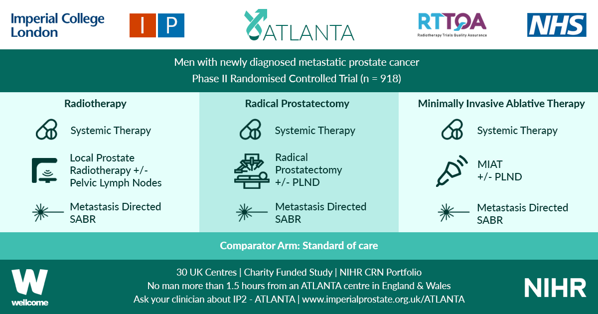
IP2 – ATLANTA is launched! ATLANTA is a phase II randomised controlled trial that will explore sequential multi-modal treatment using systemic therapy, local physical cytoreduction and metastasis directed therapy in men with newly diagnosed metastatic prostate cancer against a comparator of standard of care alone.

All men with new histologically diagnosed hormone sensitive metastatic prostate cancer, within three months of commencing androgen deprivation therapy (ADT), and of performance status 0 to 2 are eligible.  No upper limit on metastatic burden will apply, although men must be fit to undergo all trial interventions at point of randomisation.

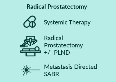
Men will be randomised to: Control (Standard of Care) OR Intervention 1 (Minimally Invasive Ablative Therapy [MIAT] +/- pelvic lymph node dissection [PLND]) OR Intervention 2 (Local Radiotherapy +/- Lymph Nodes OR Radical Prostatectomy +/- PLND). Randomisation stratified by metastatic burden (CHAARTED definition), intent to treat pelvic lymph nodes, intent to treat metastasis and intent to commence chemotherapy.

Systemic therapy in all arms includes ADT +/- Docetaxel. Radical prostatectomy will be with or without PLND. Local radiotherapy will be 60Gy/20Fr OR 74-78Gy in 2Gy per fraction over a minimum of 27 days, with or without simultaneous nodal radiotherapy. MIAT will be cryotherapy or focal HIFU. Men in both intervention arms will be eligible for metastasis directed therapy in the form of stereotactic ablative radiation (SABR) or surgery.

Men will be recruited over a two year period and followed up for a minimum of two years. Primary outcome will be progression free survival (PFS). ATLANTA is commencing in 17 UK trial centres with a target recruitment of 80 patients in the internal pilot, rising to 918 patients in full phase across 30 UK trial centres from November 2019.

ATLANTA is entirely charity funded (Wellcome Trust) and available on the NIHR CRN portfolio. Follow-up trial visits are not in excess of routine practice and extra burden is minimal. If you would like to join the main phase of ATLANTA as a site, please contact Mr Martin J. Connor ([email protected]) www.imperialprostate.org.uk/ATLANTA.

Prof. Hashim U. Ahmed (ATLANTA PI & CI),

Mr. Martin J. Connor (ATLANTA Doctoral Clinical Research Fellow)

Mr. Taimur T. Shah (Urology SpR & Research Fellow)


ATLANTA Surgeons Board: Mr Mathias Winkler, Mr Tim Dudderidge, Prof. Chris Eden, Mr Paul Cathcart, Prof. Naeem Soomro, Mr Adel Makar

ATLANTA Radiotherapy Board: Prof. John Staffurth, Dr. Alison Falconer, Dr. Stephen Mangar, Dr Olivia Naismith, RTTQA team

ATLANTA MIAT Board: Prof. Hashim U. Ahmed, Mr Stuart McCracken, Mr Raj Nigam, Mr Tim Dudderidge, Prof Iqbal Shergill

ATLANTA SABR Board: Dr Vincent Khoo, RTTQA team

ATLANTA Medical Oncologists: Dr. Naveed Sarwar, Dr Michael Gonzalez

ATLANTA Trial Sites: Imperial College Healthcare NHS Trust, The Royal Marsden Hospital, Guy’s & St Thomas’ NHS Foundation Trust, London North West Healthcare NHS Trust, Royal Surrey County (Guildford) Hospital, University Hospital Southampton, Clatterbridge Cancer Centre & Arrowe Park Hospital, Newcastle Freeman Hospital, King’s Lynn (Cambridge), Norfolk & Norwich (Cambridge), Sunderland Royal Hospital, Frimley Park Hospital, Royal Devon and Exeter Hospital, Wrexham Park Hospital, West Middlesex University Hospital, Royal United Hospital Bath, Betsi Calderwar Health Board, Lister Hospital, Hampshire (Basingstoke) Hospitals, University Hospital Coventry, Worcestershire Royal Hospital.

Trial Sponsor: Imperial College London

Trial Funder: Wellcome Trust

ClinicalTrials.gov Identifier: NCT03763253


BAUS/BJUI/USANZ Joint Session AUA 2019

British Association of Urological Surgeons/BJU International/Urological Society of Australia and New Zealand (BAUS/BJUI/USANZ) Joint Session AUA 2019

Sunday, May 5th 2:00 – 5.00 PM. McCormick Place Convention Center South Building – Room S102 BC


Registries /Smart Data /Complications – CHAIR: Duncan Summerton


1400-1420 Alan Partin

A contemporary look at biomarkers for diagnosis of Prostate Cancer

1420-1440 Chris Harding (BJUI sponsored BAUS lecture)

The Mesh Story – lessons learned and future plans

1440-1500 Nick Watkin

PROMs in Urology

1500-1520 Stephen Mark

Big Data and Urology – a pilot trial in New Zealand

1520-1540 Afternoon tea
 

Education /Training /Innovation – CHAIR: Prokar Dasgupta


1540-1600 Andrew Hung (BJUI sponsored lecture)

The emerging role of Artificial Intelligence in Surgical Science

1600-1620 Jonathan Kam

Zero learning curve Percutaneous Nephrolithotomy Access – Prone endoscopic combined intrarenal surgery and multimedia training aid to teach urology trainees

1620-1640 Madhu Koya (BJUI sponsored USANZ lecture)

Cx bladder reduces flexible cystoscopy in haematura and superficial TCC

1640-1700 Kamran Ahmad

Innovation in healthcare systems

1700-1705 BJUI Coffey-Krane Award for trainees based in The Americas presented by Prokar Dasgupta
1700-1900 BJUI Reception


Article of the week: A clinical prediction tool to determine the need for concurrent systematic sampling at the time of MRI‐guided biopsy

Every week, the Editor-in-Chief selects an Article of the Week from the current issue of BJUI. The abstract is reproduced below and you can click on the button to read the full article, which is freely available to all readers for at least 30 days from the time of this post.

In addition to the article itself, there is an editorial written by a prominent member of the urological community. These are intended to provoke comment and discussion and we invite you to use the comment tools at the bottom of each post to join the conversation. 

If you only have time to read one article this week, it should be this one.

A clinical prediction tool to determine the need for concurrent systematic sampling at the time of magnetic resonance imaging‐guided biopsy

Niranjan J. Sathianathen*, Christopher A. Warlick*, Christopher J. Weight*, Maria A. Ordonez*, Benjamin Spilseth, Gregory J. Metzger, Paari Muruganand Badrinath R. Konety*


Departments of *Urology, Radiology, and Pathology, University of Minnesota, Minneapolis, MN, USA


Read the full article

Abstract

Objective

To develop a clinical prediction tool that characterises the risk of missing significant prostate cancer by omitting systematic biopsy in men undergoing transrectal ultrasonography/magnetic resonance imaging (TRUS/MRI)‐fusion‐guided biopsy.

Patients and methods

A consecutive sample of men undergoing TRUS/MRI‐fusion‐guided biopsy with the UroNav® system (Invivo International, Best, The Netherlands) who also underwent concurrent systematic biopsy was included. By comparing the grade of cancer diagnosed on targeted and systematic biopsy cores, we identified cases where clinically significant disease (Gleason score ≥3+4) was only found on systematic and not targeted cores. Multivariable logistic regression analyses were used to identify predictive factors for finding significant cancer on systematic cores only. We then used these data to develop a nomogram and evaluated its utility using decision curve analysis.

Fig 1. Nomogram for predicting the diagnosis of clinically significant on systematic biopsy only and missed on targeted biopsy.

Results

Of the 398 men undergoing TRUS/MRI‐fusion‐guided biopsy in our study, there were 46 (11.6%) cases in which clinically significant cancer was missed on targeted biopsy and detected on systematic biopsy. The clinical setting, number of MRI lesions identified, and the highest Prostate Imaging‐Reporting and Data System (PI‐RADS) score of the lesions, were all found to be predictors of this. Our model had a good discriminative ability (concordance index = 0.70). The results from our decision curve analysis show that this model provides a higher net clinical benefit than either biopsying all men or omitting biopsy in all patients when the threshold probability is <30%.

Conclusion

We found that omitting concurrent systematic biopsy in men undergoing TRUS/MRI‐fusion‐guided biopsy would miss significant disease in more than one in 10 patients. We propose a prediction model with good discriminative ability that can be used to improve patient selection for performing concurrent systematic biopsy in order to minimise the number of missed significant cancers. It is important that our model is validated in external cohorts before being employed in routine clinical practice.

Read more Articles of the week

Editorial: Can systematic biopsy be safely avoided at the time of MRI/ultrasonography fusion biopsy?

In clinical practice, the need for maximising prostate cancer detection is often balanced against the theoretical risks of infection, bleeding, and pain associated with taking additional cores. In this novel study, Sathianathen et al. [1] provide a tool for measuring the oncological benefit of including concurrent systematic biopsy (SB) at the time of MRI‐guided targeted biopsy (TB). There were several key findings: (i) Amongst patients undergoing MRI‐guided biopsy (all biopsy settings), 11.6% were found to have significant cancers detected by SB alone; (ii) Amongst patients who had clinically significant cancers detected by SB alone, 52.2% were sampled within sextants outside the targeted regions of interest; (iii) According to the proposed nomogram, patients with prior negative biopsies, fewer MRI lesions, and lower Prostate Imaging‐Reporting and Data System (PI‐RADS) scores were at the lowest risk of missing significant cancer when SB was omitted.

Based on the present study, biopsy setting appears to be a key factor for deciding whether to omit SB. In the subset of patients undergoing primary biopsy, the authors found that 18.5% of cancers were detected by SB alone. These results are consistent with those of the MRI‐FIRST trial, which showed 14% of cancers were detected by SB only, 20% by TB only, and 66% by combining both techniques [2]. MRI‐FIRST concluded that in the primary biopsy setting, there was no difference between SB and TB in detection of clinically significant prostate cancer, although combining both techniques provided the highest detection rate.

Prior negative biopsy cohorts are generally at lower risk of harbouring significant cancer, as many cancers have already been ‘selected out’ by initial biopsies. In this setting, TB plays an important role in sampling tumour foci in difficult‐to‐reach regions of the prostate (e.g., anterior and apical) [3]. According to the authors’ nomogram, prior negative biopsy patients were least likely to benefit from concurrent SB. While the authors suggest a paradigm of selectively omitting SB, some authors have proposed omitting both TB and SB altogether in select patients. A previously reported multi‐institutional nomogram can be used to predict benign pathology after MRI‐guided biopsy, which can help reduce the number of unnecessary biopsies after MRI in the prior negative biopsy setting [4]. This clinical tool was further externally validated and optimised by Bjurlin et al. [5].

The ‘active surveillance (AS)’ setting typically refers to a confirmatory MRI‐guided biopsy in men with Grade Group 1 prostate cancer prior to enrollment in AS. Recently, the presence of cribriform morphology in Grade Group 2 patients was confirmed to be a key poor prognostic feature that would exclude patients from AS [6]. The present study, however, did not account for different Gleason pattern 4 morphologies in their analysis, as ‘significant cancer’ was defined by Grade Group alone. Studies by independent groups have found that TB combined with SB was more accurate than either modality alone for detecting cribriform at the time of MRI‐guided biopsy [78]. Therefore, concurrent SB is required to properly sample cribriform cancers in patients who are considering AS.

In this study, Sathianathen et al. [1] provide clinicians with a clinical tool for quantifying the added oncological value of concurrent SB. However, concurrent SB is probably prudent for most patients, particularly for those considering AS or focal therapy for which accurate determination of whole gland grade, cancer volume, and cribriform status are essential. As reducing the number of cores has not yet been shown to reduce biopsy‐related complications, are we willing to suboptimise cancer sampling without proven compensation?

by Matthew Truong

References

  1. Sathianathen, NJWarlick, CAWeight, CJ et al. A clinical prediction tool to determine the need for concurrent systematic sampling at the time of magnetic resonance imaging‐guided biopsy. BJU 2019123612– 7
  2. Salami, SSBen‐Levi, EYaskiv, O et al. In patients with a previous negative prostate biopsy and a suspicious lesion on magnetic resonance imaging, is a 12‐core biopsy still necessary in addition to a targeted biopsy? BJU Int 2015115562– 70
  3. Truong, MWang, BGordetsky, JB et al. Multi‐institutional nomogram predicting benign prostate pathology on magnetic resonance/ultrasound fusion biopsy in men with a prior negative 12‐core systematic biopsy. Cancer 2018124278– 85
  4. Bjurlin, MARenson, ARais‐Bahrami, S et al. Predicting benign prostate pathology on magnetic resonance imaging/ultrasound fusion biopsy in men with a prior negative 12‐core systematic biopsy: external validation of a prognostic nomogram. Eur Urol Focus 2018. [Epub ahead of print] https://doi.org/10.1016/j.euf.2018.05.005
  5. Kweldam, CFKümmerlin, IPNieboer, D et al. Presence of invasive cribriform or intraductal growth at biopsy outperforms percentage grade 4 in predicting outcome of Gleason score 3+4=7 prostate cancer. Mod Pathol 2017301126– 32
  6. Truong, MFeng, CHollenberg, G et al. A comprehensive analysis of cribriform morphology on magnetic resonance imaging/ultrasound fusion biopsy correlated with radical prostatectomy specimens. J Urol 2018199106– 13
  7. Prendeville, SGertner, MMaganti, M et al. Role of magnetic resonance imaging targeted biopsy in detection of prostate cancer harboring adverse pathological features of intraductal carcinoma and invasive cribriform carcinoma. J Urol 2018200104– 13


Article of the month: Prostate cancer mortality rates in Peru and its geographical regions

Every month, the Editor-in-Chief selects an Article of the Month from the current issue of BJUI. The abstract is reproduced below and you can click on the button to read the full article, which is freely available to all readers for at least 30 days from the time of this post.

In addition to the article itself, there is an editorial written by a prominent member of the urological community. These are intended to provoke comment and discussion and we invite you to use the comment tools at the bottom of each post to join the conversation. 

If you only have time to read one article this week, it should be this one.

Prostate cancer mortality rates in Peru and its geographical regions

Junior Smith Torres-Roman*, Eloy F. Ruiz, Jose Fabian Martinez-Herrera§, Sonia Faria Mendes Braga, Luis Taxa**, Jorge Saldaña-Gallo*, Mariela R. Pow-Sang††, Julio M. Pow-Sang‡‡ and Carlo La Vecchia§§


*Clinica de Urologia Avanzada UROZEN, Lima, Facultad de Medicina Humana, Universidad Nacional San Luis Gonzaga, Ica, CONEVID, Unidad de Conocimiento y Evidencia, Universidad Peruana Cayetano Heredia, Lima, Peru, §Cancer Center, Medical Center American British Cowdray, Mexico City, Mexico, Department of Social and Preventive Medicine, Faculty of Medicine, Universidade Federal de Minas Gerais, Belo Horizonte, Brazil, **Instituto Nacional de Enfermedades Neoplásicas, ††Department of Urology, Instituto Nacional de Enfermedades Neoplásicas, Lima, Peru, ‡‡Department of Genitourinary Oncology, Moffitt Cancer Center, Tampa, FL, USA, and §§Department of Clinical Sciences and Community Health, Universitá degli Studi di Milano, Milan, Italy


Read the full article

Abstract

Objective

To evaluate the mortality rates for prostate cancer according to geographical areas in Peru between 2005 and 2014.

Materials and Methods

Information was extracted from the Deceased Registry of the Peruvian Ministry of Health. We analysed age‐standardised mortality rates (world population) per 100 000 men. Spatial autocorrelation was determined according to the Moran Index. In addition, we used Cluster Map to explore relations between regions.

Fig. 1. Peru geographical zones by provinces. The asterisk denotes the province of Callao. Source: National Statistics Institute

Results

Mortality rates increased from 20.9 (2005–2009) to 24.1 (2010–2014) per 100 000 men, an increase of 15.2%. According to regions, during the period 2010–2014, the coast had the highest mortality rate (28.9 per 100 000), whilst the rainforest had the lowest (7.43 per 100 000). In addition, there was an increase in mortality in the coast and a decline in the rainforest over the period 2005–2014. The provinces with the highest mortality were Piura, Lambayeque, La Libertad, Callao, Lima, Ica, and Arequipa. Moreover, these provinces (except Arequipa) showed increasing trends during the years under study. The provinces with the lowest observed prostate cancer mortality rates were Loreto, Ucayali, and Madre de Dios. This study showed positive spatial autocorrelation (Moran’s I: 0.30, P= 0.01).

Conclusion

Mortality rates from prostate cancer in Peru continue to increase. These rates are higher in the coastal region compared to those in the highlands or rainforest.

Read more Articles of the week


Editorial: The burden of urological cancers in low‐ and middle‐income countries

The burden of cancer in low‐ and middle‐income countries (LMICs) continues to rise [1]. Evaluation of geographical differences in cancer mortality statistics is specifically of interest in LMICs as (inter)national guidelines are potentially less embedded in standard care, and objective measurements to assess underlying mechanisms/explanations for the burden of cancer are often lacking. Monitoring mortality statistics in these countries can thus help assess the effectiveness of national and regional health systems in treating and caring for patients with cancer [1].

Torres‐Roman et al. [2] deserve to be congratulated for their efforts to monitor mortality rates for prostate cancer at both a regional and national level in Peru. The CONCORD initiative from the WHO previously reported prostate cancer statistics for Peru, but data were limited to the capital area of Lima [1]. Torres‐Raman et al. [2] report prostate cancer mortality rates between 2005 and 2014 based on data from the Peruvian Ministry of Health, which covers ~70% of all healthcare providers in Peru. Apart from an overall increase of 15% in mortality rates, substantial variation was observed by geographical region. Mortality rates increased by 16% in the coastal region and highlands, whereas in the rainforest region the rates decreased by 19% [2]. One potential explanation for these observed differences could be the difference in ethnic and racial characteristics. The coastal region in Peru has a strong African influence and also has a larger proportion of men aged >65 years. In addition to potential differences in access to healthcare, some of the variation in prostate cancer mortality statistics most likely reflects a deficiency in reporting systems. Even though this study has its limitations due to missing data and lack of information on other important variables, such as ethnicity and socioeconomic status, it provides a first base for a critical assessment of prostate cancer care in Peru.

Studies like this one from Torres‐Roman et al. [2] show that there is a need for improvement and standardisation of (prostate) cancer care in LMICs, but also a need for improvement in data capturing, so that objective measurements can be put in place. The years of healthy life lost due to prostate cancer, as well as other urological cancers, in LMICs is increasing substantially. Even though each tumour group has its own specifications in terms of prevention and control, an epidemiological assessment of cancer burden based on the experience for urological cancers (i.e., prostate, bladder, kidney and testicular) can therefore inform future assessments of cancer burden. The urological tumour group covers both common and less common cancers (e.g. prostate vs kidney cancer), sex‐specific and cancers that affect both sexes (e.g. testicular vs bladder cancer), cancers with less known risk factors and those strongly linked with lifestyle risk factors (e.g. prostate vs bladder cancer).

It is encouraging to see an increase in the number of studies evaluating the burden of cancer in LMICs [3]; however, given the consistency in observations of an increase in mortality, there is an urgent need to further invest in prevention and management, as well as the infrastructure to collect all relevant data at a national level in these LMICs. Accurate information about cancer burden and how this varies between regions is essential to plan for an adequate health‐system response.

References

  1. Allemani, CMatsuda, TCarlo, V et al. Global surveillance of trends in cancer survival 2000‐14 (CONCORD‐3): analysis of individual records for 37 513 025 patients diagnosed with one of 18 cancers from 322 population‐based registries in 71 countries. Lancet 20183911023– 75
  2. Torres‐Roman, JRuiz, EMartinez‐Herrera, J et al. Prostate cancer mortality rates in Peru and its geographic regions. BJU Int 2019123595– 601
  3. Carioli, GVecchia, CBertuccio, P et al. Cancer mortality predictions for 2017 in Latin America. Ann Oncol 2017282286– 97


EAU19 Barcelona – Highlights from days 3-5 of the 34th Annual EAU Congress

The early Sunday morning start did not deter delegates from attending one of the three packed plenary sessions of the day. They covered a broad range of rapidly changing areas in urology from imaging in prostate cancer, an update on renal cell carcinoma (RCC) and the breaking news session discussing the potentially game changing results from the recent ARAMIS study and new research into fast bi-parametric MRI.  The role of imaging in prostate cancer is swiftly evolving, with the plenary discussion focusing on recent changes in the diagnostic pathway of localised prostate cancer, particularly with the use of MRI. Next door in the RCC plenary, the speakers debated ‘knife, needle or nothing?’ for the small renal mass in the young patient followed by an update on the very recent and potentially guideline-changing advances in systemic therapy for RCC.

The mid-morning thematic sessions covered the full spectrum of urology from semi-live surgery, the newest advances in immunotherapy, imaging and even how to run a urology office in Europe.

The 7th BJUI social media awards on Sunday night were again the social highlight of the EAU. A view of the Museu Nacional d’Art de Catalunya provided a stunning backdrop to the packed event, with the stars of #UroSoMe recognised for their outstanding work. The night kicked off with the award for the most read blog going to social media champion Professor Declan Murphy.

The awards highlighted the far reaching and valuable impact of social media, recognising a number of important achievements in the field such as Nature Reviews Urology for ‘Both sides of the scalpel: the patient and surgeon view’ with a special guest video appearance from Stephen Fry.

However, for me the most special part of the night was seeing my friend Daniel Christidis remembered and honoured with the most ‘social’ trainee award. Dan was a leader in the real and #UroSoMe world (and had personally set up my Twitter account, and those of many of the other young attendees that night) and I know would have been proud to be remembered for one of the things he did so well.

After the BJUI social media awards, it was time for a little black-tie glamour with the EAU19 Friendship Dinner at the historical Casa Llotja de Mar. The night started with a welcome from Professor Christopher Chapple underlining the importance of international partnerships in urology, followed by a fantastic night of good food, wine and enjoying the beautiful Catalan Gothic architecture.

The Monday morning plenary sessions delivered another jam-packed morning of a mix of cutting-edge science, quality of life issues in cancer survivorship and prostate cancer. The breaking news session discussed the primary results from SAUL, confirming tolerability and safety of atezolizumab in real-world mUC patients, and the results of ARCHES, which investigated the efficacy of androgen deprivation therapy with enzalutamide or placebo in metastatic hormone-sensitive prostate cancer. The controversies in prostate cancer were again debated in an interactive and diverse way between ‘jury members’ including a geriatrician, psychologist, radiation oncologist and urologist.

The last day of the thematic sessions of the congress again provided a smorgasbord of topics in urology. Later in the day, the expert-guided poster tours gave delegates a chance to navigate the huge number of posters from guidelines to local treatment of prostate cancer.

The closing plenary on Tuesday morning to a full auditorium gave a sweeping overview of the top contributions to EAU19 leaving us with a free half day to explore our generous host city and take in the stunning architecture, food and sunshine!


Bustling Barcelona provided the perfect backdrop to a well organised, action packed conference which featured world leading urologists and scientists from around the world presenting practice changing new data. Cannot wait for EAU 2020 in Amsterdam! #EAU20 #Amsterdam #UroSoMe

by Jiasian Teh, Urology Registrar, PhD Candidate, Peter MacCallum Cancer Centre

@JiasianTeh

Article of the week: Ultrasound characteristics of regions identified as suspicious by MRI predict the likelihood of clinically significant cancer on MRI–ultrasound fusion‐targeted biopsy

Every week, the Editor-in-Chief selects an Article of the Week from the current issue of BJUI. The abstract is reproduced below and you can click on the button to read the full article, which is freely available to all readers for at least 30 days from the time of this post.

In addition to the article itself, there is an editorial written by a prominent member of the urological community, and a video made by the authors. These are intended to provoke comment and discussion and we invite you to use the comment tools at the bottom of each post to join the conversation. 

If you only have time to read one article this week, it should be this one.


The ultrasound characteristics of regions identified as suspicious by magnetic resonance imaging (MRI) predict the likelihood of clinically significant cancer on MRI–ultrasound fusion‐targeted biopsy

Benjamin Press*, Andrew B. Rosenkrantz, Richard Huang and Samir S. Taneja§ 
 
*Rutgers New Jersey Medical School, Newark, NJ, Department of Radiology, Department of Urology, and §Departments of Urology and Radiology, NYU Langone Health, New York, NY, USA
 
Read the full article

Abstract

Objective

To determine whether the presence of an ultrasound hypoechoic region at the site of a region of interest (ROI) on magnetic resonance imaging (MRI) results in improved prostate cancer (PCa) detection and predicts clinically significant PCa on MRI–ultrasonography fusion‐targeted prostate biopsy (MRF‐TB).

Materials and Methods

Between July 2011 and June 2017, 1058 men who underwent MRF‐TB, with or without systematic biopsy, by a single surgeon were prospectively entered into an institutional review board‐approved database. Each MRI ROI was identified and scored for suspicion by a single radiologist, and was prospectively evaluated for presence of a hypoechoic region at the site by the surgeon and graded as 0, 1 or 2, representing none, a poorly demarcated ROI‐HyR, or a well demarcated ROI‐HyR, respectively. The interaction of MRI suspicion score (mSS) and ultrasonography grade (USG), and the prediction of cancer detection rate by USG, were evaluated through univariate and multivariate analysis.

Results

For 672 men, the overall and Gleason score (GS) ≥7 cancer detection rates were 61.2% and 39.6%, respectively. The cancer detection rates for USGs 0, 1 and 2 were 46.2%, 58.6% and 76.0% (P < 0.001) for any cancer, and 18.7%, 35.2% and 61.1% (P < 0.001) for GS ≥7 cancer, respectively. For MRF‐TB only, the GS ≥7 cancer detection rates for USG 0, 1 and 2 were 12.8%, 25.7% and 52.0%, respectively (P < 0.001). On univariate analysis, in men with mSS 2–4, USG was predictive of GS ≥7 cancer detection rate. Multivariable regression analysis showed that USG, prostate‐specific antigen density and mSS were predictive of GS ≥7 PCa on MRF‐TB.

Conclusions

Ultrasonography findings at the site of an MRI ROI independently predict the likelihood of GS ≥7 PCa, as men with a well‐demarcated ROI‐HyR at the time of MRF‐TB have a higher risk than men without.
Read more Articles of the week

Editorial: Is transrectal ultrasonography of the prostate obsolete in the MRI era?

Sampling of prostate tissue to confirm pathologically a clinical suspicion of cancer has undergone an exponential change. The random systematic prostate biopsy technique was the only method used for many decades, initially guided by the finger but, since 1989, performed with TRUS guidance. Now, within the space of only a few years, we have entered the era of performing prostate biopsies on the basis of high‐tech three‐dimensional multiparametric MRI images, including software that can track the exact course of the biopsy needle [1]. While new technical developments in general lead to better, more individually directed healthcare, there is always the risk of abandoning ‘old’ but well developed and extensively tested techniques too soon. In this issue of the BJUI, Press et al. [2] looked at the added value of the presence of an ‘old‐fashioned’ TRUS‐detected lesion in cancer‐suspicious regions on MRI to better predict the presence of clinically significant prostate cancer (csPCa) defined as Gleason score ≥7. In their study comprising 1058 men, it was shown that a well‐demarcated abnormal TRUS finding noted at the time of MRI‐TRUS fusion‐guided prostate biopsy coincides with an increased risk of csPCa detection, independent of MRI suspicion (Prostate Imaging Reporting and Data System [PI‐RADS] score).

Increasing PI‐RADS score is correlated with an increased percentage of csPCa after targeted biopsy, both at initial and repeat biopsy. In a review based on data from 8252 men, it was shown that there is a gradual increase in the detection of csPCa from PI‐RADS 3 to PI‐RADS 4 to PI‐RADS 5 index lesions. For example, at first biopsy, the overall rate of PCa detection and the percentage of csPCa were 39%, 62% and 92% and 54%, 63% and 76% for PI‐RADS 3, 4 and 5 lesions, respectively. This means that in men with PI‐RADS 3 lesions, representing approximately one‐third of men deemed eligible for further assessment, only 39% will be diagnosed with PCa and half of the PCa detected will be potentially indolent Gleason 6 PCa [3]. This makes this group of men extremely interesting for further risk stratification before biopsy. Multivariable risk stratification in which PSA density plays an important role has been shown to be of value in these men [4] but further refinement could potentially be made by including suspicious lesions identified at TRUS.

Apart from the added value of TRUS findings in terms of risk stratification, the performance of the MRI‐targeted biopsy itself could be improved by visual guidance of hypoechoic lesions. In the present study by Press et al [2], a hypoechoic TRUS lesion was present at or near the location of two‐thirds of cancer‐suspicious lesions on MRI. The authors more or less advise to direct the targeted biopsy cores not only to the MRI suspicious lesion, but also the TRUS suspicious lesion, both of which often do not fully overlay in a software‐assisted MRI‐TRUS fusion model. The extent to which this ‘correction for misregistration’ is already included during targeted biopsy in current clinical practice is unknown. Although feasible and seemingly important during software‐assisted fusion targeted biopsy, TRUS lesions in cancer‐suspicious MRI regions might be more frequently targeted during cognitive fusion‐targeted biopsy. Two recent studies underline the important message of the present study, and show that a considerable proportion of csPCa is missed in and around MRI‐suspicious lesions by targeted biopsies, as a result of sampling errors related to both misregistration and intra‐tumour heterogeneity [56]. As suggested by these studies, visual guidance by hypoechoic lesions and ‘focal saturation’ biopsy by additional (peri‐)lesional cores might improve the detection of csPCa.

In summary, ‘good old’ TRUS could be of value in those patients who are virtually always present in scenarios in which a grading system is being used, i.e. patients belonging to the so‐called grey zone. The challenge of risk stratification (i.e. personalized medicine) is to nibble at both sides of the grey zone by implementing new techniques or, more likely by implementing a combination of all available and relevant knowledge.

by Monique J. Roobol, Frank-Jan H. Drost and Arnout R. Alberts

References

  1. Verma, SChoyke, PLEberhardt, SC et al. The current state of MR imaging‐targeted biopsy techniques for detection of prostate cancer. Radiology 201728534356
  2. Press, BRosenkrantz, ABHuang, RTaneja, SSThe ultrasound characteristics of MRI suspicious regions predict the likelihood of clinically significant cancer on MRI‐ultrasound fusion targeted biopsy. BJUI 201912343946.
  3. Schoots, IGMRI in early prostate cancer detection: how to manage indeterminate or equivocal PI‐RADS 3 lesions? Transl Androl Urol 201877082
  4. Alberts, ARSchoots, IGBokhorst, LPLeenders, GJBangma, CHRoobol, MJRisk‐based patient selection for magnetic resonance imaging‐targeted prostate biopsy after negative transrectal ultrasound‐guided random biopsy avoids unnecessary magnetic resonance imaging scans. Eur Urol 201669112934
  5. Simmons, LAMKanthabalan, AArya, M et al. Accuracy of transperineal targeted prostate biopsies, visual estimation and image fusion in men needing repeat biopsy in the PICTURE trial. J Urol 2018200122734
  6. Leest, M, Cornel, EIsrael, B et al. Head‐to‐head comparison of transrectal ultrasound‐guided prostate biopsy versus multiparametric prostate resonance imaging with subsequent magnetic resonance‐guided biopsy in biopsy‐naive men with elevated prostate‐specific antigen: a large prospective multicenter clinical study. Eur Urol 2018; [Epub ahead of print]. https://doi.org/10.1016/j.eururo.2018.11.023.


© 2024 BJU International. All Rights Reserved.