Tag Archive for: #ProstateCancer

Posts

Editorial: LDR prostate brachytherapy in younger men

Langley et al.1 report on the oncological and functional outcomes of men treated with low dose rate (LDR) prostate brachytherapy in men 60 years old or younger. 597 patients with a median (range) age of 57 (44-60) years had a median follow-up of 8.9 (1.5-17.2) years. The 10- year post-implant relapse free survival using the Phoenix definition for biochemical failure (nadir plus 2 ng/ml) was 95%, 90% and 87% for low, intermediate and high-risk disease, respectively. Potency was preserved in 75% of men potent before treatment. The authors concluded that LDR brachytherapy is an efficacious treatment with excellent long-term control of prostate cancer in men ≤60 years at time of treatment. While the results from this investigation are encouraging, enthusiasm should be tempered given the short follow up, long natural history of prostate cancer and the long-life expectancy for these younger patients.

Although the overall median follow-up was 8.9 years, the calculation of PSA-free failure was derived from a median follow-up of 5.9 years. As this investigation did not identify men who may also be at risk of failure because of a rising PSA and who have not yet reached the Phoenix threshold, I anticipate longer-follow up will further reduce their favorable results. Of the 597 men, 6 (1%) died from prostate cancer. The low incidence of prostate cancer mortality, while impressive, also reflects the short follow-up. Our group has previously reported that PCSM substantially increases between the 10th and 15th year post treatment. The experience of these physicians in prostate brachytherapy is demonstrated in their favorable dosimetry outcomes-the median D90 was 106.4% of the prescription (145 Gy). These results (median D90 154.3 Gy), which were determined and computed on the day of the implant would be 10-15% higher had the CT scans been done on day 30 as most centers do. We and others have reported that patients receiving higher dose implants have improved biochemical and cancer-specific outcomes. While these data help explain their favorable oncological outcomes, they should also serve as a guide to other brachytherapy programs where implant quality should be a primary objective. Erectile function was preserved in 75% of men. These data are consistent with other reports of younger men who were treated for prostate cancer with surgery or radiation. Because this was not a randomized study, it is not possible to make direct comparisons between surgery and brachytherapy. The selection of an IIEF score of > 11 as potent might be challenged as the 12-16 group is considered to have mild to moderate ED. Nonetheless, these data are still encouraging for younger men who are considering treatment for localized prostate cancer where sexual function preservation is important.

Nelson Stone

Mount Sinai Medical Center – Urology 350 E 72nd Street, New York, New York 10021 United States

Reference

  1. Langley SM, Soares R, Uribe J, et al. Long-term oncological outcomes and toxicity in 597 men ≤60 years of age at time of low dose rate brachytherapy for localised prostate cancer. BJU Int 2017


Article of the Week: Prostate Health Index density improves detection of clinically significant prostate cancer

Every Week the Editor-in-Chief selects an Article of the Week from the current issue of BJUI. The abstract is reproduced below and you can click on the button to read the full article, which is freely available to all readers for at least 30 days from the time of this post.

In addition to the article itself, there is an accompanying editorial written by a prominent member of the urological community. This blog is intended to provoke comment and discussion and we invite you to use the comment tools at the bottom of each post to join the conversation.

Finally, the third post under the Article of the Week heading on the homepage will consist of additional material or media. This week we feature a video discussing the paper.

If you only have time to read one article this week, it should be this one.

Prostate Health Index density improves detection of clinically significant prostate cancer

Jeffrey J. Tosoian*, Sasha C. Druskin*, Darian Andreas*, Patrick Mullane*, Meera Chappidi*, Sarah Joo*, Kamyar Ghabili*, Mufaddal Mamawala*, Joseph Agostino*, Herbert B. Carter*, Alan W. Partin*, Lori J. Sokoll*§ and Ashley E. Ross*§


*Department of Urology, James Buchanan Brady Urological Institute, Johns Hopkins University School of Medicine, Baltimore, MD, Virginia Commonwealth University School of Medicine, Richmond, VA, Department of Pathology, and §Sidney Kimmel Comprehensive Cancer Center, Johns Hopkins University School of Medicine, Baltimore, MD, USA
Read the full article

Abstract

Objectives

To explore the utility of Prostate Health Index (PHI) density for the detection of clinically significant prostate cancer (PCa) in a contemporary cohort of men presenting for diagnostic evaluation of PCa.

Patients and Methods

The study cohort included patients with elevated prostate-specific antigen (PSA; >2 ng/mL) and negative digital rectal examination who underwent PHI testing and prostate biopsy at our institution in 2015. Serum markers were prospectively measured per standard clinical pathway. PHI was calculated as ([{−2}proPSA/free PSA] × [PSA]½), and density calculations were performed using prostate volume as determined by transrectal ultrasonography. Logistic regression was used to assess the ability of serum markers to predict clinically significant PCa, defined as any Gleason score ≥7 cancer or Gleason score 6 cancer in >2 cores or >50% of any positive core.

Results

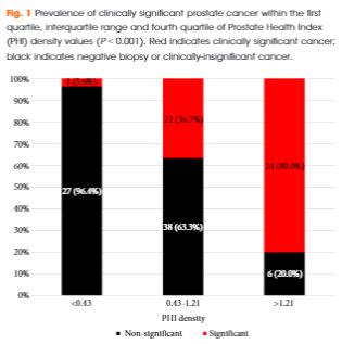
Of 118 men with PHI testing who underwent biopsy, 47 (39.8%) were found to have clinically significant PCa on biopsy. The median (interquartile range [IQR]) PHI density was 0.70 (0.43–1.21), and was 0.53 (0.36–0.75) in men with negative biopsy or clinically insignificant PCa and 1.21 (0.74–1.88) in men with clinically significant PCa (P < 0.001). Clinically significant PCa was detected in 3.6% of men in the first quartile of PHI density (<0.43), 36.7% of men in the IQR of PHI density (0.43–1.21), and 80.0% of men with PHI density >1.21 (P < 0.001). Using a threshold of 0.43, PHI density was 97.9% sensitive and 38.0% specific for clinically significant PCa, and 100% sensitive for Gleason score ≥7 disease. Compared with PSA (area under the curve [AUC] 0.52), PSA density (AUC 0.70), %free PSA (AUC 0.75), the product of %free PSA and prostate volume (AUC 0.79), and PHI (AUC 0.76), PHI density had the highest discriminative ability for clinically significant PCa (AUC 0.84).

Conclusions

Based on the present prospective single-centre experience, PHI density could be used to avoid 38% of unnecessary biopsies, while failing to detect only 2% of clinically significant cancers.

Read more articles of the week

Editorial: Prostate cancer biomarkers: new scenarios in the multi-parametric magnetic resonance imaging era

The management of prostate cancer poses difficult challenges, which is largely because we lack the necessary tools to predict its presence, and discern between indolent disease with a small chance of clinical manifestation and aggressive tumours that are more likely to be lethal.

Despite the fact that novel blood and urine tests are available, which may predict aggressive disease better than PSA; they are not routinely used due to a lack of clinical validity studies.

Tosoian et al. [1] in the present study explored the utility of prostate health index (PHI) density for detection of clinically significant prostate cancer in a contemporary cohort of men presenting for diagnostic evaluation of prostate cancer. Very interestingly the authors hypothesised that, similar to PSA density, PHI density could further improve upon the discriminative ability of PHI to detect prostate cancer. The PHI density calculation was performed using prostate volume, as determined by TRUS. Logistic regression was used to assess the ability of serum markers to predict clinically significant prostate cancer, defined as any Gleason score ≥7 cancer or Gleason score 6 cancer in >2 cores or >50% of any positive core.

They showed, albeit in a small sample size, that PHI density could further improve upon the discriminative ability of PHI and appears to be superior to PSA and other PSA derivatives for the identification of clinically significant disease [1].

However, it is noteworthy that in all studies on urine or serum biomarkers such as this, the ‘gold standard’ for cancer detection is pathological examination of multiple non-targeted systematic TRUS-guided prostate biopsies, not radical prostatectomy specimens. Intrinsically, this approach implies that no cancer predicted by the biomarker may still mean cancer missed by the biopsy.

Introducing mpMRI before prostate biopsy has the potential to improve prostate cancer sampling ink that is the most practical way to make mpMRI before biopsy economically viable for universal NHS adoption.

The aim should be the development of a clinical decision support system based on mpMRI and circulating biomarkers, as in this case PHI density evaluation, to stratify patients according to their risk of prostate cancer progression, using pathological assessment after prostatectomy as the reference standard.

Francesco Porpiglia and Stefano De Luca
Division of Urology, San Luigi Gonzaga Hospital and University of Torino, Orbassano, Italy


Read the full article


References


1 Tosoian JJDruskin SCAndreas D et al. Prostate health index density improves detection of clinically significant prostate cancer. BJU Int2017; 120: 7938.


2 Mottet NBellmunt JBolla M et al. EAU-ESTRO-SIOG guidelines on prostate cancer. Part 1: Screening, diagnosis, and local treatment with curative intent.  Eur Urol 2016; pii: S0302-2838(16)30470-5. [Epub ahead of print]. doi: 10.1016/j.eururo.2016.08.003.


3 Russo FRegge DArmando E et al. Detection of prostate cancer index lesions with multiparametric magnetic resonance imaging (mp-MRI) using whole-mount histological sections as the reference standard. BJU Int 2016; 118: 8494.


5 Porpiglia FManfredi MMele F et al. Diagnostic pathway with multiparametric magnetic resonance imaging versus standard pathway: results from a randomized prospective study in biopsy-naıve patients with suspected prostate cancer. Eur Urol 2016; pii: S0302-2838(16)30509-7. [Epub ahead of print]. doi: 10.1016/j.eururo.2016.08.041


6 Wegelin Ovan Melick HHHooft L et al. Comparing three different techniques for magnetic resonance imaging-targeted prostate biopsies: a systematic review of in-bore versus magnetic resonance imaging- transrectal ultrasound fusion versus cognitive registration. Is there a preferred technique?. Eur Urol 2016; pii: S0302-2838(16)30446-8. [Epub ahead of print]. doi: 10.1016/j.eururo.2016.07.04


Video: Prostate Health Index density improves detection of clinically significant prostate cancer

Prostate Health Index density improves detection of clinically significant prostate cancer

Read the full article

Abstract

Objectives

To explore the utility of Prostate Health Index (PHI) density for the detection of clinically significant prostate cancer (PCa) in a contemporary cohort of men presenting for diagnostic evaluation of PCa.

Patients and Methods

The study cohort included patients with elevated prostate-specific antigen (PSA; >2 ng/mL) and negative digital rectal examination who underwent PHI testing and prostate biopsy at our institution in 2015. Serum markers were prospectively measured per standard clinical pathway. PHI was calculated as ([{−2}proPSA/free PSA] × [PSA]½), and density calculations were performed using prostate volume as determined by transrectal ultrasonography. Logistic regression was used to assess the ability of serum markers to predict clinically significant PCa, defined as any Gleason score ≥7 cancer or Gleason score 6 cancer in >2 cores or >50% of any positive core.

Results

Of 118 men with PHI testing who underwent biopsy, 47 (39.8%) were found to have clinically significant PCa on biopsy. The median (interquartile range [IQR]) PHI density was 0.70 (0.43–1.21), and was 0.53 (0.36–0.75) in men with negative biopsy or clinically insignificant PCa and 1.21 (0.74–1.88) in men with clinically significant PCa (P < 0.001). Clinically significant PCa was detected in 3.6% of men in the first quartile of PHI density (<0.43), 36.7% of men in the IQR of PHI density (0.43–1.21), and 80.0% of men with PHI density >1.21 (P < 0.001). Using a threshold of 0.43, PHI density was 97.9% sensitive and 38.0% specific for clinically significant PCa, and 100% sensitive for Gleason score ≥7 disease. Compared with PSA (area under the curve [AUC] 0.52), PSA density (AUC 0.70), %free PSA (AUC 0.75), the product of %free PSA and prostate volume (AUC 0.79), and PHI (AUC 0.76), PHI density had the highest discriminative ability for clinically significant PCa (AUC 0.84).

Conclusions

Based on the present prospective single-centre experience, PHI density could be used to avoid 38% of unnecessary biopsies, while failing to detect only 2% of clinically significant cancers.

View more videos

Article of the Week: A mpMRI-based risk model to determine the risk of prostate cancer prior to biopsy

Every Week the Editor-in-Chief selects an Article of the Week from the current issue of BJUI. The abstract is reproduced below and you can click on the button to read the full article, which is freely available to all readers for at least 30 days from the time of this post.

In addition to the article itself, there is an accompanying editorial written by a prominent member of the urological community. This blog is intended to provoke comment and discussion and we invite you to use the comment tools at the bottom of each post to join the conversation.

If you only have time to read one article this week, it should be this one.

A multiparametric magnetic resonance imaging-based risk model to determine the risk of significant prostate cancer prior to biopsy

Pim J. van Leeuwen*, Andrew Hayen, James E. Thompson*†‡, Daniel Moses§Ron Shnier§, Maret Bohm, Magdaline Abuodha, Anne-Maree HaynesFrancis Ting*†‡, Jelle Barentsz, Monique Roobol**, Justin Vass††, Krishan Rasiah††Warick Delprado‡‡ and Phillip D. Stricker*†‡


*St. Vincents Prostate Cancer Centre, Garvan Institute of Medical Research/The Kinghorn Cancer Centre, Darlinghurst, School of Public Health and Community Medicine, §School of Medicine, University of New South Wales, Kensington, New South Wales, Australia, Department of Radiology and Nuclear Medicine, Radboud University Medical Centre, Nijmegen, **Department of Urology, Erasmus University Medical Center, Rotterdam, the Netherlands, ††Department of Urology, Royal North Shore Private Hospital, St Leonards, and ‡‡Douglass Hanly Moir Pathology and University of Notre Dame, Darlinghurst, New South Wales, Australia


Read the full article

Abstract

Objective

To develop and externally validate a predictive model for detection of significant prostate cancer.

Patients and Methods

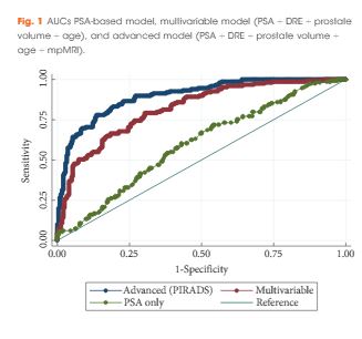
Development of the model was based on a prosp   ctive cohort including 393 men who underwent multiparametric magnetic resonance imaging (mpMRI) before biopsy. External validity of the model was then examined retrospectively in 198 men from a separate institution whom underwent mpMRI followed by biopsy for abnormal prostate-specific antigen (PSA) level or digital rectal examination (DRE). A model was developed with age, PSA level, DRE, prostate volume, previous biopsy, and Prostate Imaging Reporting and Data System (PIRADS) score, as predictors for significant prostate cancer (Gleason 7 with >5% grade 4, ≥20% cores positive or ≥7 mm of cancer in any core). Probability was studied via logistic regression. Discriminatory performance was quantified by concordance statistics and internally validated with bootstrap resampling.

Results

In all, 393 men had complete data and 149 (37.9%) had significant prostate cancer. While the variable model had good accuracy in predicting significant prostate cancer, area under the curve (AUC) of 0.80, the advanced model (incorporating mpMRI) had a significantly higher AUC of 0.88 (P < 0.001). The model was well calibrated in internal and external validation. Decision analysis showed that use of the advanced model in practice would improve biopsy outcome predictions. Clinical application of the model would reduce 28% of biopsies, whilst missing 2.6% significant prostate cancer.

Conclusions

Individualised risk assessment of significant prostate cancer using a predictive model that incorporates mpMRI PIRADS score and clinical data allows a considerable reduction in unnecessary biopsies and reduction of the risk of over-detection of insignificant prostate cancer at the cost of a very small increase in the number of significant cancers missed.

Read more articles of the week


Editorial: Novel risk stratification nomograms for counseling patients on the need for prostate biopsy

Contemporary recommendations for prostate screening incorporate the measurement of serum PSA levels into shared decision making [1]. PSA is limited by a low specificity for prostate cancer and exposes a certain number of men to unnecessary prostate biopsies. Moreover, it has been attributed to an over-diagnosis and over treatment of this disease, especially in indolent cancer that may never affect a man’s longevity [2].

Prostate cancer risk stratification and aggressiveness is necessary in both the pre-biopsy clinical counselling, as well as the decision-making process. It is clear that such an important approach cannot be accomplished based on PSA alone. In order to enhance prostate cancer screening and detection, other clinical variables such as PSA density, prostate volume, percentage free PSA, and DRE, are routinely considered in conjunction with PSA for determining the need for prostate biopsy.

Increasing evidence supports the use of MRI in prostate cancer detection when used as a localisation tool to guide MRI-targeted biopsy techniques such as MRI-ultrasonography fusion-targeted biopsy [3-5]. Pre-biopsy MRI not only allows accurate tumour localisation, but also provides an assessment of cancer suspicion using an MRI suspicion score, and thus provides accurate prediction of the likelihood of prostate cancer on prostate biopsy [6].

In this study, van Leeuwen et al. [7] developed and externally validated a set of nomograms predicting clinically significant prostate cancer by incorporating prostate MRI. The performance characteristics of the nomograms were maximised by inclusion of MRI results. The authors determined that clinical application of the model would reduce 28% of biopsies, while missing 2.6% of clinically significant prostate cancer. Ultimately incorporating these nomograms into the clinical decision-making process could result in a considerable reduction in unnecessary biopsies and reduction in the risk of over-detection of clinically insignificant disease at the cost of a small increase in the number of significant cancers missed.

The authors should be commended for their predictive nomograms, in that they may further aid in the decision to perform biopsy in men with clinical suspicion of prostate cancer. However, the findings of this study should be interpreted with caution. In formulating nomograms, the obvious clinical goal is the creation of a tool that improves the selection of men in need of biopsy. Unless nomograms are derived from general ‘at risk’ populations, including men with low- and high-risk of prostate cancer, the tool may be limited in its prediction. As an example, if all men in the training cohort have an elevated PSA level, the predictive value of PSA in the nomogram may be reduced. In this case, the training and validation cohort are not well described, but it seems to be a referral population, and the PSA range is relatively narrow. As such, it’s applicability to all men presenting for prostate cancer may be questionable.

We also have had difficulty modelling a nomogram from our MRI-targeted biopsy dataset because the power of MRI-suspicion score in predicting cancer tends to minimise the effect of other variables such as PSA, age, and gland size. The authors did not compare their multivariable predictive models to MRI alone, and this may have been the most relevant comparison. In this study, the gland size and PSA level contribute significantly to the nomogram score, but one might question the findings. For example, a man with a Prostate Imaging Reporting and Data System (PI-RADS) score of 4 or 5, a large gland and a low PSA level, has a similar or lower risk as a man with a PSA level of 15 ng/mL, a moderate gland, and a PI-RADS score of 3. This is not consistent with our clinical experience and draws concern in the reliability of the nomogram at extremes of PSA and age. Men with PI-RADS 5 have high rates of clinically significant prostate cancer, regardless of PSA or age. This also may reflect variability in the predictive accuracy of MRI depending upon MRI interpretation.

Another limitation of the study, as the authors cite, is that patients were biopsied using a transperineal mapping with a median of 30 cores. This biopsy strategy is not routinely used in most institutions, and consequently limits the generalisability of the nomograms.

Selective use of prostate biopsy among men with elevated PSA levels through further refinement of cancer risk is highly desirable. The novel risk stratification nomograms developed by van Leeuwen et al. [7] add to the tools we may use to counsel our patients on the need for prostate biopsy. Further evaluation of these nomograms on additional independent patient cohorts is warranted prior to implementation in clinical practice.

Marc A. Bjurlinand Samir S. Taneja
*Division of Urology, Department of Surgery, NYU Lutheran Medical Center, NYU Langone Health System, New York, NY, USA and Division of Urologic Oncology, Department of Urology, NYU Langone Medical Center, New York, NY, US


References


1 Carter HBAlbertsen PCBarry MJ et al. Early detection of prostate cancer: AUA Guideline. J Urol2013; 190: 41926


2 Loeb SBjurlin MANicholson J et al. Overdiagnosis and overtreatment of prostate cancer. Eur Urol
2014; 65: 104655


3 Mendhiratta NMeng XRosenkrantz AB et al. Prebiopsy MRI and MRI-ultrasound fusion-targeted prostate biopsy in men with previous negative biopsies: impact on repeat biopsy strategies. Urology 2015; 86: 11928


5 Meng XRosenkrantz ABMendhiratta N et al. Relationship between prebiopsy multiparametric magnetic resonance imaging (MRI), biopsy indication, and MRI-ultrasound fusion-targeted prostate biopsy outcomes. Eur Urol 2016; 69: 51217


7 van Leeuwen PJHayan AThompson JE et al. A multiparametric magnetic resonance imaging-based risk model to determine the risk of significant prostate cancer prior to biopsy. BJU Int 2017; 120: 7748


Article of the Week: Association of HDI with global bladder, kidney, prostate and testis cancer

Every Week the Editor-in-Chief selects an Article of the Week from the current issue of BJUI. The abstract is reproduced below and you can click on the button to read the full article, which is freely available to all readers for at least 30 days from the time of this post.

In addition to the article itself, there is an accompanying editorial written by a prominent member of the urological community. This blog is intended to provoke comment and discussion and we invite you to use the comment tools at the bottom of each post to join the conversation.

If you only have time to read one article this week, it should be this one.

Association of Human Development Index with global bladder, kidney, prostate and testis cancer incidence and mortality

Alyssa K. Greiman*, James S. Rosoff† and Sandip M. Prasad*


*Department of Urology, Medical University of South Carolina, Charleston, SC, Department of Urology, Yale School of Medicine, New Haven, CT, and Department of Surgery, Ralph M. Johnson VA Medical Center, Charleston, SC, USA


Read the full article

Abstract

Objectives

To describe contemporary worldwide age-standardized incidence and mortality rates for bladder, kidney, prostate and testis cancer and their association with development.

Materials and Methods

We obtained gender-specific, age-standardized incidence and mortality rates for 184 countries and 16 major world regions from the GLOBOCAN 2012 database. We compared the mortality-to-incidence ratios (MIRs) at national and regional levels in males and females, and assessed the association with socio-economic development using the 2014 United Nations Human Development Index (HDI).

Results

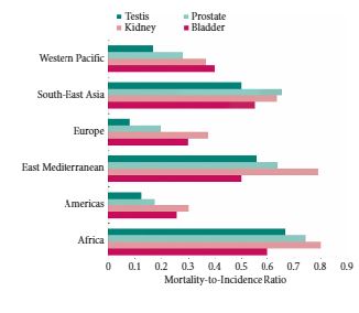
Age-standardized incidence rates were 2.9 (bladder) to 7.4 (testis) times higher for genitourinary malignancies in more developed countries compared with less developed countries. Age-standardized mortality rates were 1.5–2.2 times higher in more vs less developed countries for prostate, bladder and kidney cancer, with no variation in mortality rates observed in testis cancer. There was a strong inverse relationship between HDI and MIR in testis (regression coefficient 1.65, R2 = 0.78), prostate (regression coefficient −1.56, R2 = 0.85), kidney (regression coefficient −1.34, R2 = 0.74), and bladder cancer (regression coefficient −1.01, R2 = 0.80).

Conclusion

While incidence and mortality rates for genitourinary cancers vary widely throughout the world, the MIR is highest in less developed countries for all four major genitourinary malignancies. Further research is needed to understand whether differences in comorbidities, exposures, time to diagnosis, access to healthcare, diagnostic techniques or treatment options explain the observed inequalities in genitourinary cancer outcomes.

Read more articles of the week

Editorial: Human development and its impact on genitourinary cancers

Using the extensive data from the WHO International Agency for Research on Cancer and the United Nations Human Development Report, Greiman et al. [1] aimed to investigate how human development is associated with incidence and mortality of genitourinary cancers. Even though they generate some interesting descriptive findings, we have to remain critical of these descriptive statistics and carefully assess what needs to be investigated next.

Firstly, despite having highlighted the need for attention to indicators of longevity, education, and income per head when assessing human development, the human development index (HDI) is a rather crude measurement. As a geometric mean of normalised indices for each of these three domains, the HDI simplifies but only captures part of what human development entails. Important indicators of health care such as inequalities, poverty, human security, and empowerment are not reflected in the HDI (www.hdr.undp.org). In the context of cancer incidence and mortality this is an important limitation, as it has for instance been shown that socioeconomic status affects early phase cancer trial referrals, which can be considered as a proxy for access to health care [2]. This inequality has been hypothesised to be linked to more comorbidities and lower education in those who are most deprived – a complex interaction which may not be completely captured by the HDI.

Secondly, registration of incidence and mortality of cancers may vary substantially between countries based on both medical practice and governance. These differences are important when trying to generate hypotheses following the ecological study of Greiman et al. [1]. In the case of bladder cancer, for instance, mortality has been estimated to be 17% in the Netherlands, compared to 22% in the USA, and 50% in the UK. As cancer treatments are expected to be similar in these developed countries, it has been thought that a lower registration of non-muscle-invasive bladder cancer in the UK could explain this higher proportion [3]. Thus, discrepancies in cancer registration, even between developed countries, may limit our awareness of cancer burden.

Thirdly, the study design suffers from ‘ecological fallacy’. The latter refers to the inability to draw causal inference about the effect of the HDI on genitourinary cancer at the individual level, in conjunction with the underlying problem of heterogeneity of exposure levels [4]. This limitation was not mentioned by Greiman et al. [1], but affects their conclusions. The lack of information on, for instance, smoking data, comorbidities, and ethnicity make it difficult to understand how development is affecting cancer incidence or mortality. It would have been interesting to also investigate cancers other than genitourinary cancers because a comparison of different tumour types might have shed light on differences in medical practice or risk factors across countries and help tease out the ecological effect of human development.

Despite the aforementioned limitations, the descriptive analysis by Greiman et al. [1] can be helpful for generating hypotheses – as also outlined by the authors. This ecological effect of human development on incidence and mortality rates of genitourinary cancers is particularly relevant when evaluating the impacts of prevention and intervention programmes for these cancers. Their findings suggest that further investigation is required to examine the hypothesis regarding human development and incidence/mortality of genitourinary cancers. To further elucidate this association, methodological challenges will need to be overcome, as HDI assessment has been criticised for being too crude. Nevertheless, it should be possible to collect more detailed information to allow for an understanding of which components of a country’s collective resources affect cancer incidence and mortality the most, e.g. differences in resources used for cancer detection and treatment.

Mieke Van Hemelrijck
Division of Cancer Studies, Translational Oncology and Urology Research (TOUR), Kings College London, London, UK


References


1 Greiman AKRosoff JSPrasad SM. Association of Human Development Index with global bladder, kidney, prostate and testis cancer incidence and mortality. BJU Int2017; 120: 799-807


2 Mohd Noor A Sarker DVizor S et al. Effect of patient socioeconomic status on access to early-phase cancer trials. J Clin Oncol 2013; 31: 224– 30.


3 Boormans JLZwarthoff EC. Limited funds for bladder cancer research and what can we do about it. Bladder Cancer 2016; 2: 4951


4 Morgenstern H . Ecologic studies in epidemiology: concepts, principles, and methods. Annu Rev Public Health 1995; 16: 618


Article of the Week: Evaluation of targeted and systematic biopsies using MRI and US image-fusion guided transperineal prostate biopsy

Every Week the Editor-in-Chief selects an Article of the Week from the current issue of BJUI. The abstract is reproduced below and you can click on the button to read the full article, which is freely available to all readers for at least 30 days from the time of this post.

In addition to the article itself, there is an accompanying editorial written by a prominent member of the urological community. This blog is intended to provoke comment and discussion and we invite you to use the comment tools at the bottom of each post to join the conversation.

If you only have time to read one article this week, it should be this one.

Multicentre evaluation of targeted and systematic biopsies using magnetic resonance and ultrasound image-fusion guided transperineal prostate biopsy in patients with a previous negative biopsy


Nienke L. Hansen*†‡, Claudia Kesch§, Tristan Barrett, Brendan Koo, Jan P. Radtke§**, David Bonekamp** , Heinz-Peter Schlemmer**, Anne Y. Warren‡††, Kathrin Wieczorek‡‡Markus Hohenfellner§, Christof Kastner§§ and Boris Hadaschik§


*Department of Diagnostic and Interventional Radiology, University Hospital RWTH Aachen, Aachen, Germany, CamPARI Clinic, Addenbrookes Hospital and University of Cambridge, Cambridge, UK, Department of Diagnostic and Interventional Radiology, University Hospital Cologne, Cologne§Department of Urology, University Hospital Heidelberg, Heidelberg, Germany, Department of Radiology, Addenbrookes Hospital and University of Cambridge, Cambridge, UK, **Department of Radiology, DKFZ, Heidelberg, Germany, ††Department of Pathology, AddenbrookeHospital and University of Cambridge, Cambridge, UK, ‡‡Institute of Pathology, University of Heidelberg, Heidelberg, Germany, and §§Department of Urology, Addenbrookes Hospital and University of Cambridge, Cambridge, UK


Read the full article

Abstract

Objectives

To evaluate the detection rates of targeted and systematic biopsies in magnetic resonance imaging (MRI) and ultrasound (US) image-fusion transperineal prostate biopsy for patients with previous benign transrectal biopsies in two high-volume centres.

Patients and Methods

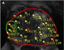
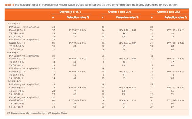
A two centre prospective outcome study of 487 patients with previous benign biopsies that underwent transperineal MRI/US fusion-guided targeted and systematic saturation biopsy from 2012 to 2015. Multiparametric MRI (mpMRI) was reported according to Prostate Imaging Reporting and Data System (PI-RADS) Version 1. Detection of Gleason score 7–10 prostate cancer on biopsy was the primary outcome. Positive (PPV) and negative (NPV) predictive values including 95% confidence intervals (95% CIs) were calculated. Detection rates of targeted and systematic biopsies were compared using McNemar’s test.

Results

The median (interquartile range) PSA level was 9.0 (6.7–13.4) ng/mL. PI-RADS 3–5 mpMRI lesions were reported in 343 (70%) patients and Gleason score 7–10 prostate cancer was detected in 149 (31%). The PPV (95% CI) for detecting Gleason score 7–10 prostate cancer was 0.20 (±0.07) for PI-RADS 3, 0.32 (±0.09) for PI-RADS 4, and 0.70 (±0.08) for PI-RADS 5. The NPV (95% CI) of PI-RADS 1–2 was 0.92 (±0.04) for Gleason score 7–10 and 0.99 (±0.02) for Gleason score ≥4 + 3 cancer. Systematic biopsies alone found 125/138 (91%) Gleason score 7–10 cancers. In patients with suspicious lesions (PI-RADS 4–5) on mpMRI, systematic biopsies would not have detected 12/113 significant prostate cancers (11%), while targeted biopsies alone would have failed to diagnose 10/113 (9%). In equivocal lesions (PI-RADS 3), targeted biopsy alone would not have diagnosed 14/25 (56%) of Gleason score 7–10 cancers, whereas systematic biopsies alone would have missed 1/25 (4%). Combination with PSA density improved the area under the curve of PI-RADS from 0.822 to 0.846.

Conclusion

In patients with high probability mpMRI lesions, the highest detection rates of Gleason score 7–10 cancer still required combined targeted and systematic MRI/US image-fusion; however, systematic biopsy alone may be sufficient in patients with equivocal lesions. Repeated prostate biopsies may not be needed at all for patients with a low PSA density and a negative mpMRI read by experienced radiologists.

Read more articles of the week

Editorial: Getting to the right biopsy in the right patient at the right time

Guidelines now recommend performing multiparametric MRI (mpMRI) and targeted prostate biopsies in men with a history of prior negative biopsy and continued concern for significant cancer. This new approach to prostate re-biopsy is aimed at improving prostate cancer detection. However, several important clinical factors may help clinicians’ fine-tune the process of repeated prostate biopsy. In this month’s issue of the BJUI, Hansen et al. [1] present a multicentre study of patients with prior negative TRUS biopsy undergoing MRI/TRUS-fusion transperineal biopsy.

In the study, 487 men undergo mpMRI and transperineal biopsy with detection of clinically significant (Gleason score 7–10) cancer as the primary outcome. Several factors are evaluated to compare cancer detection rates, including systematic biopsies, targeted biopsies, PSA density (PSAD), and Prostate Imaging Reporting and Data System (PI-RADS) version 1 score. From their cohort, a suspicious lesion (PIRADS 3–5) was identified in 343 (70%) patients. Prostate cancer was detected in 249 (51%), with 149 (31%) having Gleason score 7–10 cancer. Potentially missed significant cancers from the anterior prostate were found in 27% (40/149). Cancer was detected in 28% (40/144) of patients with PI-RADS 1–2 lesions, with 8% (11/144) being Gleason score 7–10. For patients with PI-RADS 3–5 lesions, cancer was identified in 61% (209/343) with 40% (138/343) being Gleason score 7–10. For patients with PI-RADS 3–5 lesions, systematic biopsies alone failed to detect 13/138 significant cancers, while targeted biopsies missed 24/138 cancers. The combination of systematic and targeted biopsies was significantly better for Gleason score 7–10 prostate cancer detection than either alone. The addition of a PSAD threshold of 0.15 ng/mL/mL for the detection of Gleason score 7–10 resulted in a significant improvement in the area under the curve (0.846) of the receiver operating characteristic curve for PSAD groups and PI-RADS score.

Getting the right biopsy: In this study [1], patients with a prior negative TRUS biopsy underwent TRUS-fusion transperineal biopsy. Having two approaches to prostate biopsy can be advantageous when evaluating men with prior negative biopsies. Historical studies have found comparable prostate cancer detection between transrectal and transperineal biopsies for men undergoing both initial biopsy [2] and saturation re-biopsy [3]. However, the detection of anterior lesions has remained a persistent challenge from the transrectal approach. As in the current study [1], use of transperineal biopsy can detect cancer in up to 30% of tumours that would otherwise be missed on extended template TRUS biopsy [4]. Although attempts to reach anterior lesions from the transrectal approach may be feasible [5], the transperineal approach is felt to provide better sampling in comparison [6].

Getting the right patient: Patient-specific factors such as PI-RADS lesions 3–5 and PSAD have become increasing utilised for stratifying patients who may benefit from additional biopsies using image guidance. As the authors suggest, patients with negative imaging may consider deferring repeat biopsy, particularly those with reassuring PSADs (<0.15 ng/mL/mL). In their study [1], only 4% (6/144) of men with negative mpMRI and a PSAD of <0.15 ng/mL/mL harboured clinically significant cancer (five Gleason score 3 + 4 and one Gleason score 8). Patients with concerning PSAD, but negative mpMRI and those with lesions identified in the peripheral zone could have the option to undergo repeated, fusion-directed TRUS or transperineal biopsy. For patients with lesions identified in the anterior prostate, a transperineal prostate biopsy may provide the highest detection rate.

At the right time: Now that high quality prostate MRI is becoming more widely available; men with a prior negative biopsy should strongly consider the benefit of repeated biopsy after prostate imaging. In addition to identifying suspicious lesions, calculating PSAD has been found to improve the likelihood of detecting clinically significant prostate cancer. Without additional testing, a personalised biopsy plan can be created.

A thorough discussion of the prescribed biopsy approach and the likelihood of detecting a significant cancer is the final step to the right biopsy in the right patient at the right time.

Kelly Stratton


Department of Urology, University of Oklahoma College of Medicine, Oklahoma City, OK, USA


Read the full article


1 Hansen NL, Kesch C, Barrett T et al. Multicentre evaluation of target and systematic biopsies using magnetic resonance and ultrasound image-fusion guided transperineal prostate biopsy in patients with a previous negative biopsy. BJU Int 2016; 120: 6318


2 Hara R, Jo Y, Fujii T et al. Optimal approach for prostate cancer detection as initial biopsy: prospective randomized study comparing transperineal versus transrectal systematic 12-core biopsy. Urology 2008; 71: 1915


3 Abdollah F, Novara G, Briganti A et al. Trans-rectal versus trans- perineal saturation rebiopsy of the prostate: is there a difference in cancer detection rate? Urology 2011; 77: 9215


4 KomaiY, Numao N, Yoshida S et al. High diagnostic ability of multiparametric magnetic re onance imaging to detect anterior prostate cancer miss ed by transrectal 1 2-core biopsy. JUrol2013; 190: 867 7


5 Volkin D, Turkbey B, Hoang AN et al. Multiparametric magnetic resonance imaging (MRI) and subsequent MRI/ultrasonography fusion-guided biopsy increase the detection of anteriorly located prostate cancers. BJU Int 2014; 114: E439


6 Borkowetz A, Platzek I, Toma M et al. Direct comparison of multiparametric magnetic resonance imaging (MRI) results with nal histopathology in patients with proven prostate cancer in MRI/ ultrasonography-fusion biopsy. BJU Int 2016; 118: 21320


© 2024 BJU International. All Rights Reserved.