Tag Archive for: #ProstateCancer

Posts

Article of the Week: Effect of MetS on serum PSA levels is concealed by enlarged prostate

Every Week the Editor-in-Chief selects an Article of the Week from the current issue of BJUI. The abstract is reproduced below and you can click on the button to read the full article, which is freely available to all readers for at least 30 days from the time of this post.

In addition to the article itself, there is an accompanying editorial written by a prominent member of the urological community. This blog is intended to provoke comment and discussion and we invite you to use the comment tools at the bottom of each post to join the conversation.

Finally, the third post under the Article of the Week heading on the homepage will consist of additional material or media. This week we feature a video discussing the paper.

If you only have time to read one article this week, it should be this one.

Actual lowering effect of metabolic syndrome on serum prostate-specific antigen levels is partly concealed by enlarged prostate: results from a large-scale population-based study

Sicong Zhao*, Ming Xia*, Jianchun Tang† and Yong Yan*


*Department of Urology, and Department of Cardiology, Beijing Shijitan Hospital, Capital Medical University, Beijing, China


Abstract

Objectives

To clarify the lowering effect of metabolic syndrome (MetS) on serum prostate-specific antigen (PSA) levels in a Chinese screened population.

Subjects and Methods

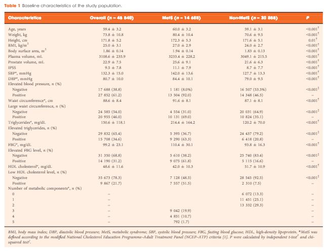
A total of 45 540 ostensibly healthy men aged 55–69 years who underwent routine health check-ups at Beijing Shijitan Hospital between 2008 and 2015 were included in the study. All the men underwent detailed clinical evaluations. PSA mass density was calculated (serum PSA level × plasma volume ÷ prostate volume) for simultaneously adjusting plasma volume and prostate volume. According to the modified National Cholesterol Education Programme–Adult Treatment Panel (NCEP-ATP) III criteria, patients were dichotomized by the presence of MetS, and differences in PSA density and PSA mass density were compared between groups. Linear regression analysis was used to evaluate the effect of MetS on serum PSA levels.

Results

When larger prostate volume in men with MetS was adjusted for, both PSA density and PSA mass density in men with MetS were significantly lower than in men without MetS, and the estimated difference in mean serum PSA level between men with and without MetS was greater than that before adjusting for prostate volume. In the multivariate regression model, the presence of MetS was independently associated with an 11.3% decline in serum PSA levels compared with the absence of MetS. In addition, increasing number of positive MetS components was significantly and linearly associated with decline in serum PSA levels.

Conclusion

The actual lowering effect of MetS on serum PSA levels was partly concealed by the enlarged prostate in men with MetS, and the presence of MetS was independently associated with lower serum PSA levels. Urologists need to be aware of the effect of MetS on serum PSA levels and should discuss this subject with their patients.

Article of the Week: 11C-acetate PET/CT imaging for detection of recurrent disease after RP or RT in patients with PCa

Every Week the Editor-in-Chief selects an Article of the Week from the current issue of BJUI. The abstract is reproduced below and you can click on the button to read the full article, which is freely available to all readers for at least 30 days from the time of this post.

In addition to the article itself, there is an accompanying editorial written by a prominent member of the urological community. This blog is intended to provoke comment and discussion and we invite you to use the comment tools at the bottom of each post to join the conversation.

If you only have time to read one article this week, it should be this one.

11C-acetate positron-emission tomography/computed tomography imaging for detection of recurrent disease after radical prostatectomy or radiotherapy in patients with prostate cancer

Lukas Hendrik Esch*, Melanie Fahlbusch, Peter Albers‡, Hubertus Hautzel§ and Volker Muller-Mattheis


*Department of Urology, St Antonius Hospital, Gronau, Department of Gynaecology, Bethesda Hospital, Duisburg, Departments of‡ Urology, and §Nuclear Medicine, Medical Faculty, Heinrich-Heine University, Dusseldorf, Germany


Read the full article

Abstract

Objectives

To evaluate, in a prospective study, the effectiveness of computed tomography (CT)-matched 11C-acetate (AC) positron-emission tomography (PET) in patients with prostate cancer (PCa) who had prostate-specific antigen (PSA) relapse after radical prostatectomy (RP) or radiotherapy (RT).

Patients and Methods

In 103 relapsing patients after RP (n = 97) or RT (n = 6) AC-PET images and CT scans were obtained. In patients with AC-PET-positive results with localized PCa recurrence, detected lesions were resected and histologically verified or, after local RT, followed-up by PSA testing. Patients with distant disease on AC-PET were treated with androgen deprivation/chemotherapy.

Results

Of 103 patients, 42 were AC-PET-positive. PSA levels were <1.0, <2.0 and <4.0 ng/mL in six, 16 and 20 patients, respectively. In 25/42 patients AC-PET suggested lymph node metastases: 16/25 patients underwent surgery (10/16 metastasis, 6/16 inflammation); 9/25 patients underwent RT of lymph node metastases, which was followed by decreasing PSA level. In 17/42 patients who had distant disease, systemic treatment was commenced. Combining patients who underwent surgery and those who underwent RT, 19/25 patients were true-positive in terms of AC-PET (positive predictive value 76%). In 5/19 patients, PSA level was <2.0 ng/mL, in 2/19 patients it was <1.0 ng/mL and in 14/19 patients it was 5.4–23.1 ng/mL. In AC-PET-positive patients after surgery or RT (without androgen deprivation), median (range) time to renewed PSA increase was 6 (5–9) months.

Conclusions

Only a minority of patients with relapsing PCa appear to benefit from AC-PET for guiding potential local treatment. False-positive results show that factors other than tumour metabolism induce increased AC uptake. The time free of recurrence after local treatment was shorter than expected.

Editorial: Is choline-based PET imaging still relevant in recurrent prostate cancer?

The search for the ideal imaging method to detect small metastatic deposits has largely remained elusive in the field of prostate cancer. However, functional positron emission tomography (PET)/CT is now guiding us in different directions. 11C-acetate PET/CT imaging was one of the first molecular imaging probes showing promise in clinical studies, along with 11C-choline, and more recently challenged by 68Ga-prostate-specific membrane antigen (PSMA) PET/CT [1-3]. So against this background, what does this new study by Esch et al. [4] presented here offer us?

Let us rewind and focus on where molecular imaging may have an impact in prostate cancer. In primary staging, the potential to accurately identify oligometastatic disease or even widespread metastatic disease before primary gland treatment is clearly advantageous. It can allow for wider treatment fields (extended lymphadenectomy or radiation fields), directed treatment of oligometastatic disease, placement into appropriate cytoreductive trials, and in some instances consideration of the use of earlier chemo-hormonal therapy. This study did not address this important clinical scenario [4]. Nor did it focus on primary diagnosis, another ‘holy grail’ for imaging more recently dominated by MRI [5].

The clinical question addressed in this study was whether 11C-acetate PET is able to detect distant metastatic disease in men with PSA relapse after having undergone radical prostatectomy or prostate bed radiotherapy [4]. In other words, if distant metastatic disease is found then prostate bed radiation may be futile, although this assumption has not been subjected to high quality trials, but would seem logical. Also, in the era of oligometastatic disease, treatment (surgery and/or radiation) may be directed at nodal and other deposits to prolong time of systemic therapy such as androgen-deprivation therapy. So what is the ‘sweet spot’? Some would state for men having undergone surgery a PSA level of 0.2 ng/mL places the patient at risk of recurrence, although in clinical practice the level at which investigations are warranted can be as high as 1.0 ng/mL. This study found 11C-acetate PET had few positive results below a PSA level of 1.0 ng/mL. This is in contrast to a recent meta-analysis of 68Ga-PSMA PET/CT reporting positive studies at PSA levels of ≤0.2 ng/mL in 42% of patients [6].

The other group studied the detection of recurrent disease in men having undergone primary radiotherapy, although the numbers were low (six patients). We know this group of men are often undertreated for salvage treatment, and frequently placed on hormonal therapy. Again, early knowledge of locoregional or distant recurrence is required to allow best case selection and thus avoid futile salvage surgery, and also to know who may benefit from salvage lymphadenectomy and treatment of distant oligometastatic disease, if required.

The false-positive rate result for 11C-acetate PET/CT of 24% is notable and concerning. This may lead to unnecessary intervention, or even withholding prostate bed radiation that may have been of benefit. In contrast the false-positive rate for 11C-choline and 68Ga-PSMA PET/CT is far lower, although false-positive PSMA studies may occur in benign conditions and non-prostate cancer tumours.

It should be acknowledged that a strength of this study was comparative histology in a proportion of patients, allowing true sensitivity and specificity to be determined [4]. This is important information, as it guides correct decision-making, and such information is lacking for many other prostate cancer imaging probes, including 68Ga-PSMA, where these data are urgently required.

Overall, this study is important and adds to the rich milieu of available molecular imaging data on staging of possible recurrent or metastatic prostate cancer to date [4]. Current prospective trials exploring 11C-choline, 18F-FACBC (anti-1-amino-3-18F-fluorocyclobutane-1-carboxylic acid), 18F-choline and 68Ga-PSMA with MRI and clinical outcomes should provide further insight into the most appropriate molecular imaging technique for staging prostate cancer.

Nathan Lawrentschuk, BJUI USANZ Editor*,,, Julia M. Coreldand Andrew Scott,§,¶
*Department of Surgery, University of Melbourne and Olivia Newton-John Cancer Research Institute, Austin Hospital , Peter MacCallum Cancer Centre, La Trobe University, Departments of


Medicine, § Molecular Imaging and Therapy
Read the full article


References


1 Afshar-Oromieh A, Zechmann CM, Malcher A et al. Comparison of PET imaging with a (68)Ga-labelled PSMA ligand and (18)F-choline- based PET/CT for the diagnosis of recurrent prostate cancer. Eur J Nucl Med Mol Imaging 2014; 41: 1120


2 Silver DA, Pellicer I, Fair WR, Heston WD, Cordon-Cardo C. Prostate- specic membrane antigen expression in normal and malignant human tissues. Clin Cancer Res 1997; 3: 815


5 Johnson LM, Turkbey B, Figg WD, Choyke PL. Multiparametric MRI in prostate cancer management. Nat Rev Clin Oncol 2014; 11: 34653


Residents’ Podcast: sRPLND+PLND for ‘node-only’ recurrent PCa

Jesse Ory, Kyle Lehmann and Jeff Himmelman

Department of Urology, Dalhousie University
Halifax, NS, Canada

Read the full article

Abstract

Objectives

To describe the technique of robot-assisted high-extended salvage retroperitoneal and pelvic lymphadenectomy (sRPLND+PLND) for ‘node-only’ recurrent prostate cancer.

Patients and Methods

In all, 10 patients underwent robot-assisted sRPLND+PLND (09/2015–03/2016) for ‘node-only’ recurrent prostate cancer, as identified by 11C-acetate positron emission tomography/computed tomography imaging. Our anatomical template extends from bilateral renal artery/vein cranially up to Cloquet’s node caudally, completely excising lymphatic-fatty tissue from aorto-caval and iliac vascular trees; RPLND precedes PLND. Meticulous node-mapping assessed nodes at four prospectively assigned anatomical zones.

Results

The median operative time was 4.8 h, estimated blood loss 100 mL and hospital stay 1 day. No patient had an intraoperative complication, open conversion or blood transfusion. Three patients had spontaneously resolving Clavien–Dindo grade II postoperative complications. The mean (range) number of nodes excised per patient was 83 (41–132) and mean (range) number of positive nodes per patient was 23 (0–109). Seven patients (70%) had positive nodes on final pathology. Node-positive rates per anatomical level I, II, III and IV were 28%, 32%, 33% and 33%, respectively. In patients with positive nodes, the median PSA level had decreased by 83% at the 2-month follow-up.

Conclusion

The initial series of robot-assisted sRPLND+PLND is presented, wherein we duplicate open surgery with superior nodal counts and decreased morbidity. Robot-assisted technical details for an anatomical LND template up to the renal vessels are presented. Longer follow-up is necessary to assess oncological outcomes.

BJUI Podcasts now available on iTunes, subscribe here https://itunes.apple.com/gb/podcast/bju-international/id1309570262

Article of the Week: sRPLND+PLND for ‘node-only’ recurrent PCa

Every Week the Editor-in-Chief selects an Article of the Week from the current issue of BJUI. The abstract is reproduced below and you can click on the button to read the full article, which is freely available to all readers for at least 30 days from the time of this post.

In addition to the article itself, there is an accompanying editorial written by a prominent member of the urological community. This blog is intended to provoke comment and discussion and we invite you to use the comment tools at the bottom of each post to join the conversation.

If you only have time to read one article this week, it should be this one.

Robotic salvage retroperitoneal and pelvic lymph node dissection for ‘node-only’ recurrent prostate cancer: technique and initial series

Andre Abreu*, Carlos Fay*, Daniel Park*, David Quinn*, Tanya Dorff*,John Carpten*, Peter Kuhn*, Parkash Gill*, Fabio Almeida* and Inderbir Gill*


*University of Southern California (USC) Institute of Urology, Catherine & Joseph Aresty Department of Urology, Keck School of Medicine, USC, Los Angeles, CA, USA, and Pontical Catholic University of Rio Grande do Sul, Porto Alegre, Brazil


Read the full article

Abstract

Objectives

To describe the technique of robot-assisted high-extended salvage retroperitoneal and pelvic lymphadenectomy (sRPLND+PLND) for ‘node-only’ recurrent prostate cancer.

Patients and Methods

In all, 10 patients underwent robot-assisted sRPLND+PLND (09/2015–03/2016) for ‘node-only’ recurrent prostate cancer, as identified by 11C-acetate positron emission tomography/computed tomography imaging. Our anatomical template extends from bilateral renal artery/vein cranially up to Cloquet’s node caudally, completely excising lymphatic-fatty tissue from aorto-caval and iliac vascular trees; RPLND precedes PLND. Meticulous node-mapping assessed nodes at four prospectively assigned anatomical zones.

Results

The median operative time was 4.8 h, estimated blood loss 100 mL and hospital stay 1 day. No patient had an intraoperative complication, open conversion or blood transfusion. Three patients had spontaneously resolving Clavien–Dindo grade II postoperative complications. The mean (range) number of nodes excised per patient was 83 (41–132) and mean (range) number of positive nodes per patient was 23 (0–109). Seven patients (70%) had positive nodes on final pathology. Node-positive rates per anatomical level I, II, III and IV were 28%, 32%, 33% and 33%, respectively. In patients with positive nodes, the median PSA level had decreased by 83% at the 2-month follow-up.

Conclusion

The initial series of robot-assisted sRPLND+PLND is presented, wherein we duplicate open surgery with superior nodal counts and decreased morbidity. Robot-assisted technical details for an anatomical LND template up to the renal vessels are presented. Longer follow-up is necessary to assess oncological outcomes.


Editorial: sLND – if yes, Robotics?

The manuscript in this issue of the BJUI by de Castro Abreu et al. [1] reports the results of the first series of patients to undergo robotic-assisted salvage lymph node dissection (sLND) for nodal recurrence of prostate cancer. Despite the absence of a high level of evidence, sLND has been gaining attention in recent years. Indeed, several series have shown promising results of such an approach, especially in terms of PSA response to surgery and delay in clinical recurrence [2-4]. However, sLND is a complex surgery and is not devoid of complications, as in up to 13.8% of patients Clavien–Dindo ≥IIIa complications occur [5]. When analysing the results of the current manuscript [1], it is impressive to read that the mean number of LNs removed was 83, ranging from 41 to 132, which is significantly higher than the reported figures of open sLND series. Moreover, despite the long median operative time (4.8 h), complication rates and postoperative course were excellent as compared to previously published series, although a direct comparison between the open and robot-assisted approach should be only addressed in prospective studies. The authors should be congratulated on the superb results obtained during the learning curve of such complex surgery, but some issues need to be discussed.

First, it is difficult to understand whether such results apply only to very expert surgeons. In other words, is it possible to translate such surgery to less experienced robotic surgeons? Second, is it necessary to extend the LND to a similar extent in all cases? Previous reports have shown that patients with retroperitoneal involvement may not benefit from sLND as much as their counterparts with only pelvic involvement [2]. The authors [1] show no significant impact of such an extended approach on complications and postoperative course, but the invasiveness of such an extended approach needs to be justified in each case. Third, the introduction of new tracer methods, such as prostate-specific membrane antigen (PSMA) positron emission tomography/CT, with higher specificity for prostate cancer may reduce the need for such extended templates, without compromising the oncological results [6]. Fourth, is the robotic approach feasible and safe in patients previously submitted to radical prostatectomy independently from the approach (open vs laparoscopic/robotic), from the extent of the previous LND, as well as from the previous administration of adjuvant/salvage radiotherapy? All these answers will need to be addressed in future studies on subgroups of patients undergoing sLND. Most importantly, until a high level of evidence is available, sLND should still be considered experimental and should be performed by highly experienced surgeons.

Read the full article
Nazareno Suardi and Francesco Montorsi
Department of Urology, Urological Research Institute, Vita Salute San Raffaele University, Milan, Italy


References

Article of the Week: Sentinel node biopsy for prostate cancer: report from a consensus panel meeting

Every Week the Editor-in-Chief selects an Article of the Week from the current issue of BJUI. The abstract is reproduced below and you can click on the button to read the full article, which is freely available to all readers for at least 30 days from the time of this post.

In addition to the article itself, there is an accompanying editorial written by a prominent member of the urological community. This blog is intended to provoke comment and discussion and we invite you to use the comment tools at the bottom of each post to join the conversation.

If you only have time to read one article this week, it should be this one.

Sentinel node biopsy for prostate cancer: report from a consensus panel meeting

Henk G. van der Poel* , Esther M. Wit*, Cenk Acar, Nynke S. van den Berg,Fijs W. B. van Leeuwen, Renato A. Valdes Olmos, Alexander Winter§,Friedhelm Wawroschek§, Fredrik Liedberg**, Steven Maclennan††and Thomas Lam†† On behalf of the Sentinel Node Prostate Cancer Consensus Panel Group members 


*Department of Urology, Netherlands Cancer Institute, Amsterdam, The Netherlands, Department of Urology, Eryaman Hospital, Ankara, Turkey, Department of Radiology, University of Leiden Medical Centre, Leiden, The Netherlands, §Klinikum Oldenburg, School of Medicine and Health Sciences, University Hospital for Urology, Oldenburg, Germany, Department of Urology, Skane University Hospital, Malmo, **Department of Translational Medicine Lund University, Lund, Sweden, and ††Academic Urology Unit, University of Aberdeen, Aberdeen, UK


Read the full article

Abstract

Objective

To explore the evidence and knowledge gaps in sentinel node biopsy (SNB) in prostate cancer through a consensus panel of experts.

Methods

A two-round Delphi survey among experts was followed by a consensus panel meeting of 16 experts in February 2016. Agreement voting was performed using the research and development project/University of California, Los Angeles Appropriateness Methodology on 150 statements in nine domains. The disagreement index based on the interpercentile range, adjusted for symmetry score, was used to assess consensus and non-consensus among panel members.

aotw-aug-2017-4

Results

Consensus was obtained on 91 of 150 statements (61%). The main outcomes were: (1) the results from an extended lymph node dissection (eLND) are still considered the ‘gold standard’, and sentinel node (SN) detection should be combined with eLND, at least in patients with intermediate- and high-risk prostate cancer; (2) the role of SN detection in low-risk prostate cancer is unclear; and (3) future studies should contain oncological endpoints as number of positive nodes outside the eLND template, false-negative and false-positive SN procedures, and recurrence-free survival. A high rate of consensus was obtained regarding outcome measures of future clinical trials on SNB (89%). Consensus on tracer technology was only obtained in 47% of statements, reflecting a need for further research and standardization in this area. The low-level evidence in the available literature and the composition of mainly SNB users in the panel constitute the major limitations of the study.

Conclusions

Consensus on a majority of elementary statements on SN detection in prostate cancer was obtained.; therefore, the results from this consensus report will provide a basis for the design of further studies in the field. A group of experts identified evidence and knowledge gaps on SN detection in prostate cancer and its application in daily practice. Information from the consensus statements can be used to direct further studies.

Editorial: Sentinel nodes in prostate cancer– are we chasing a ghost?

Van der Poel et al. [1] report the findings of a consensus panel meeting on sentinel node (SN) biopsy at the time of radical prostatectomy. The consensus process was based on a two-round Delphi survey on the current evidence and knowledge gaps on SN detection. Then, a number of experts among those who answered the survey were invited to further discuss these issues during a consensus meeting.

The authors are to be complimented on their effort to establish standard definitions, techniques, and reporting of outcomes of SN detection. Their results push the field forward and will make it possible to compare future data between institutions, individual surgeons, and techniques of SN detection. Nevertheless, although the authors admittedly represented a group of potentially biased experts, current limitations of SN detection were fairly acknowledged.

One thorny issue remains, the definition of SN and whether a true SN exists or not. The SN concept implies that one should reliably find tumour in the first echelon of drainage when tumour is present. The SN concept further assumes that if the SN is free of tumour, then so are the next drainage stations. Does that really happen in prostate cancer? We know from previous mapping studies that primary lymphatic landing sites of the prostate are heterogeneously localised, that drainage varies from site to site of tracer injection (and thus tumour location), and that drainage varies from patient to patient [2, 3]. Thus, prostate cancer does not fit the Halstedian paradigm of a pre-defined, stepwise, uniform pathway of metastatic spread. An additional setback of SN detection is that lymph nodes that contain a large tumour burden often fail to take up the tracer [2, 4]. One might critically argue that this limitation may be dependent on the tracer used. The consensus group could not conclusively agree on which tracer technology to use. Looking forward, imaging probes that target tumour-specific molecules may improve tumour detection during pelvic lymph node dissection (PLND). The prostate-specific membrane antigen (PSMA) represents a particularly promising marker in prostate cancer imaging. However, using 68Ga-PSMA-positron emission tomography/CT for the detection of lymph node metastases before radical prostatectomy, one group failed to observe the expected improvement and reported only 33% sensitivity [5]. Future studies will determine whether these advances in prostate cancer detection will translate into more precise targeted dissection of lymph node metastases.

Altogether, the consensus group concluded that extended PLND should remain the standard of care, at least in patients with intermediate- and high-risk prostate cancer. This conclusion is laudable, but to be fair, we have no level 1 evidence for this either. However, every oncology-oriented surgeon would agree that each potential positive lymph node should be found and removed during surgery with curative intent. In case of true low-risk prostate cancer, this is probably less of an issue, but it should be mentioned that this population is at very low risk of dying from prostate cancer, even if left untreated, and the indication for radical prostatectomy should be questioned rather than that for PLND. Furthermore, pathological Gleason score is underestimated in preoperative biopsies in ~30% of all cases [6], making the decision to perform PLND or not in low-risk disease difficult.

Again, the authors should be complimented for their thorough work. We still do not know whether the SN exists, but performing surgery with image-guidance provides quality control for completeness of resection and might help detect (positive?) lymph nodes outside of the extended PLND template. Indeed, up to 35% of prostate lymphatic drainage sites may remain outside the extended anatomical template [3]. SN detection might also teach us when and where to stop dissection to decrease potential morbidity of PLND without leaving tumour behind. Thus, chasing the ghost of the SN has made and makes us better and more meticulous cancer surgeons. At the same time, it is humbling that we have been chasing a ghost for so long without bringing up level 1 evidence.

George N. Thalmann and Daniel P. Nguyen


Department of Urology, University Hospital Bern, Bern, Switzerland


Read the full article


References

1 van der Poel HG, Wit EM, Acar C et al. Sentinel node biopsy for prostate cancer: report from a consensus panel meeting. BJU Int 2017; 120: 20411


4 Weckermann D, Dorn R, Holl G, Wagner T, Harzmann R. Limitations of radioguided surgery in high-risk prostate cancer. Eur Urol 2007; 51: 154958


Article of the Week: Impact of 68Ga-PSMA PET/CT in PCa with rising PSA

Every Week the Editor-in-Chief selects an Article of the Week from the current issue of BJUI. The abstract is reproduced below and you can click on the button to read the full article, which is freely available to all readers for at least 30 days from the time of this post.

In addition to the article itself, there is an accompanying editorial written by a prominent member of the urological community. This blog is intended to provoke comment and discussion and we invite you to use the comment tools at the bottom of each post to join the conversation.

If you only have time to read one article this week, it should be this one.

Clinical impact of 68Ga-prostate-specific membrane antigen (PSMA) positron emission tomography/computed tomography (PET/CT) in patients with prostate cancer with rising prostate-specific antigen after treatment with curative intent: preliminary analysis of a multidisciplinary approach


Simone Albisinni*, Carlos Artigas, Fouad Aoun*, Ibrahim Biaou*, Julien Grosman*, Thierry Gil, Eric Hawaux*, Ksenija Limani*, Francois-Xavier Otte§, Alexandre Peltier*, Spyridon Sideris, Nicolas Sirtaine, Patrick Flamen† and Roland van Velthoven*


Departments of *Urology, Nuclear Medicine, Oncology, §Radiation Oncology, and Pathology, Institut Jules Bordet, Universite Libre de Bruxelles, Brussels, Belgium


Read the full article

How to Cite

Albisinni, S., Artigas, C., Aoun, F., Biaou, I., Grosman, J., Gil, T., Hawaux, E., Limani, K., Otte, F.-X., Peltier, A., Sideris, S., Sirtaine, N., Flamen, P. and van Velthoven, R. (2017), Clinical impact of 68Ga-prostate-specific membrane antigen (PSMA) positron emission tomography/computed tomography (PET/CT) in patients with prostate cancer with rising prostate-specific antigen after treatment with curative intent: preliminary analysis of a multidisciplinary approach. BJU International, 120: 197–203. doi: 10.1111/bju.13739

Abstract

Objective

To assess the impact of a novel molecular imaging technique, 68Ga-(HBED-CC)-prostate-specific membrane antigen (PSMA) positron emission tomography/computed tomography (PET/CT), in the clinical management of patients with prostate cancer with rising prostate-specific antigen (PSA) after treatment with curative intent.

Patients and Methods

In all, 131 consecutive patients were referred to our centre for a 68Ga-PSMA PET/CT in the setting of recurring prostate cancer. Of these patients, 11/131(8%) presented with persistent PSA after radical prostatectomy, while 120/131 (92%) were referred for biochemical recurrence after surgery, radiotherapy or both. The images where taken 1 h after injection of 2 MBq/kg of the 68Ga-(HBED-CC)-PSMA ligand. All examinations were interpreted by two experienced nuclear medicine specialists. Using the results of the examination, a multidisciplinary oncology committee (MOC) reported on the treatment strategy. A positive impact on clinical management was considered if the examination determined a modification in the treatment strategy compared to the MOC decision before PSMA imaging.

aotw-aug-2017-3

Results

All patients completed the examination with no adverse reactions. The median (interquartile range) PSA level at the time of the examination was 2.2 (0.72–6.7) ng/mL. Overall, 68Ga-PSMA PET/CT detected at least one lesion suspicious for prostate cancer in 98/131 (75%) patients. There was an impact on subsequent management in 99/131 patients (76%). The main modifications included continuing surveillance (withholding hormonal therapy), hormonal manipulations, stereotaxic radiotherapy, salvage radiotherapy, salvage node dissection or salvage local treatment (prostatectomy, high-intensity focussed ultrasound).

Conclusion

Our preliminary experience suggests that performing 68Ga-PSMA PET/CT in patients with prostate cancer with rising PSA after treatment with curative intent can be clinically useful as it changes the treatment strategy in a significant proportion of patients. However, larger prospective trials are needed to validate our present findings.


Editorial: Defining the clinical utility of PSMA-targeted PET imaging of prostate cancer

In the field of oncology, positron emission tomography (PET) is most commonly performed using 2-deoxy-2-[18F]fluoro-d-glucose (18F-FDG), a radiofluorinated glucose analogue that accumulates in cells undergoing aerobic glycolysis. Unfortunately, because of the low glycolytic activity of hormone-naïve prostate cancer cells, 18F-FDG PET has been of little value in imaging men with this malignancy [1]. Instead, clinicians have been left to rely mostly on 99mTc-methylene diphosphonate bone scan, CT, and MRI to stage and follow patients. Recently, however, the development of multiple urea-based small molecules targeting the type II transmembrane glycoprotein prostate-specific membrane antigen (PSMA) has allowed for the highly sensitive and specific detection of prostate cancer using PET imaging [2]. To date, the majority of clinical data with PSMA-targeted PET have been generated with the 68Ga-PSMA-11 radiotracer (also known as 68Ga-PSMA-HBED-CC). Notably, studies evaluating PSMA-targeted PET have mostly focused on establishing the diagnostic performance characteristics of the various radiotracers (e.g. sensitivity and specificity), with relatively few reports exploring the clinical impact or utility of this form of molecular imaging.

In this month’s edition of BJUI, Albisinni et al. [3] aimed to look beyond the performance characteristics of 68Ga-PSMA-11 PET/CT and retrospectively analysed the impact of this imaging test on the management of 131 men with a persistently elevated PSA level or biochemical recurrence after local treatment of their prostate cancer with curative intent. Of these patients, 106 (81%) had undergone a previous radical prostatectomy. The authors defined clinical utility as any imaging finding (or lack thereof) leading to a change in a patient’s pre-PET treatment plan. In total, 68Ga-PSMA-11 PET/CT demonstrated clinical utility in 76% of imaged patients. Most commonly, the results of this imaging test led to avoidance of androgen deprivation therapy (44% of all patients imaged) in place of an alternative management strategy, such as surveillance or salvage radiation therapy. Another notable finding was that among men who had planned to undergo salvage radiation therapy prior to 68Ga-PSMA-11 PET/CT, the majority (19 of 32 [59%]) were managed with an alternative approach after undergoing imaging.

Albisinni et al. [3] are not alone in their observations regarding the high clinical utility of 68Ga-PSMA-11 PET. For example, van Leeuwen et al. [4] previously reported that nearly 30% of men who were felt to be candidates for post-prostatectomy salvage radiation therapy had findings on 68Ga-PSMA-11 PET/CT that led to a major change in management. Additionally, Sterzing et al. [5] found that approximately 50% of patients undergoing radiation therapy planning for primary or recurrent prostate cancer experienced a change to their treatment concept after imaging with 68Ga-PSMA-11 PET/CT. Combined, these data suggest that a substantial proportion of men with prostate cancer stand to have their management altered by undergoing PSMA-targeted PET imaging.

While encouraging, the study by Albisinni et al. is somewhat limited by its retrospective design [3]. An outstanding example of how data on the clinical utility of an imaging test can be prospectively collected comes to us from the National Oncology PET Registry (NOPR) in the USA. Working in collaboration with the Centers for Medicare and Medicaid Services (CMS), NOPR was established to assess the question of clinical utility related to 18F-FDG PET/CT. To measure clinical utility, NOPR required physicians to complete questionnaires assessing the indication for imaging as well as pre- and post-PET treatment plans. In a 2008 study from NOPR incorporating data from 40 863 18F-FDG studies performed at 1368 centres, it was reported that 38% of patients experienced a change in intended management as a result of this imaging test [6]. In light of these and other data from NOPR, 18F-FDG PET/CT is now widely used across a range of tumour histologies. Moreover, this imaging study is readily reimbursed by both the CMS and private insurers.

In summary, we are delighted by the results of Albisinni et al. [3] and look forward to other prospective studies (for example ClinicalTrials.gov identifier NCT02825875) that aim to define the clinical utility of PSMA-targeted PET imaging of prostate cancer.

Michael A. Gorin,* Martin G. PomperKenneth J. Pienta* and Steven P. Rowe


*The James Buchanan Brady Urological Institute and Department of Urology, and Russell H. Morgan Department of Radiology and Radiological Science, Johns Hopkins University School of Medicine, Baltimore, MD, USA


Read the full article


References


© 2024 BJU International. All Rights Reserved.